Department of Pathology, Kunming Children's Hospital, 288 Qianxing Road, Yunnan, 650028, Kunming, China.
Second People's Hospital of Yunnan Province, 176 Qingnian Road, Yunnan, 650034, Kunming, China.
BMC Pediatr. 2021 Mar 11;21(1):121. doi: 10.1186/s12887-021-02590-7.
Malignant melanoma (MM) arises predominantly after adolescence and is uncommon in children. Congenital MM in newborns is even rarer with a dearth of published literature; as a consequence, there is no uniform standard for the pathogenesis and treatment for neonatal malignant melanoma. Herein we report a case of giant congenital nodular MM in a newborn, including its clinical, imaging, pathological and molecular pathological features. This case is the largest giant congenital primary nodular malignant melanoma in utero in neonates currently reported in China.
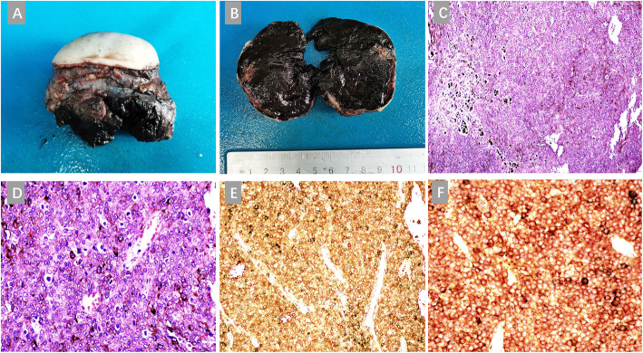
A female neonatal patient was found to have a 2.97 cm× 1.82 cm×1.50 cm mass with a clear boundary at the right acromion in color Doppler ultrasound examination at 24 weeks of gestation. The mass increased to 3.0 cm×5.0 cm×9.0 cm at birth, and local ulceration was seen. MRI demonstrated that the mass was located on the right shoulder and underarm in a lobulated appearance, and surrounded the right scapula which was deformed. Clinical stage:IV(AJCC 8th Edition (2017)). α-Fetoprofein (AFP) by hematological examination: 1210ng/ml, NSE: 21.28ng/ml, LDH: 842U/L. The patient underwent surgical resection of the tumor, and was pathologically diagnosed as neonatal congenital malignant melanoma; immunohistochemistry (IHC): S-100 (+), HMB45 (+), Melan A (+), and Tyrosinase (+). Molecular pathological examination for BRAF V600E showed no mutations (Quantitative Real-time PCR, qPCR); And so were NRAS, C-kit (exons 9,11,13,14,17,18), and TERT (promoter locus, C228T and C250T) (Sanger sequencing). Non-surgical therapies were not carried out after the surgical resection of the tumor. After 6 months of follow-up, the child developed normally, and color Doppler ultrasound showed no obvious tumor growth or abnormality in the original tumor site.
It is extremely rare to see giant congenital primary nodular MM in utero in neonates. The pathogenesis, treatment and prognosis of congenital MM need further research. The diagnosis mainly depends on histopathology and immunohistochemistry, and it needs to be differentiated from malignant lymphoma and primitive neuroectodermal tumor. The current treatment strategy for MM relies on the surgical excision of the mass. Research directed at molecular detection for genetic mutations would contribute to targeted therapy and better prognosis.
恶性黑色素瘤(MM)主要发生在青春期后,在儿童中罕见。新生儿先天性 MM 更为罕见,文献报道也很少;因此,新生儿恶性黑色素瘤的发病机制和治疗尚无统一标准。本文报道了一例新生儿巨大先天性结节性 MM,包括其临床、影像学、病理学和分子病理学特征。该病例是目前中国报道的最大的先天性原发性结节性恶性黑色素瘤。
一名女性新生儿在妊娠 24 周的彩色多普勒超声检查中发现右侧肩峰处有一个 2.97cm×1.82cm×1.50cm 的边界清晰的肿块。出生时肿块增大至 3.0cm×5.0cm×9.0cm,局部出现溃疡。MRI 显示肿块位于右肩部和腋窝,呈分叶状,包绕变形的右肩胛骨。临床分期:IV(AJCC 8 版(2017 年))。血液检查甲胎蛋白(AFP):1210ng/ml,NSE:21.28ng/ml,LDH:842U/L。患者行肿瘤切除术,病理诊断为新生儿先天性恶性黑色素瘤;免疫组化(IHC):S-100(+),HMB45(+),Melan A(+),酪氨酸酶(+)。BRAF V600E 分子病理学检查未见突变(实时荧光定量 PCR,qPCR);NRAS、C-kit(外显子 9、11、13、14、17、18)和 TERT(启动子位点,C228T 和 C250T)也未见突变(Sanger 测序)。肿瘤切除后未进行非手术治疗。术后 6 个月随访,患儿发育正常,彩色多普勒超声未见原肿瘤部位明显肿瘤生长或异常。
新生儿先天性巨大原发性结节性 MM 极为罕见。先天性 MM 的发病机制、治疗和预后需要进一步研究。诊断主要依赖于组织病理学和免疫组织化学,需要与恶性淋巴瘤和原始神经外胚层肿瘤相鉴别。目前 MM 的治疗策略依赖于肿块的手术切除。针对基因突变的分子检测研究将有助于靶向治疗和改善预后。