• 文献检索
  • 文档翻译
  • 深度研究
  • 学术资讯
  • Suppr Zotero 插件Zotero 插件
  • 邀请有礼
  • 套餐&价格
  • 历史记录
应用&插件
Suppr Zotero 插件Zotero 插件浏览器插件Mac 客户端Windows 客户端微信小程序
定价
高级版会员购买积分包购买API积分包
服务
文献检索文档翻译深度研究API 文档MCP 服务
关于我们
关于 Suppr公司介绍联系我们用户协议隐私条款
关注我们

Suppr 超能文献

核心技术专利:CN118964589B侵权必究
粤ICP备2023148730 号-1Suppr @ 2026

文献检索

告别复杂PubMed语法,用中文像聊天一样搜索,搜遍4000万医学文献。AI智能推荐,让科研检索更轻松。

立即免费搜索

文件翻译

保留排版,准确专业,支持PDF/Word/PPT等文件格式,支持 12+语言互译。

免费翻译文档

深度研究

AI帮你快速写综述,25分钟生成高质量综述,智能提取关键信息,辅助科研写作。

立即免费体验

主动脉狭窄的超声心动图评估:英国超声心动图学会实用指南

Echocardiographic assessment of aortic stenosis: a practical guideline from the British Society of Echocardiography.

作者信息

Ring Liam, Shah Benoy N, Bhattacharyya Sanjeev, Harkness Allan, Belham Mark, Oxborough David, Pearce Keith, Rana Bushra S, Augustine Daniel X, Robinson Shaun, Tribouilloy Christophe

机构信息

West Suffolk Hospital NHS Foundation Trust, Bury St Edmunds, UK.

University Hospital Southampton NHS Foundation Trust, Southampton, UK.

出版信息

Echo Res Pract. 2021 Apr 28;8(1):G19-G59. doi: 10.1530/ERP-20-0035.

DOI:10.1530/ERP-20-0035
PMID:33709955
原文链接:https://pmc.ncbi.nlm.nih.gov/articles/PMC8115410/
Abstract

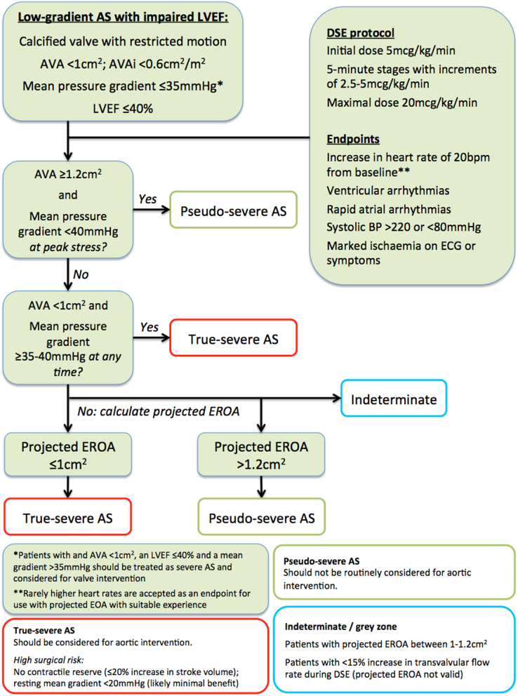
The guideline provides a practical step-by-step guide in order to facilitate high-quality echocardiographic studies of patients with aortic stenosis. In addition, it addresses commonly encountered yet challenging clinical scenarios and covers the use of advanced echocardiographic techniques, including TOE and Dobutamine stress echocardiography in the assessment of aortic stenosis.

摘要

该指南提供了一份实用的循序渐进指南,以促进对主动脉瓣狭窄患者进行高质量的超声心动图检查。此外,它还涉及常见但具有挑战性的临床情况,并涵盖了先进超声心动图技术的应用,包括经食管超声心动图(TOE)和多巴酚丁胺负荷超声心动图在主动脉瓣狭窄评估中的应用。

https://cdn.ncbi.nlm.nih.gov/pmc/blobs/8865/8115410/7fc5be4a607a/ERP-20-0035fig17.jpg
https://cdn.ncbi.nlm.nih.gov/pmc/blobs/8865/8115410/699449f5298b/ERP-20-0035fig1.jpg
https://cdn.ncbi.nlm.nih.gov/pmc/blobs/8865/8115410/aa9ce9af8d2c/ERP-20-0035fig2.jpg
https://cdn.ncbi.nlm.nih.gov/pmc/blobs/8865/8115410/ef567c646aab/ERP-20-0035fig3.jpg
https://cdn.ncbi.nlm.nih.gov/pmc/blobs/8865/8115410/94faac3afd9c/ERP-20-0035fig4.jpg
https://cdn.ncbi.nlm.nih.gov/pmc/blobs/8865/8115410/baf5ae456d56/ERP-20-0035fig5.jpg
https://cdn.ncbi.nlm.nih.gov/pmc/blobs/8865/8115410/8779c99791b4/ERP-20-0035fig6.jpg
https://cdn.ncbi.nlm.nih.gov/pmc/blobs/8865/8115410/094c08a37322/ERP-20-0035fig7.jpg
https://cdn.ncbi.nlm.nih.gov/pmc/blobs/8865/8115410/fb359f8d4c48/ERP-20-0035fig8.jpg
https://cdn.ncbi.nlm.nih.gov/pmc/blobs/8865/8115410/b6ceae1f0287/ERP-20-0035fig9.jpg
https://cdn.ncbi.nlm.nih.gov/pmc/blobs/8865/8115410/6e8a838b4db9/ERP-20-0035fig10.jpg
https://cdn.ncbi.nlm.nih.gov/pmc/blobs/8865/8115410/a52c7de25c78/ERP-20-0035fig11.jpg
https://cdn.ncbi.nlm.nih.gov/pmc/blobs/8865/8115410/6adc84937e6c/ERP-20-0035fig12.jpg
https://cdn.ncbi.nlm.nih.gov/pmc/blobs/8865/8115410/cb22df2b3738/ERP-20-0035fig13.jpg
https://cdn.ncbi.nlm.nih.gov/pmc/blobs/8865/8115410/e2c12108ff62/ERP-20-0035fig14.jpg
https://cdn.ncbi.nlm.nih.gov/pmc/blobs/8865/8115410/4041cf9bca77/ERP-20-0035fig15.jpg
https://cdn.ncbi.nlm.nih.gov/pmc/blobs/8865/8115410/13effa3b581f/ERP-20-0035fig16.jpg
https://cdn.ncbi.nlm.nih.gov/pmc/blobs/8865/8115410/7fc5be4a607a/ERP-20-0035fig17.jpg

相似文献

1
Echocardiographic assessment of aortic stenosis: a practical guideline from the British Society of Echocardiography.主动脉狭窄的超声心动图评估:英国超声心动图学会实用指南
Echo Res Pract. 2021 Apr 28;8(1):G19-G59. doi: 10.1530/ERP-20-0035.
2
Echocardiographic Evaluation of Aortic Stenosis - Normal Flow and Low Flow Scenarios.主动脉瓣狭窄的超声心动图评估——正常血流和低血流情况
Eur Cardiol. 2014 Dec;9(2):92-99. doi: 10.15420/ecr.2014.9.2.92.
3
Role of echocardiography in the diagnosis and treatment of patients with aortic stenosis.超声心动图在主动脉瓣狭窄患者诊断和治疗中的作用。
Curr Opin Cardiol. 2003 Sep;18(5):327-33. doi: 10.1097/00001573-200309000-00001.
4
The assessment of mitral valve disease: a guideline from the British Society of Echocardiography.二尖瓣疾病的评估:英国超声心动图学会指南
Echo Res Pract. 2021 Sep 27;8(1):G87-G136. doi: 10.1530/ERP-20-0034.
5
Clinical Value of Stress Transaortic Flow Rate During Dobutamine Echocardiography in Reduced Left Ventricular Ejection Fraction, Low-Gradient Aortic Stenosis: A Multicenter Study.多巴酚丁胺超声心动图跨主动脉血流速度储备在射血分数降低、低梯度主动脉瓣狭窄中的临床价值:一项多中心研究。
Circ Cardiovasc Imaging. 2021 Nov;14(11):e012809. doi: 10.1161/CIRCIMAGING.121.012809. Epub 2021 Nov 8.
6
[Echocardiography in aortic stenosis: new insights into challenging scenarios].[主动脉瓣狭窄的超声心动图:对具有挑战性情况的新见解]
Ital Heart J Suppl. 2004 Jun;5(6):457-65.
7
Contemporary Imaging of Aortic Stenosis.当代主动脉瓣狭窄的影像学评估
Heart Lung Circ. 2019 Sep;28(9):1310-1319. doi: 10.1016/j.hlc.2019.05.177. Epub 2019 Jun 12.
8
Moderate Aortic Stenosis and Heart Failure With Reduced Ejection Fraction: Can Imaging Guide Us to Therapy?中度主动脉瓣狭窄与射血分数降低的心力衰竭:影像学检查能为我们的治疗提供指导吗?
JACC Cardiovasc Imaging. 2019 Jan;12(1):172-184. doi: 10.1016/j.jcmg.2018.10.021.
9
Resting echocardiographic predictors for true-severe aortic stenosis in patients with low-gradient severe aortic stenosis: A dobutamine stress echocardiography study.静息超声心动图预测低梯度重度主动脉瓣狭窄患者真严重主动脉瓣狭窄的指标:多巴酚丁胺负荷超声心动图研究。
Echocardiography. 2021 Oct;38(10):1731-1740. doi: 10.1111/echo.15201. Epub 2021 Sep 23.
10
[Stress echocardiography with dobutamine in patients with aortic stenosis].[多巴酚丁胺负荷超声心动图在主动脉瓣狭窄患者中的应用]
Przegl Lek. 2002;59(8):665-7.

引用本文的文献

1
Importance of Imaging Assessment Criteria in Predicting the Need for Post-Dilatation in Transcatheter Aortic Valve Implantation with a Self-Expanding Bioprosthesis.影像评估标准在预测经导管主动脉瓣置换术使用自膨胀生物假体后扩张需求中的重要性
J Cardiovasc Dev Dis. 2025 Aug 1;12(8):296. doi: 10.3390/jcdd12080296.
2
Tracking progression of aortic stenosis with echocardiography.用超声心动图追踪主动脉瓣狭窄的进展
Echo Res Pract. 2025 Aug 4;12(1):19. doi: 10.1186/s44156-025-00086-z.
3
Flow Characteristics by Blood Speckle Imaging in Non-Stenotic Congenital Aortic Root Disease Surrounding Valve-Preserving Operations.

本文引用的文献

1
2020 ACC/AHA Guideline for the Management of Patients With Valvular Heart Disease: Executive Summary: A Report of the American College of Cardiology/American Heart Association Joint Committee on Clinical Practice Guidelines.2020 ACC/AHA 瓣膜性心脏病患者管理指南:执行摘要:美国心脏病学会/美国心脏协会临床实践指南联合委员会的报告。
J Am Coll Cardiol. 2021 Feb 2;77(4):450-500. doi: 10.1016/j.jacc.2020.11.035. Epub 2020 Dec 17.
2
A practical guideline for performing a comprehensive transthoracic echocardiogram in adults: the British Society of Echocardiography minimum dataset.成人经胸超声心动图全面检查实用指南:英国超声心动图学会最小数据集
Echo Res Pract. 2020 Dec;7(4):G59-G93. doi: 10.1530/ERP-20-0026.
3
保留瓣膜手术周围非狭窄性先天性主动脉根部疾病的血流斑点成像血流特征
Bioengineering (Basel). 2025 Jul 17;12(7):776. doi: 10.3390/bioengineering12070776.
4
Impact of cardiovascular comorbidities on echocardiographic parameters in aortic stenosis.心血管合并症对主动脉瓣狭窄患者超声心动图参数的影响。
Postepy Kardiol Interwencyjnej. 2025 Jun 4;21(2):203-210. doi: 10.5114/aic.2025.151822. eCollection 2025 Jun.
5
Balloon Aortic Valvuloplasty Prior to Self-Expanding TAVI: The BAVSE-TAVI Registry.自膨胀经导管主动脉瓣置入术之前的球囊主动脉瓣成形术:BAVSE-TAVI注册研究
Catheter Cardiovasc Interv. 2025 Sep;106(3):1674-1681. doi: 10.1002/ccd.31731. Epub 2025 Jul 1.
6
Aortic Valve Calcium: A Narrative Review of its Role in the Assessment of Aortic Stenosis and as a Predictor of Post-transcatheter Aortic Valve Implantation Outcomes.主动脉瓣钙化:关于其在主动脉瓣狭窄评估中的作用以及作为经导管主动脉瓣植入术后结果预测指标的叙述性综述
Interv Cardiol. 2025 May 2;20:e16. doi: 10.15420/icr.2024.17. eCollection 2025.
7
Racial and ethnic disparities in aortic stenosis within a universal healthcare system characterized by natural language processing for targeted intervention.在一个以自然语言处理进行靶向干预为特征的全民医疗体系中,主动脉瓣狭窄的种族和民族差异。
Eur Heart J Digit Health. 2025 Mar 18;6(3):392-403. doi: 10.1093/ehjdh/ztaf018. eCollection 2025 May.
8
Doppler echocardiography: The noninvasive method for accurately estimating stroke volume in ischaemic cardiogenic shock.多普勒超声心动图:准确估算缺血性心源性休克患者每搏输出量的无创方法。
J Intensive Care Soc. 2025 May 8:17511437251338609. doi: 10.1177/17511437251338609.
9
Four-dimensional flow provides incremental diagnostic value over echocardiography in aortic stenosis.在主动脉瓣狭窄的诊断中,四维血流相对于超声心动图具有更高的诊断价值。
Open Heart. 2025 May 7;12(1):e003081. doi: 10.1136/openhrt-2024-003081.
10
Integrating Echocardiographic Findings to Improve Diagnostic and Prognostic Assessments in Moderate Aortic Valve Stenosis: A Comprehensive Review.整合超声心动图检查结果以改善中度主动脉瓣狭窄的诊断和预后评估:一项综述
J Tehran Heart Cent. 2024 Jul;19(3):153-161.
Transvalvular Flow Rate Determines Prognostic Value of Aortic Valve Area in Aortic Stenosis.
跨瓣流量决定主动脉瓣狭窄主动脉瓣面积的预后价值。
J Am Coll Cardiol. 2020 Apr 21;75(15):1758-1769. doi: 10.1016/j.jacc.2020.02.046.
4
Left Ventricular Global Longitudinal Strain Is Associated With Long-Term Outcomes in Moderate Aortic Stenosis.左心室整体纵向应变与中度主动脉瓣狭窄的长期预后相关。
Circ Cardiovasc Imaging. 2020 Apr;13(4):e009958. doi: 10.1161/CIRCIMAGING.119.009958. Epub 2020 Apr 9.
5
Prevalence and outcome of dual aortic stenosis and cardiac amyloid pathology in patients referred for transcatheter aortic valve implantation.接受经导管主动脉瓣植入术患者的双主动脉瓣狭窄与心脏淀粉样变病理的患病率及预后
Eur Heart J. 2020 Aug 1;41(29):2759-2767. doi: 10.1093/eurheartj/ehaa170.
6
Hypertension in aortic stenosis: a focused review and recommendations for clinical practice.主动脉瓣狭窄中的高血压:重点综述及临床实践建议。
J Hypertens. 2020 Jul;38(7):1211-1219. doi: 10.1097/HJH.0000000000002426.
7
Decline in Left Ventricular Ejection Fraction During Follow-Up in Patients With Severe Aortic Stenosis.严重主动脉瓣狭窄患者随访过程中左心室射血分数下降。
JACC Cardiovasc Interv. 2019 Dec 23;12(24):2499-2511. doi: 10.1016/j.jcin.2019.09.015.
8
Differences in Flow-Gradient Patterns Between Severe Bicuspid Aortic Stenosis and Severe Tricuspid Aortic Stenosis - Mechanistic Insight From Multimodal Imaging.重度二叶式主动脉瓣狭窄与重度三尖瓣主动脉瓣狭窄间血流梯度模式的差异——多模态影像学的机制见解。
Circ J. 2019 Dec 25;84(1):119-126. doi: 10.1253/circj.CJ-19-0702. Epub 2019 Nov 30.
9
Antihypertensive treatment with calcium channel blockers in patients with moderate or severe aortic stenosis: Relationship with all-cause mortality.钙通道阻滞剂在中重度主动脉瓣狭窄患者中的降压治疗:与全因死亡率的关系。
Int J Cardiol. 2020 Jan 1;298:122-125. doi: 10.1016/j.ijcard.2019.09.007. Epub 2019 Sep 7.
10
Outcome of Normal-Flow Low-Gradient Severe Aortic Stenosis With Preserved Left Ventricular Ejection Fraction: A Propensity-Matched Study.正常血流低梯度重度主动脉瓣狭窄伴左心室射血分数保留患者的转归:一项倾向评分匹配研究。
J Am Heart Assoc. 2019 Oct;8(19):e012301. doi: 10.1161/JAHA.119.012301. Epub 2019 Sep 25.