• 文献检索
  • 文档翻译
  • 深度研究
  • 学术资讯
  • Suppr Zotero 插件Zotero 插件
  • 邀请有礼
  • 套餐&价格
  • 历史记录
应用&插件
Suppr Zotero 插件Zotero 插件浏览器插件Mac 客户端Windows 客户端微信小程序
定价
高级版会员购买积分包购买API积分包
服务
文献检索文档翻译深度研究API 文档MCP 服务
关于我们
关于 Suppr公司介绍联系我们用户协议隐私条款
关注我们

Suppr 超能文献

核心技术专利:CN118964589B侵权必究
粤ICP备2023148730 号-1Suppr @ 2026

文献检索

告别复杂PubMed语法,用中文像聊天一样搜索,搜遍4000万医学文献。AI智能推荐,让科研检索更轻松。

立即免费搜索

文件翻译

保留排版,准确专业,支持PDF/Word/PPT等文件格式,支持 12+语言互译。

免费翻译文档

深度研究

AI帮你快速写综述,25分钟生成高质量综述,智能提取关键信息,辅助科研写作。

立即免费体验

光学相干断层扫描血管造影术显示玻璃体切除术后黄斑无血管区缩小

Reduction of Foveal Avascular Zone After Vitrectomy Demonstrated by Optical Coherence Tomography Angiography.

作者信息

Petrou Petros, Angelidis Constantine D, Andreanos Konstantinos, Kanakis Menelaos, Kandarakis Stylianos, Karamaounas Aristotelis, Papakonstantinou Evangelia, Mamas Nikolaos, Droutsas Konstantinos, Georgalas Ilias

机构信息

Ophthalmology, "G. Gennimatas" General Hospital of Athens, Athens, GRC.

Ophthalmology, "G. Gennimatas" General Hospital of Athens, National and Kapodistrian University of Athens School of Medicine, Athens, GRC.

出版信息

Cureus. 2021 Mar 7;13(3):e13757. doi: 10.7759/cureus.13757.

DOI:10.7759/cureus.13757
PMID:33717769
原文链接:https://pmc.ncbi.nlm.nih.gov/articles/PMC7943050/
Abstract

PURPOSE

To investigate the effect of pars plana vitrectomy on foveal circulation, and in particular the foveal avascular zone (FAZ), using optical coherence tomography angiography (OCTA).

METHODS

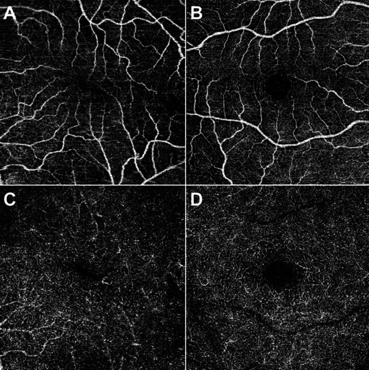
This was a prospective, non-randomized, comparative case series of patients that underwent vitrectomy. Twenty-six eyes of 26 patients that underwent vitrectomy were studied postoperatively by OCTA. Our patients underwent 23 or 25G pars plana vitrectomy (PPV) for any posterior segment pathology. Three-dimensional OCTAs (DRI Triton Swept Source OCT; Topcon) of the capillary plexus were obtained three months post-operatively. The FAZ measurements of the fellow eyes were used as controls.

MAIN OUTCOME MEASURES

Change in FAZ area between vitrectomized eyes and controls.

RESULTS

From a total of 26 patients, 17 underwent vitrectomy due to retinal detachment (RD). Almost all patients demonstrated a statistically significant reduction in FAZ size based on the OCTA measurements. Τhe mean difference in FAZ size for the superficial capillary plexus (SCP) was -93.77 ± 71.73 μm and for the deep capillary plexus (DCP) -88.87 ± 75.41 μm, both statistically significant (p=0.000), while the amount of reduction in μm was the same for both SCP and DCP.

CONCLUSION

The foveal avascular zone seems to be reduced following vitrectomy as shown by optical coherence tomography angiography. It is postulated that this may be the result of changes in the physiology of the vitrectomized eye, and that this change should be attributed to the removal of the vitreous itself rather than other structures such as the internal limiting membrane.

摘要

目的

使用光学相干断层扫描血管造影(OCTA)研究玻璃体切除术对黄斑循环,尤其是黄斑无血管区(FAZ)的影响。

方法

这是一个对接受玻璃体切除术患者的前瞻性、非随机、对比病例系列研究。对26例接受玻璃体切除术患者的26只眼进行了术后OCTA研究。我们的患者因任何后段病变接受了23G或25G的玻璃体切除术(PPV)。术后三个月获得了毛细血管丛的三维OCTA(DRI Triton扫频源OCT;拓普康)。对侧眼的FAZ测量值用作对照。

主要观察指标

玻璃体切除眼与对照眼之间FAZ面积的变化。

结果

在总共26例患者中,17例因视网膜脱离(RD)接受了玻璃体切除术。几乎所有患者根据OCTA测量显示FAZ大小有统计学意义的减小。浅表毛细血管丛(SCP)的FAZ大小平均差异为-93.77±71.73μm,深部毛细血管丛(DCP)为-88.87±75.41μm,两者均具有统计学意义(p=0.000),而SCP和DCP在μm的减小量相同。

结论

光学相干断层扫描血管造影显示,玻璃体切除术后黄斑无血管区似乎减小。据推测,这可能是玻璃体切除眼生理变化的结果,并且这种变化应归因于玻璃体本身的切除,而不是其他结构,如内界膜。

https://cdn.ncbi.nlm.nih.gov/pmc/blobs/b927/7943050/4989f29146b5/cureus-0013-00000013757-i01.jpg

相似文献

1
Reduction of Foveal Avascular Zone After Vitrectomy Demonstrated by Optical Coherence Tomography Angiography.光学相干断层扫描血管造影术显示玻璃体切除术后黄斑无血管区缩小
Cureus. 2021 Mar 7;13(3):e13757. doi: 10.7759/cureus.13757.
2
Functional and Structural Outcomes at the Foveal Avascular Zone with Optical Coherence Tomography Following Macula off Retinal Detachment Repair.黄斑脱离视网膜脱离修复术后经光学相干断层扫描检测的黄斑无血管区的功能和结构转归
Clin Ophthalmol. 2020 Oct 13;14:3261-3270. doi: 10.2147/OPTH.S271944. eCollection 2020.
3
Microvascular changes after pars plana vitrectomy for rhegmatogenous retinal detachment repair: A comparative study based on gas tamponade agent.孔源性视网膜脱离修复术后行睫状体平坦部玻璃体切割术后的微血管变化:基于气体填充剂的对比研究。
Eur J Ophthalmol. 2024 Jul;34(4):1247-1254. doi: 10.1177/11206721231218656. Epub 2023 Dec 3.
4
Foveal microvasculature features of surgically closed macular hole using optical coherence tomography angiography.使用光学相干断层扫描血管造影术观察手术封闭黄斑裂孔后的黄斑中心凹微血管特征
BMC Ophthalmol. 2017 Nov 28;17(1):217. doi: 10.1186/s12886-017-0607-z.
5
Foveal Avascular Zone Area Changes Analyzed Using OCT Angiography after Successful Rhegmatogenous Retinal Detachment Repair.视网膜脱离复位成功后使用光学相干断层扫描血管造影分析黄斑无血管区面积变化
Curr Eye Res. 2018 May;43(5):674-678. doi: 10.1080/02713683.2018.1437922. Epub 2018 Feb 16.
6
Assessment of Vascular Changes in Patients after Pars Plana Vitrectomy Surgery Due to Macula-Off Rhegmatogenous Retinal Detachment.黄斑脱离性孔源性视网膜脱离患者玻璃体切割术后血管变化的评估
J Clin Med. 2021 Oct 28;10(21):5054. doi: 10.3390/jcm10215054.
7
Inner retinal layers' alterations and microvasculature changes after vitrectomy for rhegmatogenous retinal detachment.孔源性视网膜脱离玻璃体切除术后视网膜内层改变和微血管变化。
Int Ophthalmol. 2020 Dec;40(12):3349-3356. doi: 10.1007/s10792-020-01521-2. Epub 2020 Jul 29.
8
Changes in Outcomes of Macular Optical Coherence Tomography Angiography Following Surgery for Optic Disc Pit Maculopathy.视盘小凹黄斑病变手术后黄斑光学相干断层扫描血管造影结果的变化
Diagnostics (Basel). 2024 Apr 23;14(9):874. doi: 10.3390/diagnostics14090874.
9
Evolution of macular microvasculature and retinal layers alterations in patients with macula off retinal detachment after vitrectomy.黄斑脱离视网膜复位术后黄斑区微循环和视网膜层改变的演变。
Eur J Ophthalmol. 2022 Jan;32(1):520-526. doi: 10.1177/1120672121992984. Epub 2021 Feb 7.
10
[Correlation of capillary plexus with visual acuity in idiopathic macular epiretinal membrane eyes using optical coherence tomography angiography].[利用光学相干断层扫描血管造影术研究特发性黄斑视网膜前膜眼中毛细血管丛与视力的相关性]
Zhonghua Yan Ke Za Zhi. 2019 Oct 11;55(10):757-762. doi: 10.3760/cma.j.issn.0412-4081.2019.10.006.

引用本文的文献

1
Multimodal Imaging of Diabetic Retinopathy: Insights from Optical Coherence Tomography Angiography and Adaptive Optics.糖尿病视网膜病变的多模态成像:光学相干断层扫描血管造影和自适应光学的见解
Diagnostics (Basel). 2025 Jul 8;15(14):1732. doi: 10.3390/diagnostics15141732.
2
Analyses of Foveal Avascular Zone in Patients with General Blunt Ocular Trauma Using Optical Coherence Tomography Angiography.应用光学相干断层血管成像技术分析钝挫伤性眼外伤患者的中心凹无血管区。
Korean J Ophthalmol. 2023 Feb;37(1):62-69. doi: 10.3341/kjo.2022.0081. Epub 2023 Feb 3.

本文引用的文献

1
REGIONAL DENSITIES OF RETINAL CAPILLARIES AND RETINAL SENSITIVITIES AFTER MACULAR HOLE SURGERY WITH INTERNAL LIMITING MEMBRANE PEELING.黄斑裂孔手术联合内界膜剥除术后视网膜毛细血管的区域密度和视网膜敏感性
Retina. 2020 Aug;40(8):1585-1591. doi: 10.1097/IAE.0000000000002637.
2
Decrease of the foveal avascular zone area after internal limiting membrane peeling: single case study.内界膜剥除术后黄斑无血管区面积减小:单病例研究
Int Med Case Rep J. 2017 Mar 8;10:81-85. doi: 10.2147/IMCRJ.S129174. eCollection 2017.
3
SUPERFICIAL FOVEAL AVASCULAR ZONE DETERMINED BY OPTICAL COHERENCE TOMOGRAPHY ANGIOGRAPHY BEFORE AND AFTER MACULAR HOLE SURGERY.
黄斑裂孔手术前后通过光学相干断层扫描血管造影术确定的浅表性黄斑血管区
Retina. 2017 Mar;37(3):444-450. doi: 10.1097/IAE.0000000000001205.
4
The Influence of Age and Central Foveal Thickness on Foveal Zone Size in Healthy People.年龄和中央凹厚度对健康人群黄斑区大小的影响。
Ophthalmic Surg Lasers Imaging Retina. 2016 Feb;47(2):142-8. doi: 10.3928/23258160-20160126-07.
5
CORRELATION OF FOVEAL AVASCULAR ZONE SIZE WITH FOVEAL MORPHOLOGY IN NORMAL EYES USING OPTICAL COHERENCE TOMOGRAPHY ANGIOGRAPHY.使用光学相干断层扫描血管造影术对正常眼黄斑中心凹血管区大小与黄斑中心凹形态的相关性研究
Retina. 2015 Nov;35(11):2188-95. doi: 10.1097/IAE.0000000000000847.
6
Optical Coherence Tomography Angiography of Type 1 Neovascularization in Age-Related Macular Degeneration.年龄相关性黄斑变性1型新生血管的光学相干断层扫描血管造影
Am J Ophthalmol. 2015 Oct;160(4):739-48.e2. doi: 10.1016/j.ajo.2015.06.030. Epub 2015 Jul 9.
7
Macular perfusion in healthy Chinese: an optical coherence tomography angiogram study.健康中国人的黄斑灌注:一项光学相干断层扫描血管造影研究。
Invest Ophthalmol Vis Sci. 2015 May;56(5):3212-7. doi: 10.1167/iovs.14-16270.
8
Quantitative optical coherence tomography angiography of vascular abnormalities in the living human eye.活体人眼血管异常的定量光学相干断层扫描血管造影术。
Proc Natl Acad Sci U S A. 2015 May 5;112(18):E2395-402. doi: 10.1073/pnas.1500185112. Epub 2015 Apr 20.
9
Optical Coherence Tomography Angiography in Diabetic Retinopathy: A Prospective Pilot Study.光学相干断层扫描血管造影在糖尿病视网膜病变中的应用:一项前瞻性试点研究。
Am J Ophthalmol. 2015 Jul;160(1):35-44.e1. doi: 10.1016/j.ajo.2015.04.021. Epub 2015 Apr 18.
10
The association between the foveal avascular zone and retinal thickness.黄斑无血管区与视网膜厚度之间的关联。
Invest Ophthalmol Vis Sci. 2014 Sep 30;55(10):6870-7. doi: 10.1167/iovs.14-15446.