Kaibori Masaki, Kosaka Hisashi, Matsui Kosuke, Ishizaki Morihiko, Matsushima Hideyuki, Tsuda Takumi, Hishikawa Hidehiko, Okumura Tadayoshi, Sekimoto Mitsugu
Department of Surgery, Kansai Medical University, Osaka, Japan.
Front Oncol. 2021 Feb 25;11:638327. doi: 10.3389/fonc.2021.638327. eCollection 2021.
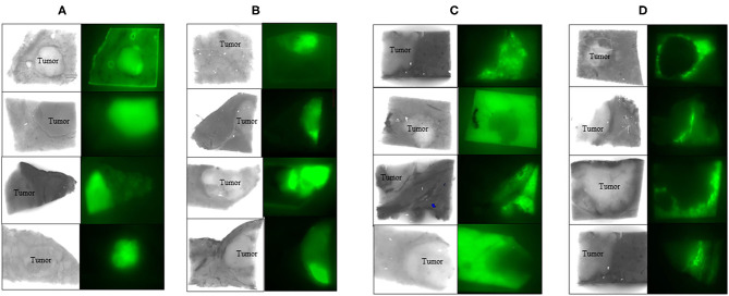
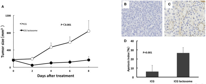
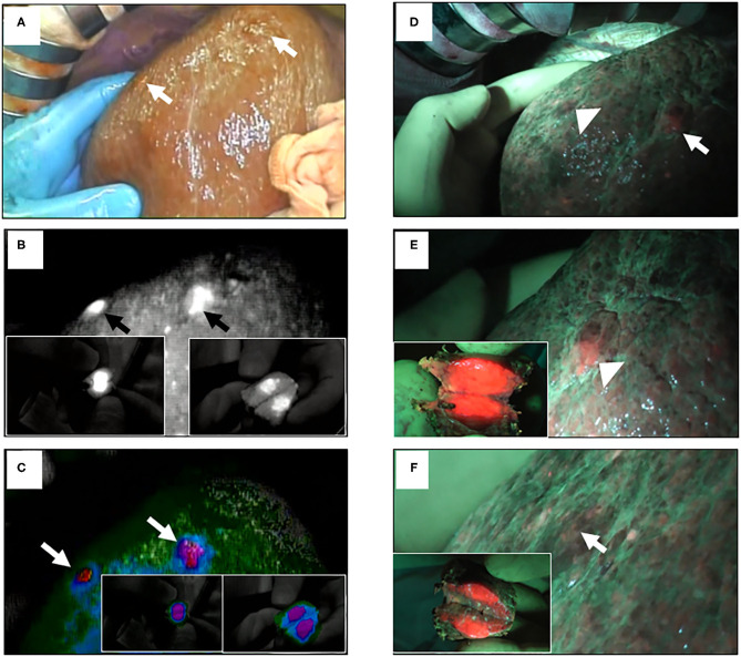
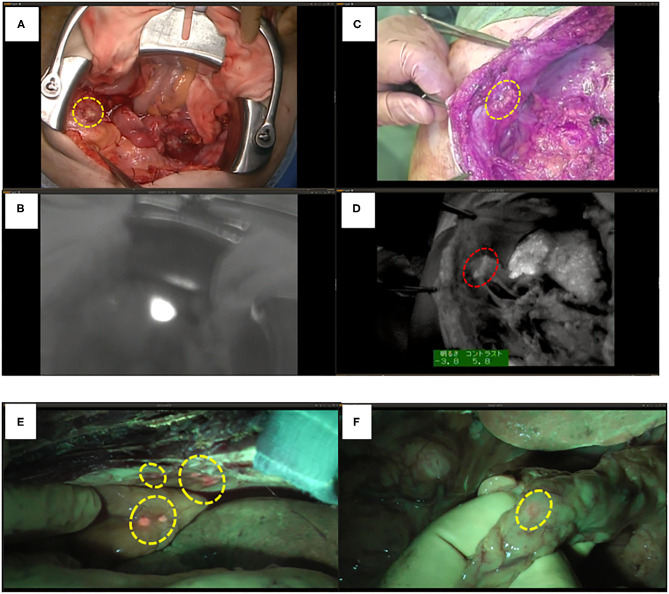
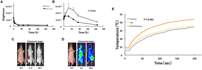
Surgery with fluorescence equipment has improved to treat the malignant viscera, including hepatobiliary and pancreatic neoplasms. In both open and minimally invasive surgeries, optical imaging using near-infrared (NIR) fluorescence is used to assess anatomy and function in real time. Here, we review a variety of publications related to clinical applications of NIR fluorescence imaging in liver surgery. We have developed a novel nanoparticle (indocyanine green lactosome) that is biocompatible and can be used for imaging cancer tissues and also as a drug delivery system. To date, stable particles are formed in blood and have an ~10-20 h half-life. Particles labeled with a NIR fluorescent agent have been applied to cancer tissues by the enhanced permeability and retention effect in animals. Furthermore, this article reviews recent developments in photodynamic therapy with NIR fluorescence imaging, which may contribute and accelerate the innovative treatments for liver tumors.
使用荧光设备的手术已得到改进,用于治疗包括肝胆和胰腺肿瘤在内的恶性内脏器官。在开放手术和微创手术中,使用近红外(NIR)荧光的光学成像用于实时评估解剖结构和功能。在此,我们综述了一系列与NIR荧光成像在肝脏手术中的临床应用相关的出版物。我们开发了一种新型纳米颗粒(吲哚菁绿脂质体),它具有生物相容性,可用于癌症组织成像,也可作为药物递送系统。迄今为止,稳定的颗粒在血液中形成,半衰期约为10 - 20小时。用NIR荧光剂标记的颗粒已通过动物体内的增强渗透和滞留效应应用于癌症组织。此外,本文还综述了NIR荧光成像在光动力治疗方面的最新进展,这可能有助于并加速肝脏肿瘤的创新治疗。