• 文献检索
  • 文档翻译
  • 深度研究
  • 学术资讯
  • Suppr Zotero 插件Zotero 插件
  • 邀请有礼
  • 套餐&价格
  • 历史记录
应用&插件
Suppr Zotero 插件Zotero 插件浏览器插件Mac 客户端Windows 客户端微信小程序
定价
高级版会员购买积分包购买API积分包
服务
文献检索文档翻译深度研究API 文档MCP 服务
关于我们
关于 Suppr公司介绍联系我们用户协议隐私条款
关注我们

Suppr 超能文献

核心技术专利:CN118964589B侵权必究
粤ICP备2023148730 号-1Suppr @ 2026

文献检索

告别复杂PubMed语法,用中文像聊天一样搜索,搜遍4000万医学文献。AI智能推荐,让科研检索更轻松。

立即免费搜索

文件翻译

保留排版,准确专业,支持PDF/Word/PPT等文件格式,支持 12+语言互译。

免费翻译文档

深度研究

AI帮你快速写综述,25分钟生成高质量综述,智能提取关键信息,辅助科研写作。

立即免费体验

严重急性呼吸综合征冠状病毒2刺突蛋白/血管紧张素转换酶2结合界面的计算诱变:与实验证据的比较

Computational Mutagenesis at the SARS-CoV-2 Spike Protein/Angiotensin-Converting Enzyme 2 Binding Interface: Comparison with Experimental Evidence.

作者信息

Laurini Erik, Marson Domenico, Aulic Suzana, Fermeglia Alice, Pricl Sabrina

机构信息

Molecular Biology and Nanotechnology Laboratory (MolBNL@UniTS), DEA, University of Trieste, 34127 Trieste, Italy.

Department of General Biophysics, Faculty of Biology and Environmental Protection, University of Lodz, 90-136 Lodz, Poland.

出版信息

ACS Nano. 2021 Apr 27;15(4):6929-6948. doi: 10.1021/acsnano.0c10833. Epub 2021 Mar 18.

DOI:10.1021/acsnano.0c10833
PMID:33733740
原文链接:https://pmc.ncbi.nlm.nih.gov/articles/PMC8009103/
Abstract

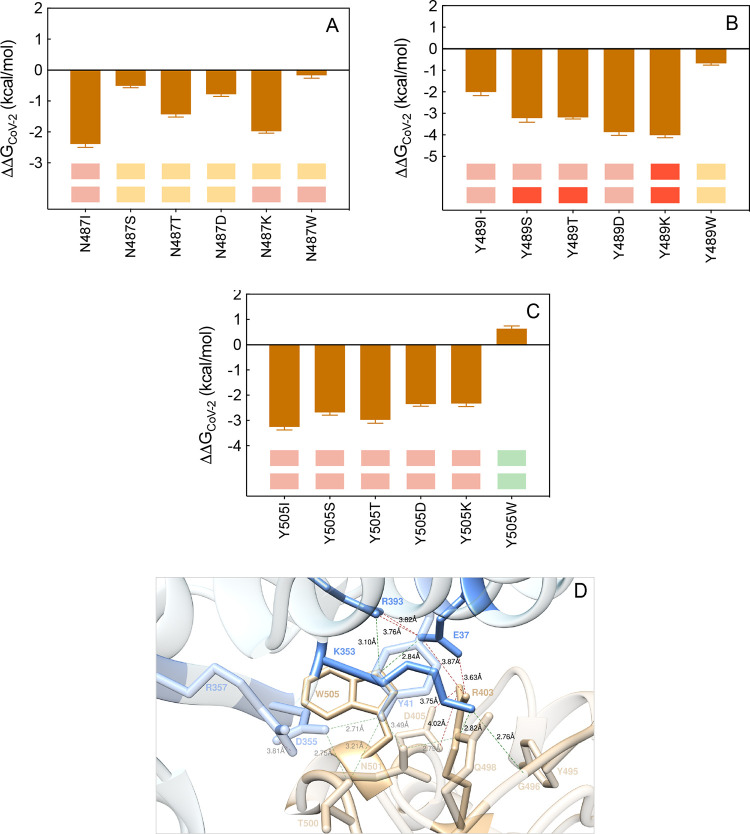
The coronavirus disease-2019 (COVID-19) pandemic, caused by the pathogen severe acute respiratory syndrome coronavirus 2 (SARS-CoV-2), started in China during late 2019 and swiftly spread worldwide. Since COVID-19 emergence, many therapeutic regimens have been relentlessly explored, and although two vaccines have just received emergency use authorization by different governmental agencies, antiviral therapeutics based neutralizing antibodies and small-drug inhibitors can still be vital viable options to prevent and treat SARS-CoV-2 infections. The viral spike glycoprotein (S-protein) is the key molecular player that promotes human host cellular invasion via recognition of and binding to the angiotensin-converting enzyme 2 gene (ACE2). In this work, we report the results obtained by mutating the 18 ACE2 residues and the 14 S-protein receptor binding domain (S-RBD) residues that contribute to the receptor/viral protein binding interface. Specifically, each wild-type protein-protein interface residue was replaced by a hydrophobic (isoleucine), polar (serine and threonine), charged (aspartic acid/glutamic acid and lysine/arginine), and bulky (tryptophan) residue, respectively, in order to study the different effects exerted by nature, shape, and dimensions of the mutant amino acids on the structure and strength of the resulting binding interface. The computational results were next validated against the corresponding experimental data, yielding an overall agreement of 92%. Interestingly, a non-negligible number of mis-sense variations were predicted to enhance ACE2/S-RBD binding, including the variants Q24T, T27D/K/W, D30E, H34S7T/K, E35D, Q42K, L79I/W, R357K, and R393K on ACE2 and L455D/W, F456K/W, Q493K, N501T, and Y505W on S-RBD, respectively.

摘要

2019年冠状病毒病(COVID-19)大流行由病原体严重急性呼吸综合征冠状病毒2(SARS-CoV-2)引起,于2019年末在中国爆发,并迅速蔓延至全球。自COVID-19出现以来,人们一直在不懈探索多种治疗方案,尽管已有两种疫苗刚刚获得不同政府机构的紧急使用授权,但基于中和抗体和小分子药物抑制剂的抗病毒疗法对于预防和治疗SARS-CoV-2感染而言,仍然是至关重要的可行选择。病毒刺突糖蛋白(S蛋白)是通过识别并结合血管紧张素转换酶2基因(ACE2)来促进侵入人类宿主细胞的关键分子。在本研究中,我们报告了对18个ACE2残基和14个S蛋白受体结合域(S-RBD)残基进行突变的结果,这些残基构成了受体/病毒蛋白结合界面。具体而言,分别将每个野生型蛋白-蛋白界面残基替换为一个疏水(异亮氨酸)、极性(丝氨酸和苏氨酸)、带电荷(天冬氨酸/谷氨酸和赖氨酸/精氨酸)和体积较大(色氨酸)的残基,以研究突变氨基酸的性质、形状和尺寸对所得结合界面的结构和强度产生的不同影响。接下来,将计算结果与相应的实验数据进行验证,总体一致性达92%。有趣的是,预测有相当数量的错义变异会增强ACE2/S-RBD结合,包括ACE2上的Q24T、T27D/K/W、D30E、H34S7T/K、E35D、Q42K、L79I/W、R357K和R393K变异,以及S-RBD上的L455D/W、F456K/W、Q493K、N501T和Y505W变异。

https://cdn.ncbi.nlm.nih.gov/pmc/blobs/9690/8155338/b947c230bc58/nn0c10833_0021.jpg
https://cdn.ncbi.nlm.nih.gov/pmc/blobs/9690/8155338/7e60502875a2/nn0c10833_0001.jpg
https://cdn.ncbi.nlm.nih.gov/pmc/blobs/9690/8155338/7dee6bc570e8/nn0c10833_0002.jpg
https://cdn.ncbi.nlm.nih.gov/pmc/blobs/9690/8155338/74c9be3de4bf/nn0c10833_0003.jpg
https://cdn.ncbi.nlm.nih.gov/pmc/blobs/9690/8155338/30d9198f45c9/nn0c10833_0004.jpg
https://cdn.ncbi.nlm.nih.gov/pmc/blobs/9690/8155338/939c2a2dd5cc/nn0c10833_0005.jpg
https://cdn.ncbi.nlm.nih.gov/pmc/blobs/9690/8155338/1305954972c2/nn0c10833_0006.jpg
https://cdn.ncbi.nlm.nih.gov/pmc/blobs/9690/8155338/184e35bd9d35/nn0c10833_0007.jpg
https://cdn.ncbi.nlm.nih.gov/pmc/blobs/9690/8155338/330eea17a91b/nn0c10833_0008.jpg
https://cdn.ncbi.nlm.nih.gov/pmc/blobs/9690/8155338/ad85724a765c/nn0c10833_0009.jpg
https://cdn.ncbi.nlm.nih.gov/pmc/blobs/9690/8155338/11be666d12be/nn0c10833_0010.jpg
https://cdn.ncbi.nlm.nih.gov/pmc/blobs/9690/8155338/5f84e7f6b428/nn0c10833_0011.jpg
https://cdn.ncbi.nlm.nih.gov/pmc/blobs/9690/8155338/ff2192ce8980/nn0c10833_0012.jpg
https://cdn.ncbi.nlm.nih.gov/pmc/blobs/9690/8155338/2c395627b383/nn0c10833_0013.jpg
https://cdn.ncbi.nlm.nih.gov/pmc/blobs/9690/8155338/f572898c8bd5/nn0c10833_0014.jpg
https://cdn.ncbi.nlm.nih.gov/pmc/blobs/9690/8155338/a9702a668dcd/nn0c10833_0015.jpg
https://cdn.ncbi.nlm.nih.gov/pmc/blobs/9690/8155338/b383462f7463/nn0c10833_0016.jpg
https://cdn.ncbi.nlm.nih.gov/pmc/blobs/9690/8155338/bc245e544c9a/nn0c10833_0017.jpg
https://cdn.ncbi.nlm.nih.gov/pmc/blobs/9690/8155338/90359689c354/nn0c10833_0018.jpg
https://cdn.ncbi.nlm.nih.gov/pmc/blobs/9690/8155338/6abf0c443d94/nn0c10833_0019.jpg
https://cdn.ncbi.nlm.nih.gov/pmc/blobs/9690/8155338/747e5db32a1f/nn0c10833_0020.jpg
https://cdn.ncbi.nlm.nih.gov/pmc/blobs/9690/8155338/b947c230bc58/nn0c10833_0021.jpg

相似文献

1
Computational Mutagenesis at the SARS-CoV-2 Spike Protein/Angiotensin-Converting Enzyme 2 Binding Interface: Comparison with Experimental Evidence.严重急性呼吸综合征冠状病毒2刺突蛋白/血管紧张素转换酶2结合界面的计算诱变:与实验证据的比较
ACS Nano. 2021 Apr 27;15(4):6929-6948. doi: 10.1021/acsnano.0c10833. Epub 2021 Mar 18.
2
Computational Alanine Scanning and Structural Analysis of the SARS-CoV-2 Spike Protein/Angiotensin-Converting Enzyme 2 Complex.基于 SARS-CoV-2 刺突蛋白/血管紧张素转化酶 2 复合物的丙氨酸扫描计算和结构分析。
ACS Nano. 2020 Sep 22;14(9):11821-11830. doi: 10.1021/acsnano.0c04674. Epub 2020 Aug 26.
3
Coevolution, Dynamics and Allostery Conspire in Shaping Cooperative Binding and Signal Transmission of the SARS-CoV-2 Spike Protein with Human Angiotensin-Converting Enzyme 2.新冠病毒刺突蛋白与人血管紧张素转化酶 2 协同结合和信号转导的共进化、动力学和变构协同作用。
Int J Mol Sci. 2020 Nov 4;21(21):8268. doi: 10.3390/ijms21218268.
4
Different compounds against Angiotensin-Converting Enzyme 2 (ACE2) receptor potentially containing the infectivity of SARS-CoV-2: an in silico study.针对血管紧张素转化酶 2(ACE2)受体的不同化合物可能含有 SARS-CoV-2 的感染力:一项计算机研究。
J Mol Model. 2022 Mar 5;28(4):82. doi: 10.1007/s00894-022-05059-1.
5
Identification of potential SARS-CoV-2 entry inhibitors by targeting the interface region between the spike RBD and human ACE2.通过针对刺突 RBD 和人 ACE2 之间的界面区域,鉴定潜在的 SARS-CoV-2 进入抑制剂。
J Infect Public Health. 2021 Feb;14(2):227-237. doi: 10.1016/j.jiph.2020.12.014. Epub 2020 Dec 21.
6
Effect of mutation on structure, function and dynamics of receptor binding domain of human SARS-CoV-2 with host cell receptor ACE2: a molecular dynamics simulations study.突变对人 SARS-CoV-2 受体结合域与宿主细胞受体 ACE2 结构、功能和动力学的影响:分子动力学模拟研究。
J Biomol Struct Dyn. 2021 Nov;39(18):7231-7245. doi: 10.1080/07391102.2020.1802348. Epub 2020 Aug 7.
7
Investigation of the genetic variation in ACE2 on the structural recognition by the novel coronavirus (SARS-CoV-2).新型冠状病毒(SARS-CoV-2)对血管紧张素转换酶2(ACE2)结构识别的遗传变异研究。
J Transl Med. 2020 Aug 24;18(1):321. doi: 10.1186/s12967-020-02486-7.
8
Impact of Genetic Variability in ACE2 Expression on the Evolutionary Dynamics of SARS-CoV-2 Spike D614G Mutation.ACE2 表达中的遗传变异性对 SARS-CoV-2 刺突 D614G 突变进化动态的影响。
Genes (Basel). 2020 Dec 24;12(1):16. doi: 10.3390/genes12010016.
9
Q493K and Q498H substitutions in Spike promote adaptation of SARS-CoV-2 in mice.S 蛋白 493 位和 498 位的 Q 突变为 SARS-CoV-2 在小鼠体内的适应性进化提供了条件。
EBioMedicine. 2021 May;67:103381. doi: 10.1016/j.ebiom.2021.103381. Epub 2021 May 14.
10
A molecular docking study revealed that synthetic peptides induced conformational changes in the structure of SARS-CoV-2 spike glycoprotein, disrupting the interaction with human ACE2 receptor.一项分子对接研究表明,合成肽诱导了 SARS-CoV-2 刺突糖蛋白结构的构象变化,破坏了与人类 ACE2 受体的相互作用。
Int J Biol Macromol. 2020 Dec 1;164:66-76. doi: 10.1016/j.ijbiomac.2020.07.174. Epub 2020 Jul 18.

引用本文的文献

1
A Computational Approach: The Functional Effects of Thyroid Peroxidase Variants in Thyroid Cancer and Genetic Disorders.计算方法:甲状腺过氧化物酶变体在甲状腺癌和遗传疾病中的功能影响。
JCO Clin Cancer Inform. 2024 Jan;8:e2300140. doi: 10.1200/CCI.23.00140.
2
Genomic Determinants Potentially Associated with Clinical Manifestations of Human-Pathogenic Tick-Borne Flaviviruses.与人类致病蜱传黄病毒临床表现相关的基因组决定因素。
Int J Mol Sci. 2022 Nov 2;23(21):13404. doi: 10.3390/ijms232113404.
3
Large interfacial relocation in RBD-ACE2 complex may explain fast-spreading property of Omicron.
RBD-ACE2复合物中的大量界面重排可能解释了奥密克戎的快速传播特性。
J Mol Struct. 2022 Dec 15;1270:133842. doi: 10.1016/j.molstruc.2022.133842. Epub 2022 Jul 31.
4
Dynamics of the interaction between the receptor-binding domain of SARS-CoV-2 Omicron (B.1.1.529) variant and human angiotensin-converting enzyme 2.SARS-CoV-2 奥密克戎(B.1.1.529)变体受体结合域与人血管紧张素转化酶 2 相互作用的动力学。
PeerJ. 2022 Jul 5;10:e13680. doi: 10.7717/peerj.13680. eCollection 2022.
5
Mutational analysis of SARS-CoV-2 variants of concern reveals key tradeoffs between receptor affinity and antibody escape.对关注的 SARS-CoV-2 变体的突变分析揭示了受体亲和力和抗体逃逸之间的关键权衡。
PLoS Comput Biol. 2022 May 31;18(5):e1010160. doi: 10.1371/journal.pcbi.1010160. eCollection 2022 May.
6
An Electrostatically-steered Conformational Selection Mechanism Promotes SARS-CoV-2 Spike Protein Variation.静电导向的构象选择机制促进了 SARS-CoV-2 刺突蛋白的变异。
J Mol Biol. 2022 Jul 15;434(13):167637. doi: 10.1016/j.jmb.2022.167637. Epub 2022 May 17.
7
Methodology-Centered Review of Molecular Modeling, Simulation, and Prediction of SARS-CoV-2.基于方法的 SARS-CoV-2 分子建模、模拟和预测综述。
Chem Rev. 2022 Jul 13;122(13):11287-11368. doi: 10.1021/acs.chemrev.1c00965. Epub 2022 May 20.
8
Comprehensive role of SARS-CoV-2 spike glycoprotein in regulating host signaling pathway.SARS-CoV-2 刺突糖蛋白在调节宿主信号通路中的全面作用。
J Med Virol. 2022 Sep;94(9):4071-4087. doi: 10.1002/jmv.27820. Epub 2022 May 9.
9
A CNN model for predicting binding affinity changes between SARS-CoV-2 spike RBD variants and ACE2 homologues.一种用于预测严重急性呼吸综合征冠状病毒2(SARS-CoV-2)刺突受体结合域(RBD)变体与血管紧张素转换酶2(ACE2)同源物之间结合亲和力变化的卷积神经网络(CNN)模型。
bioRxiv. 2022 Mar 23:2022.03.22.485413. doi: 10.1101/2022.03.22.485413.
10
Computational Design of Miniproteins as SARS-CoV-2 Therapeutic Inhibitors.作为 SARS-CoV-2 治疗抑制剂的小型蛋白的计算设计。
Int J Mol Sci. 2022 Jan 13;23(2):838. doi: 10.3390/ijms23020838.