Institute of Clinical Medicine, School of Medicine, National Yang Ming Chiao Tung University, 2F, Shou-Ren Bldg., No.155, Sec.2, Li-Nong St., Beitou Dist, Taipei, 11221, Taiwan.
Faculty of Medicine, School of Medicine, National Yang Ming Chiao Tung University, Taipei, 11221, Taiwan.
Stem Cell Res Ther. 2021 Mar 19;12(1):193. doi: 10.1186/s13287-021-02270-4.
Life-long peritoneal dialysis (PD) as a renal replacement therapy is limited by peritoneal fibrosis. Previous studies showed immunomodulatory and antifibrotic effects of adipose-derived mesenchymal stem cells (ADSCs) on peritoneal fibrosis. However, the role of the peritoneal macrophage in this process remains uninvestigated.
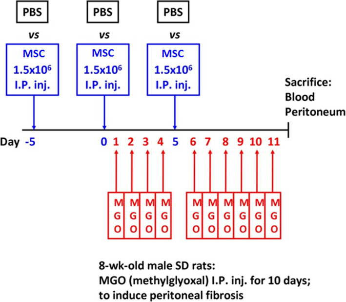
We examined the therapeutic effects of ADSC and bone marrow-derived mesenchymal stem cells (BM-MSC) in the rat model of dialysis-induced peritoneal fibrosis using methylglyoxal. In addition, treatment of macrophages with the conditioned medium of ADSC and BM-MSC was performed individually to identify the beneficial component of the stem cell secretome.
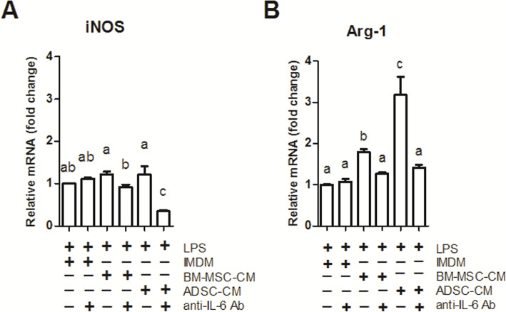
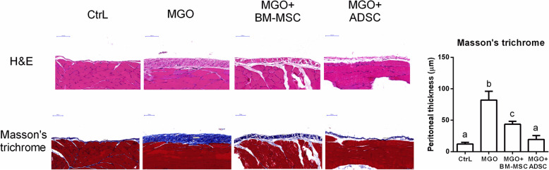
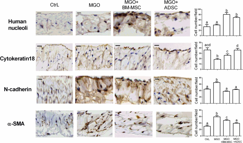
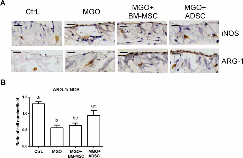
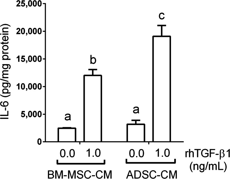
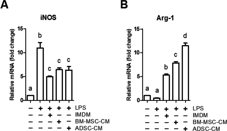
In the in vivo experiments, we found dialysis-induced rat peritoneal fibrosis was attenuated by both ADSC and BM-MSC. Interestingly, ADSC possessed a more prominent therapeutic effect than BM-MSC in ameliorating peritoneal membrane thickening while also upregulating epithelial cell markers in rat peritoneal tissues. The therapeutic effects of ADSC were positively associated with M2 macrophage polarization. In the in vitro experiments, we confirmed that interleukin-6 (IL-6) secreted by MSCs upon transforming growth factor-β1 stimulation promotes M2 macrophage polarization.
In dialysis-induced peritoneal fibrosis, MSCs are situated in an inflammatory environment of TGF-β1 and secrete IL-6 to polarize macrophages into the M2 phenotype. Our findings reveal a previously unidentified role of tissue macrophage in this antifibrotic process. ADSC has the advantage of abundance and accessibility, making the application values extremely promising. In dialysis-induced peritoneal fibrosis, peritoneal mesothelial cells secrete transforming growth factor-β1 (TGF-β1) when exposed to methylglyoxal (MGO)-containing peritoneal dialysate. When situated in TGF-β1, the inflammatory environment induces mesenchymal stem cells to secrete interleukin-6 (IL-6), IL-6 polarizes macrophages into the M2 phenotype. The dominant peritoneal tissue M2 macrophages, marked by upregulated Arg-1 expression, account for the attenuation of MGO-induced dedifferentiation of peritoneal mesothelial cells to maintain epithelial integrity.
终身腹膜透析(PD)作为肾脏替代疗法受到腹膜纤维化的限制。先前的研究表明脂肪间充质干细胞(ADSCs)对腹膜纤维化具有免疫调节和抗纤维化作用。然而,这一过程中腹膜巨噬细胞的作用仍未被研究。
我们使用乙二醛检查 ADSC 和骨髓间充质干细胞(BM-MSC)在透析诱导的腹膜纤维化大鼠模型中的治疗效果。此外,单独用 ADSC 和 BM-MSC 的条件培养基处理巨噬细胞,以鉴定干细胞分泌组的有益成分。
在体内实验中,我们发现 ADSC 和 BM-MSC 均可减轻诱导的大鼠腹膜纤维化。有趣的是,与 BM-MSC 相比,ADSC 在改善腹膜组织增厚方面具有更显著的治疗效果,同时也上调了上皮细胞标志物。ADSC 的治疗效果与 M2 巨噬细胞极化呈正相关。在体外实验中,我们证实转化生长因子-β1 刺激下 MSC 分泌的白细胞介素-6(IL-6)促进 M2 巨噬细胞极化。
在透析诱导的腹膜纤维化中,MSC 处于 TGF-β1 的炎症环境中,并分泌 IL-6 将巨噬细胞极化为 M2 表型。我们的研究结果揭示了组织巨噬细胞在这一抗纤维化过程中的一个先前未知的作用。ADSC 具有丰富和易获得的优势,应用价值极具潜力。在透析诱导的腹膜纤维化中,腹膜间皮细胞在接触含有乙二醛(MGO)的腹膜透析液时会分泌转化生长因子-β1(TGF-β1)。处于 TGF-β1 炎症环境中,间充质干细胞分泌白细胞介素-6(IL-6),IL-6 将巨噬细胞极化为 M2 表型。M2 型巨噬细胞是主要的腹膜组织巨噬细胞,Arg-1 表达上调,有助于减轻 MGO 诱导的腹膜间皮细胞去分化,维持上皮完整性。