• 文献检索
  • 文档翻译
  • 深度研究
  • 学术资讯
  • Suppr Zotero 插件Zotero 插件
  • 邀请有礼
  • 套餐&价格
  • 历史记录
应用&插件
Suppr Zotero 插件Zotero 插件浏览器插件Mac 客户端Windows 客户端微信小程序
定价
高级版会员购买积分包购买API积分包
服务
文献检索文档翻译深度研究API 文档MCP 服务
关于我们
关于 Suppr公司介绍联系我们用户协议隐私条款
关注我们

Suppr 超能文献

核心技术专利:CN118964589B侵权必究
粤ICP备2023148730 号-1Suppr @ 2026

文献检索

告别复杂PubMed语法,用中文像聊天一样搜索,搜遍4000万医学文献。AI智能推荐,让科研检索更轻松。

立即免费搜索

文件翻译

保留排版,准确专业,支持PDF/Word/PPT等文件格式,支持 12+语言互译。

免费翻译文档

深度研究

AI帮你快速写综述,25分钟生成高质量综述,智能提取关键信息,辅助科研写作。

立即免费体验

间充质干细胞通过抑制炎症和 TGF-β1 信号通路改善实验性腹膜纤维化。

Mesenchymal stem cells ameliorate experimental peritoneal fibrosis by suppressing inflammation and inhibiting TGF-β1 signaling.

机构信息

Department of Nephrology, Hiroshima University Hospital, Hiroshima, Japan.

出版信息

Kidney Int. 2013 Aug;84(2):297-307. doi: 10.1038/ki.2013.81. Epub 2013 Mar 13.

DOI:10.1038/ki.2013.81
PMID:23486522
原文链接:https://pmc.ncbi.nlm.nih.gov/articles/PMC3731556/
Abstract

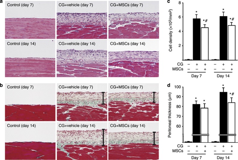
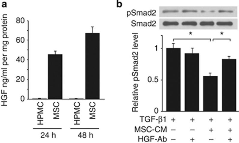
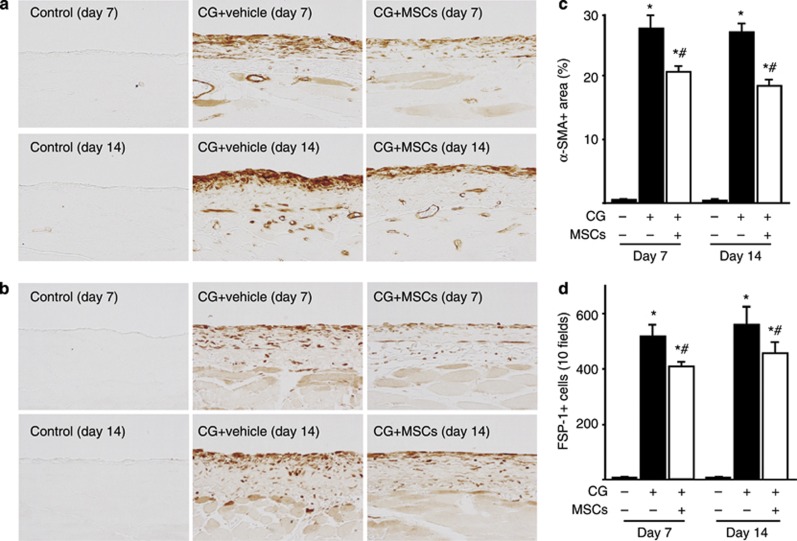
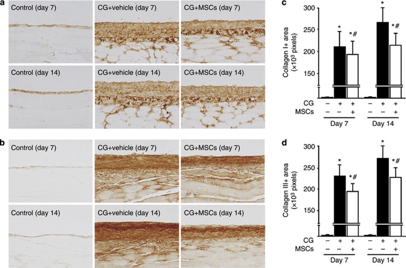
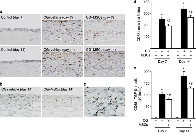
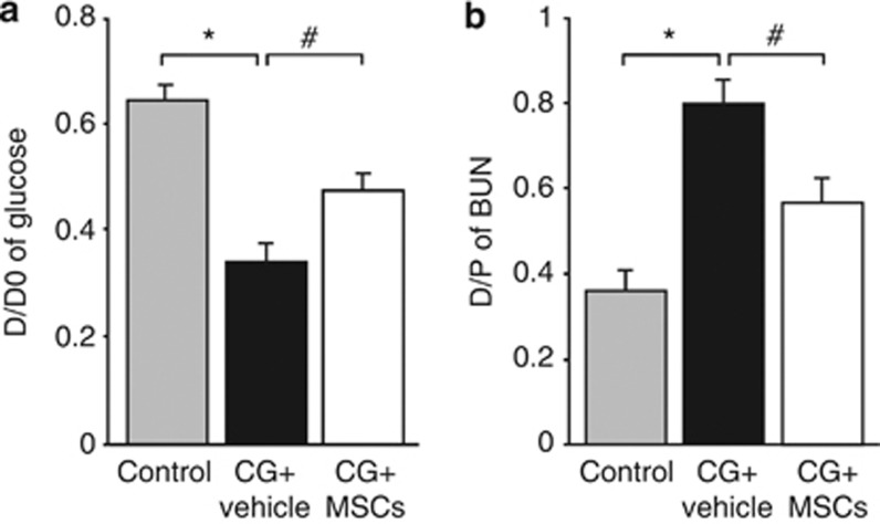
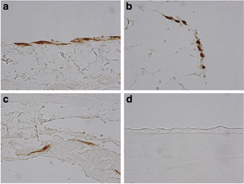
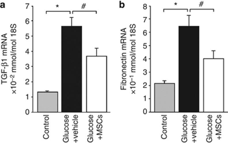
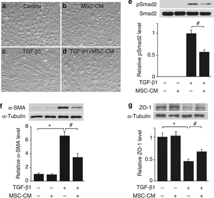
Mesenchymal stem cells (MSCs) are multipotent adult stem cells that have regenerative capability and exert paracrine actions on damaged tissues. Since peritoneal fibrosis is a serious complication of peritoneal dialysis, we tested whether MSCs suppress this using a chlorhexidine gluconate model in rats. Although MSCs isolated from green fluorescent protein-positive rats were detected for only 3 days following their injection, immunohistochemical staining showed that MSCs suppressed the expression of mesenchymal cells, their effects on the deposition of extracellular matrix proteins, and the infiltration of macrophages for 14 days. Moreover, MSCs reduced the functional impairment of the peritoneal membrane. Cocultures of MSCs and human peritoneal mesothelial cells using a Transwell system indicated that the beneficial effects of MSCs on the glucose-induced upregulation of transforming growth factor-β1(TGF-β1) and fibronectin mRNA expression in the human cells were likely due to paracrine actions. Preincubation in MSC-conditioned medium suppressed TGF-β1-induced epithelial-to-mesenchymal transition, α-smooth muscle actin, and the decrease in zonula occludens-1 in cultured human peritoneal mesothelial cells. Although bone morphogenic protein 7 was not detected, MSCs secreted hepatocyte growth factor and a neutralizing antibody to this inhibited TGF-β1 signaling. Thus, our findings imply that MSCs ameliorate experimental peritoneal fibrosis by suppressing inflammation and TGF-β1 signaling in a paracrine manner.

摘要

间充质干细胞(MSCs)是具有再生能力的多能成体干细胞,对受损组织具有旁分泌作用。由于腹膜纤维化是腹膜透析的严重并发症,我们使用葡萄糖酸氯己定模型在大鼠中测试了 MSCs 是否能抑制这种情况。尽管从绿色荧光蛋白阳性大鼠中分离的 MSCs 在注射后仅能检测到 3 天,但免疫组织化学染色显示 MSCs 抑制了间充质细胞的表达及其对细胞外基质蛋白沉积和巨噬细胞浸润的作用,持续了 14 天。此外,MSCs 减少了腹膜功能的损伤。通过 Transwell 系统共培养 MSCs 和人腹膜间皮细胞表明,MSCs 对葡萄糖诱导的转化生长因子-β1(TGF-β1)和纤连蛋白 mRNA 表达在人细胞中的上调的有益作用可能是由于旁分泌作用。在 MSC 条件培养基中孵育可抑制 TGF-β1 诱导的上皮细胞向间充质细胞转化、α-平滑肌肌动蛋白和培养的人腹膜间皮细胞中封闭蛋白-1 的减少。虽然未检测到骨形态发生蛋白 7,但 MSCs 分泌肝细胞生长因子,并且针对该因子的中和抗体抑制了 TGF-β1 信号。因此,我们的研究结果表明,MSCs 通过旁分泌方式抑制炎症和 TGF-β1 信号转导来改善实验性腹膜纤维化。

https://cdn.ncbi.nlm.nih.gov/pmc/blobs/c0c5/3731556/06efb6e828d5/ki201381f10.jpg
https://cdn.ncbi.nlm.nih.gov/pmc/blobs/c0c5/3731556/c7cc491c437c/ki201381f1.jpg
https://cdn.ncbi.nlm.nih.gov/pmc/blobs/c0c5/3731556/293029a1e2ac/ki201381f2.jpg
https://cdn.ncbi.nlm.nih.gov/pmc/blobs/c0c5/3731556/f0aad79f537d/ki201381f3.jpg
https://cdn.ncbi.nlm.nih.gov/pmc/blobs/c0c5/3731556/da98d9d785d3/ki201381f4.jpg
https://cdn.ncbi.nlm.nih.gov/pmc/blobs/c0c5/3731556/581742f28b0f/ki201381f5.jpg
https://cdn.ncbi.nlm.nih.gov/pmc/blobs/c0c5/3731556/afc19f37d35c/ki201381f6.jpg
https://cdn.ncbi.nlm.nih.gov/pmc/blobs/c0c5/3731556/b18d9442c4f4/ki201381f7.jpg
https://cdn.ncbi.nlm.nih.gov/pmc/blobs/c0c5/3731556/19dd25ae6b3e/ki201381f8.jpg
https://cdn.ncbi.nlm.nih.gov/pmc/blobs/c0c5/3731556/cbf3a72a1982/ki201381f9.jpg
https://cdn.ncbi.nlm.nih.gov/pmc/blobs/c0c5/3731556/06efb6e828d5/ki201381f10.jpg

相似文献

1
Mesenchymal stem cells ameliorate experimental peritoneal fibrosis by suppressing inflammation and inhibiting TGF-β1 signaling.间充质干细胞通过抑制炎症和 TGF-β1 信号通路改善实验性腹膜纤维化。
Kidney Int. 2013 Aug;84(2):297-307. doi: 10.1038/ki.2013.81. Epub 2013 Mar 13.
2
Mesenchymal stem cells attenuate peritoneal injury through secretion of TSG-6.间充质干细胞通过分泌 TSG-6 减轻腹膜损伤。
PLoS One. 2012;7(8):e43768. doi: 10.1371/journal.pone.0043768. Epub 2012 Aug 17.
3
Mesenchymal stem cells cultured in serum-free medium ameliorate experimental peritoneal fibrosis.无血清培养基中培养的间充质干细胞可改善实验性腹膜纤维化。
Stem Cell Res Ther. 2021 Mar 23;12(1):203. doi: 10.1186/s13287-021-02273-1.
4
Nitro-oleic acid inhibits the high glucose-induced epithelial-mesenchymal transition in peritoneal mesothelial cells and attenuates peritoneal fibrosis.硝异山梨醇抑制高糖诱导的腹膜间皮细胞上皮间质转化并减轻腹膜纤维化。
Am J Physiol Renal Physiol. 2020 Feb 1;318(2):F457-F467. doi: 10.1152/ajprenal.00425.2019. Epub 2019 Nov 25.
5
Blockade of thrombospondin-1 ameliorates high glucose-induced peritoneal fibrosis through downregulation of TGF-β1/Smad3 signaling pathway.阻断血小板反应蛋白-1 可通过下调 TGF-β1/Smad3 信号通路改善高糖诱导的腹膜纤维化。
J Cell Physiol. 2020 Jan;235(1):364-379. doi: 10.1002/jcp.28976. Epub 2019 Jun 24.
6
TGF-β1-VEGF-A pathway induces neoangiogenesis with peritoneal fibrosis in patients undergoing peritoneal dialysis.TGF-β1-VEGF-A 通路诱导接受腹膜透析患者的新生血管形成和腹膜纤维化。
Am J Physiol Renal Physiol. 2018 Feb 1;314(2):F167-F180. doi: 10.1152/ajprenal.00052.2017. Epub 2017 Oct 4.
7
Conditioned mesenchymal stem cells attenuate progression of chronic kidney disease through inhibition of epithelial-to-mesenchymal transition and immune modulation.条件化间充质干细胞通过抑制上皮-间质转化和免疫调节来减轻慢性肾脏病的进展。
J Cell Mol Med. 2012 Dec;16(12):2935-49. doi: 10.1111/j.1582-4934.2012.01610.x.
8
Adipose-derived mesenchymal stem cells transplantation facilitate experimental peritoneal fibrosis repair by suppressing epithelial-mesenchymal transition.脂肪来源的间充质干细胞移植通过抑制上皮-间质转化促进实验性腹膜纤维化修复。
J Nephrol. 2014 Oct;27(5):507-14. doi: 10.1007/s40620-014-0133-5. Epub 2014 Aug 22.
9
Effects of TGF-β1 Receptor Inhibitor GW788388 on the Epithelial to Mesenchymal Transition of Peritoneal Mesothelial Cells.TGF-β1 受体抑制剂 GW788388 对腹膜间皮细胞上皮-间充质转化的影响。
Int J Mol Sci. 2021 Apr 29;22(9):4739. doi: 10.3390/ijms22094739.
10
Protective Effects of Transforming Growth Factor-β1 Knockdown in Human Umbilical Cord Mesenchymal Stem Cells against Subarachnoid Hemorrhage in a Rat Model.转化生长因子-β1基因敲低对大鼠蛛网膜下腔出血模型中人类脐带间充质干细胞的保护作用
Cerebrovasc Dis. 2020;49(1):79-87. doi: 10.1159/000505311. Epub 2020 Jan 15.

引用本文的文献

1
Liu Wei Di Huang Decoction Alleviates Renal Fibrosis by Inhibiting Endothelial Mesenchymal Transitions via Upregulating Sirt1 Expression and Inhibiting the Wnt/β-Catenin Signaling Pathway.六味地黄丸通过上调Sirt1表达并抑制Wnt/β-连环蛋白信号通路来抑制内皮-间充质转化,从而减轻肾纤维化。
Drug Des Devel Ther. 2025 Jul 30;19:6587-6603. doi: 10.2147/DDDT.S517938. eCollection 2025.
2
Non-steroidal mineralocorticoid receptor antagonist finerenone inhibits peritoneal fibrosis induced by high-glucose dialysate via regulating enhancer of zeste homolog 2 (EZH2) expression.非甾体类盐皮质激素受体拮抗剂非奈利酮通过调节zeste同源物2(EZH2)的表达来抑制高糖透析液诱导的腹膜纤维化。
Ren Fail. 2025 Dec;47(1):2491156. doi: 10.1080/0886022X.2025.2491156. Epub 2025 May 13.
3

本文引用的文献

1
Differentiation of bone marrow-derived cells into regenerated mesothelial cells in peritoneal remodeling using a peritoneal fibrosis mouse model.利用腹膜纤维化小鼠模型研究骨髓来源细胞在腹膜重塑过程中向再生间皮细胞的分化。
J Artif Organs. 2012 Sep;15(3):272-82. doi: 10.1007/s10047-012-0648-2. Epub 2012 May 24.
2
Blocking TGF-β1 protects the peritoneal membrane from dialysate-induced damage.阻断 TGF-β1 可保护腹膜免受透析液损伤。
J Am Soc Nephrol. 2011 Sep;22(9):1682-95. doi: 10.1681/ASN.2010111197. Epub 2011 Jul 8.
3
Klotho inhibits transforming growth factor-beta1 (TGF-beta1) signaling and suppresses renal fibrosis and cancer metastasis in mice.
Targeting MLL1/WDR5-Mediated Epigenetic Regulation Mitigates Peritoneal Fibrosis by Reducing p16.靶向MLL1/WDR5介导的表观遗传调控通过降低p16减轻腹膜纤维化。
FASEB J. 2025 Apr 30;39(8):e70543. doi: 10.1096/fj.202402382R.
4
Adipose Tissue Stem Cells (ASCs) and ASC-Derived Extracellular Vesicles Prevent the Development of Experimental Peritoneal Fibrosis.脂肪组织干细胞(ASCs)和ASC衍生的细胞外囊泡可预防实验性腹膜纤维化的发展。
Cells. 2025 Mar 14;14(6):436. doi: 10.3390/cells14060436.
5
Kidney Fibrosis In Vitro and In Vivo Models: Path Toward Physiologically Relevant Humanized Models.肾纤维化的体外和体内模型:迈向生理相关人源化模型的路径。
Adv Healthc Mater. 2025 Apr;14(9):e2403230. doi: 10.1002/adhm.202403230. Epub 2025 Feb 5.
6
Efficacy of mesenchymal stem cells in the treatment of peritoneal fibrosis in animal models: a systematic review and meta-analysis.间充质干细胞治疗动物模型腹膜纤维化的疗效:系统评价与荟萃分析
Ren Fail. 2024 Dec;46(2):2438863. doi: 10.1080/0886022X.2024.2438863. Epub 2024 Dec 16.
7
The mechanisms behind heatstroke-induced intestinal damage.中暑诱导肠道损伤背后的机制。
Cell Death Discov. 2024 Oct 28;10(1):455. doi: 10.1038/s41420-024-02210-0.
8
Impact of immunosuppressive drugs on efficacy of mesenchymal stem cell therapy for suppressing renal fibrosis.免疫抑制药物对间充质干细胞抑制肾纤维化治疗效果的影响。
Stem Cells Transl Med. 2024 Nov 12;13(11):1067-1085. doi: 10.1093/stcltm/szae073.
9
Selective activation of PPARα by pemafibrate mitigates peritoneal inflammation and fibrosis through suppression of NLRP3 inflammasome and modulation of inflammation.佩马弗他酯通过抑制 NLRP3 炎症小体和调节炎症选择性激活 PPARα,减轻腹膜炎和纤维化。
Sci Rep. 2024 Oct 11;14(1):23816. doi: 10.1038/s41598-024-74340-5.
10
Deletion of p38 MAPK in macrophages ameliorates peritoneal fibrosis and inflammation in peritoneal dialysis.巨噬细胞中 p38MAPK 的缺失可改善腹膜透析中的腹膜纤维化和炎症。
Sci Rep. 2024 Sep 11;14(1):21220. doi: 10.1038/s41598-024-71859-5.
Klotho 抑制转化生长因子-β1(TGF-β1)信号通路,抑制小鼠的肾纤维化和癌症转移。
J Biol Chem. 2011 Mar 11;286(10):8655-8665. doi: 10.1074/jbc.M110.174037. Epub 2011 Jan 5.
4
15-Deoxy-Delta12,14-prostaglandin J2 inhibits angiotensin II-induced fibronectin expression via hepatocyte growth factor induction in human peritoneal mesothelial cells.15-脱氧-Δ12,14-前列腺素J2通过诱导人腹膜间皮细胞产生肝细胞生长因子来抑制血管紧张素II诱导的纤连蛋白表达。
Ther Apher Dial. 2010 Feb;14(1):43-51. doi: 10.1111/j.1744-9987.2009.00702.x.
5
Mesenchymal stromal cells: current understanding and clinical status.间质基质细胞:当前的认识和临床状况。
Stem Cells. 2010 Mar 31;28(3):585-96. doi: 10.1002/stem.269.
6
Human umbilical cord mesenchymal stem cells reduce fibrosis of bleomycin-induced lung injury.人脐带间充质干细胞减轻博来霉素诱导的肺损伤纤维化。
Am J Pathol. 2009 Jul;175(1):303-13. doi: 10.2353/ajpath.2009.080629. Epub 2009 Jun 4.
7
Mizoribine suppresses the progression of experimental peritoneal fibrosis in a rat model.咪唑立宾抑制大鼠实验性腹膜纤维化的进展。
Nephron Exp Nephrol. 2009;112(2):e59-69. doi: 10.1159/000213896. Epub 2009 Apr 23.
8
HGF and BMP-7 ameliorate high glucose-induced epithelial-to-mesenchymal transition of peritoneal mesothelium.肝细胞生长因子和骨形态发生蛋白-7可改善高糖诱导的腹膜间皮细胞上皮-间质转化。
J Am Soc Nephrol. 2009 Mar;20(3):567-81. doi: 10.1681/ASN.2008040424. Epub 2009 Feb 4.
9
Cyclooxygenase-2 mediates dialysate-induced alterations of the peritoneal membrane.环氧化酶-2介导透析液引起的腹膜变化。
J Am Soc Nephrol. 2009 Mar;20(3):582-92. doi: 10.1681/ASN.2008020211. Epub 2009 Jan 21.
10
Bone marrow stromal cells attenuate sepsis via prostaglandin E(2)-dependent reprogramming of host macrophages to increase their interleukin-10 production.骨髓基质细胞通过前列腺素E(2)依赖的宿主巨噬细胞重编程来减轻脓毒症,从而增加其白细胞介素-10的产生。
Nat Med. 2009 Jan;15(1):42-9. doi: 10.1038/nm.1905. Epub 2008 Nov 21.