Department of Nephrology, Hiroshima University Hospital, 1-2-3 Kasumi, Minami-ku, Hiroshima, Hiroshima, 734-8551, Japan.
Department of Stem Cell Biology and Medicine, Graduate School of Biomedical & Health Sciences, Hiroshima University, 1-2-3 Kasumi, Minami-ku, Hiroshima, Hiroshima, 734-8553, Japan.
Stem Cell Res Ther. 2021 Mar 23;12(1):203. doi: 10.1186/s13287-021-02273-1.
Mesenchymal stem cells (MSCs) provide potential treatments for peritoneal fibrosis. However, MSCs cultured in media containing serum bring risks of infection and other problems. In this study, we compared the effect of human MSCs in serum-free medium (SF-MSCs) on peritoneal fibrosis with that of MSCs cultured in medium containing 10% fetal bovine serum (10%MSCs).
Peritoneal fibrosis was induced by intraperitoneally injecting 0.1% chlorhexidine gluconate (CG). SF-MSCs or 10%MSCs were intraperitoneally administered 30 min after the CG injection. Ten days after the CG and MSC injections, we performed histological analyses and peritoneal equilibrium testing. In the in vitro experiments, we used transforming growth factor (TGF)-β1-stimulated human peritoneal mesothelial cells incubated in conditioned medium from MSCs to examine whether the SF-MSCs showed enhanced ability to produce antifibrotic humoral factors.
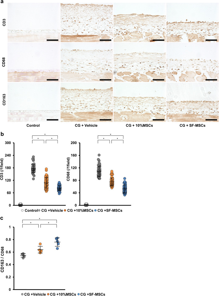
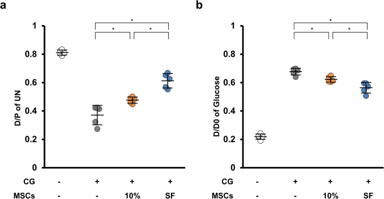
Histological staining showed that the SF-MSCs significantly suppressed CG-induced cell accumulation and thickening compared with that of the 10%MSCs. Additionally, the SF-MSCs significantly inhibited mesenchymal cell expression, extracellular matrix protein deposition and inflammatory cell infiltration. Peritoneal equilibration testing showed that compared with administering 10%MSCs, administering SF-MSCs significantly reduced the functional impairments of the peritoneal membrane. The in vitro experiments showed that although the conditioned medium from MSCs suppressed TGF-β1 signaling, the suppression did not significantly differ between the SF-MSCs and 10%MSCs.
Serum-free culture conditions can enhance the antifibrotic abilities of MSCs by suppressing inflammation. Administering ex vivo expanded SF-MSCs may be a potential therapy for preventing peritoneal fibrotic progression.
间充质干细胞(MSCs)为腹膜纤维化提供了潜在的治疗方法。然而,在含有血清的培养基中培养的 MSCs 会带来感染和其他问题的风险。在这项研究中,我们比较了无血清培养基(SF-MSCs)中培养的人 MSCs 与含 10%胎牛血清的培养基中培养的 MSCs 对腹膜纤维化的影响。
通过腹腔内注射 0.1%葡萄糖酸氯己定(CG)诱导腹膜纤维化。CG 注射后 30 分钟,腹腔内给予 SF-MSCs 或 10%MSCs。CG 和 MSC 注射后 10 天,进行组织学分析和腹膜平衡试验。在体外实验中,我们使用转化生长因子(TGF)-β1 刺激的人腹膜间皮细胞,孵育于 MSC 来源的条件培养基中,以检测 SF-MSCs 是否显示出增强产生抗纤维化体液因子的能力。
组织学染色显示,与 10%MSCs 相比,SF-MSCs 显著抑制 CG 诱导的细胞积聚和增厚。此外,SF-MSCs 显著抑制间充质细胞表达、细胞外基质蛋白沉积和炎症细胞浸润。腹膜平衡试验表明,与给予 10%MSCs 相比,给予 SF-MSCs 可显著降低腹膜膜的功能损伤。体外实验表明,尽管 MSC 来源的条件培养基抑制了 TGF-β1 信号,但 SF-MSCs 与 10%MSCs 之间的抑制作用无显著差异。
无血清培养条件可通过抑制炎症增强 MSCs 的抗纤维化能力。给予体外扩增的 SF-MSCs 可能是预防腹膜纤维化进展的一种潜在治疗方法。