Department of Emergency Medicine, San Luigi Gonzaga University Hospital, Regione Gonzole 10, Orbassano, 10024, Turin, Italy.
Institute of Clinical Physiology, National Research Council, Via Moruzzi 1, 56124, Pisa, Italy.
Intensive Care Med. 2021 Apr;47(4):444-454. doi: 10.1007/s00134-021-06373-7. Epub 2021 Mar 20.
To analyze the application of a lung ultrasound (LUS)-based diagnostic approach to patients suspected of COVID-19, combining the LUS likelihood of COVID-19 pneumonia with patient's symptoms and clinical history.
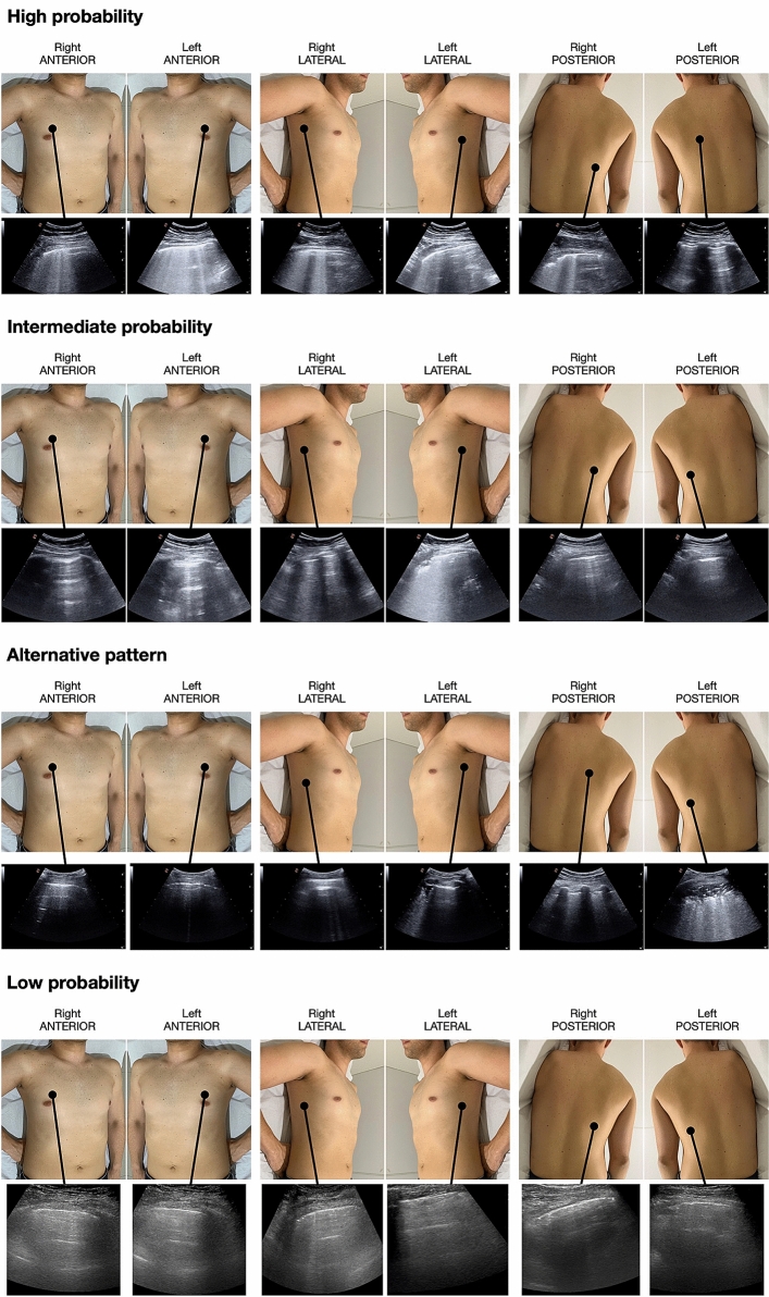
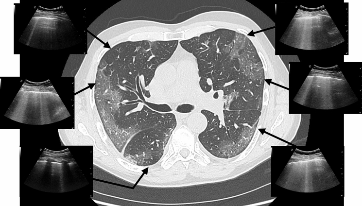
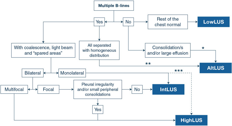
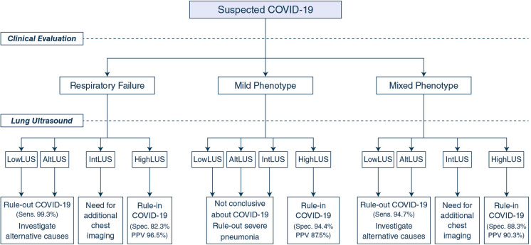
This is an international multicenter observational study in 20 US and European hospitals. Patients suspected of COVID-19 were tested with reverse transcription-polymerase chain reaction (RT-PCR) swab test and had an LUS examination. We identified three clinical phenotypes based on pre-existing chronic diseases (mixed phenotype), and on the presence (severe phenotype) or absence (mild phenotype) of signs and/or symptoms of respiratory failure at presentation. We defined the LUS likelihood of COVID-19 pneumonia according to four different patterns: high (HighLUS), intermediate (IntLUS), alternative (AltLUS), and low (LowLUS) probability. The combination of patterns and phenotypes with RT-PCR results was described and analyzed.
We studied 1462 patients, classified in mild (n = 400), severe (n = 727), and mixed (n = 335) phenotypes. HighLUS and IntLUS showed an overall sensitivity of 90.2% (95% CI 88.23-91.97%) in identifying patients with positive RT-PCR, with higher values in the mixed (94.7%) and severe phenotype (97.1%), and even higher in those patients with objective respiratory failure (99.3%). The HighLUS showed a specificity of 88.8% (CI 85.55-91.65%) that was higher in the mild phenotype (94.4%; CI 90.0-97.0%). At multivariate analysis, the HighLUS was a strong independent predictor of RT-PCR positivity (odds ratio 4.2, confidence interval 2.6-6.7, p < 0.0001).
Combining LUS patterns of probability with clinical phenotypes at presentation can rapidly identify those patients with or without COVID-19 pneumonia at bedside. This approach could support and expedite patients' management during a pandemic surge.
分析一种基于肺部超声(LUS)的诊断方法在疑似 COVID-19 患者中的应用,将 LUS 对 COVID-19 肺炎的可能性与患者的症状和临床病史相结合。
这是一项在 20 家美国和欧洲医院进行的国际多中心观察性研究。疑似 COVID-19 的患者接受逆转录-聚合酶链反应(RT-PCR)拭子检测,并进行 LUS 检查。我们根据预先存在的慢性疾病(混合表型)以及出现时呼吸衰竭的体征和/或症状的存在(严重表型)或不存在(轻度表型),确定了三种临床表型。我们根据四种不同的模式定义了 COVID-19 肺炎的 LUS 可能性:高(HighLUS)、中(IntLUS)、替代(AltLUS)和低(LowLUS)概率。描述和分析了模式和表型与 RT-PCR 结果的组合。
我们研究了 1462 名患者,分为轻度(n=400)、重度(n=727)和混合(n=335)表型。HighLUS 和 IntLUS 在识别 RT-PCR 阳性患者方面的总体敏感性为 90.2%(95%CI 88.23-91.97%),在混合(94.7%)和重度表型(97.1%)中更高,在有客观呼吸衰竭的患者中甚至更高(99.3%)。HighLUS 的特异性为 88.8%(CI 85.55-91.65%),在轻度表型中更高(94.4%;CI 90.0-97.0%)。多变量分析显示,HighLUS 是 RT-PCR 阳性的强独立预测因子(比值比 4.2,置信区间 2.6-6.7,p<0.0001)。
将 LUS 概率模式与就诊时的临床表型相结合,可以快速识别出有或没有 COVID-19 肺炎的患者。这种方法可以支持并加快大流行期间患者的管理。