Scholefield Melissa, Church Stephanie J, Xu Jingshu, Patassini Stefano, Roncaroli Federico, Hooper Nigel M, Unwin Richard D, Cooper Garth J S
Division of Cardiovascular Sciences, Faculty of Biology, Medicine and Health, Centre for Advanced Discovery & Experimental Therapeutics, School of Medical Sciences, The University of Manchester, Manchester Academic Health Science Centre, Manchester, United Kingdom.
Faculty of Science, School of Biological Sciences, University of Auckland, Auckland, New Zealand.
Front Aging Neurosci. 2021 Mar 3;13:641222. doi: 10.3389/fnagi.2021.641222. eCollection 2021.
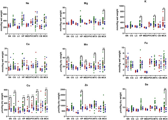
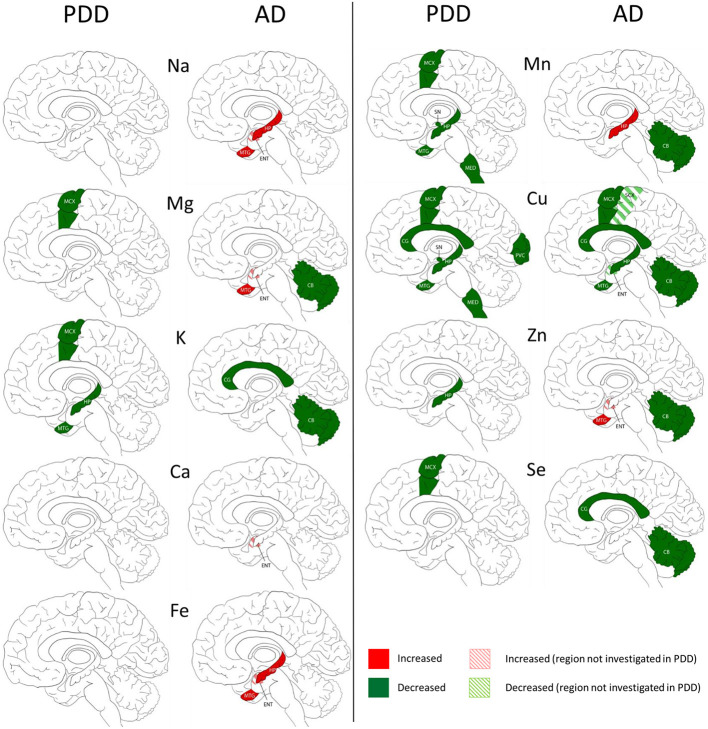
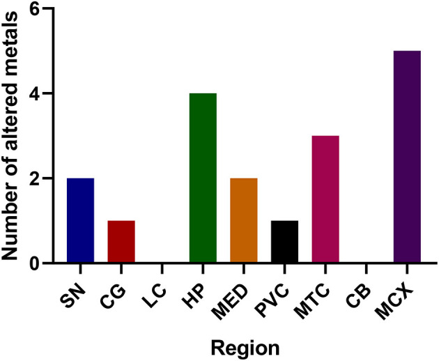
Several studies of Parkinson's disease (PD) have reported dysregulation of cerebral metals, particularly decreases in copper and increases in iron in substantia nigra (SN). However, few studies have investigated regions outside the SN, fewer have measured levels of multiple metals across different regions within the same brains, and there are no currently-available reports of metal levels in Parkinson's disease dementia (PDD). This study aimed to compare concentrations of nine essential metals across nine different brain regions in cases of PDD and controls. Investigated were: primary motor cortex (MCX); cingulate gyrus (CG); primary visual cortex (PVC); hippocampus (HP); cerebellar cortex (CB); SN; locus coeruleus (LC); medulla oblongata (MED); and middle temporal gyrus (MTG), thus covering regions with severe, moderate, or low levels of neuronal loss in PDD. Levels of eight essential metals and selenium were determined using an analytical methodology involving the use of inductively-coupled plasma mass spectrometry (ICP-MS), and compared between cases and controls, to better understand the extent and severity of metal perturbations. Findings were also compared with those from our previous study of sporadic Alzheimer's disease dementia (ADD), which employed equivalent methods, to identify differences and similarities between these conditions. Widespread copper decreases occurred in PDD in seven of nine regions (exceptions being LC and CB). Four PDD-affected regions showed similar decreases in ADD: CG, HP, MTG, and MCX. Decreases in potassium and manganese were present in HP, MTG and MCX; decreased manganese was also found in SN and MED. Decreased selenium and magnesium were present in MCX, and decreased zinc in HP. There was no evidence for increased iron in SN or any other region. These results identify alterations in levels of several metals across multiple regions of PDD brain, the commonest being widespread decreases in copper that closely resemble those in ADD, pointing to similar disease mechanisms in both dementias.
多项关于帕金森病(PD)的研究报告称,大脑中的金属元素失调,尤其是黑质(SN)中铜含量降低、铁含量升高。然而,很少有研究对黑质以外的区域进行调查,更少有人在同一大脑的不同区域测量多种金属的含量,目前也没有关于帕金森病痴呆(PDD)患者金属含量的报告。本研究旨在比较PDD患者和对照组九个不同脑区中九种必需金属的浓度。研究的脑区包括:初级运动皮层(MCX);扣带回(CG);初级视觉皮层(PVC);海马体(HP);小脑皮层(CB);黑质(SN);蓝斑(LC);延髓(MED);以及颞中回(MTG),从而覆盖了PDD中神经元损失程度严重、中度或轻度的区域。使用电感耦合等离子体质谱法(ICP-MS)的分析方法测定了八种必需金属和硒的含量,并在病例组和对照组之间进行比较,以更好地了解金属紊乱的程度和严重性。研究结果还与我们之前采用相同方法对散发性阿尔茨海默病痴呆(ADD)的研究结果进行了比较,以确定这两种疾病之间的异同。PDD患者九个区域中的七个区域出现了广泛的铜含量降低(LC和CB除外)。四个受PDD影响的区域在ADD中也出现了类似的降低:CG、HP、MTG和MCX。HP、MTG和MCX中钾和锰含量降低;SN和MED中也发现锰含量降低。MCX中硒和镁含量降低,HP中锌含量降低。没有证据表明SN或任何其他区域的铁含量增加。这些结果表明,PDD患者大脑多个区域的几种金属含量发生了变化,最常见的是广泛的铜含量降低,这与ADD中的情况非常相似,表明这两种痴呆症存在相似的疾病机制。