McDermott Conor, Łącki Maciej, Sainsbury Ben, Henry Jessica, Filippov Mihail, Rossa Carlos
Faculty of Engineering and Applied Science, Ontario Tech University, Oshawa, ON, Canada.
Marion Surgical, Toronto, ON, Canada.
Front Big Data. 2021 Mar 9;4:612561. doi: 10.3389/fdata.2021.612561. eCollection 2021.
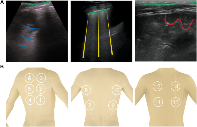
The sustained increase in new cases of COVID-19 across the world and potential for subsequent outbreaks call for new tools to assist health professionals with early diagnosis and patient monitoring. Growing evidence around the world is showing that lung ultrasound examination can detect manifestations of COVID-19 infection. Ultrasound imaging has several characteristics that make it ideally suited for routine use: small hand-held systems can be contained inside a protective sheath, making it easier to disinfect than X-ray or computed tomography equipment; lung ultrasound allows triage of patients in long term care homes, tents or other areas outside of the hospital where other imaging modalities are not available; and it can determine lung involvement during the early phases of the disease and monitor affected patients at bedside on a daily basis. However, some challenges still remain with routine use of lung ultrasound. Namely, current examination practices and image interpretation are quite challenging, especially for unspecialized personnel. This paper reviews how lung ultrasound (LUS) imaging can be used for COVID-19 diagnosis and explores different image processing methods that have the potential to detect manifestations of COVID-19 in LUS images. Then, the paper reviews how general lung ultrasound examinations are performed before addressing how COVID-19 manifests itself in the images. This will provide the basis to study contemporary methods for both segmentation and classification of lung ultrasound images. The paper concludes with a discussion regarding practical considerations of lung ultrasound image processing use and draws parallels between different methods to allow researchers to decide which particular method may be best considering their needs. With the deficit of trained sonographers who are working to diagnose the thousands of people afflicted by COVID-19, a partially or totally automated lung ultrasound detection and diagnosis tool would be a major asset to fight the pandemic at the front lines.
全球新冠肺炎新病例持续增加以及后续爆发的可能性,需要新的工具来协助卫生专业人员进行早期诊断和患者监测。世界各地越来越多的证据表明,肺部超声检查可以检测出新冠肺炎感染的表现。超声成像具有几个使其非常适合常规使用的特点:小型手持系统可以装在保护套内,比X射线或计算机断层扫描设备更容易消毒;肺部超声可以对长期护理机构、帐篷或医院外其他无法使用其他成像方式的区域的患者进行分流;它可以在疾病早期确定肺部受累情况,并每天在床边对受影响的患者进行监测。然而,肺部超声的常规使用仍然存在一些挑战。也就是说,目前的检查方法和图像解读颇具挑战性,尤其是对于非专业人员而言。本文回顾了肺部超声(LUS)成像如何用于新冠肺炎诊断,并探讨了有可能在LUS图像中检测出新冠肺炎表现的不同图像处理方法。然后,在阐述新冠肺炎在图像中的表现之前,本文先回顾了一般肺部超声检查是如何进行的。这将为研究肺部超声图像分割和分类的当代方法提供基础。本文最后讨论了肺部超声图像处理应用的实际考虑因素,并对不同方法进行了比较,以便研究人员根据自身需求决定哪种特定方法可能是最佳选择。鉴于致力于诊断数千名新冠肺炎患者的训练有素的超声检查人员短缺,一种部分或完全自动化的肺部超声检测和诊断工具将成为抗击疫情前线的一项重要资产。