Jalil Bilal A, Khan Ahsan, Kugasia Irfanali R, Ijaz Mohsin
Department of Pulmonary and Critical Care Medicine, Baylor Scott and White Medical Center, Waxahachie, Texas.
Proc (Bayl Univ Med Cent). 2020 Oct 26;34(1):1-4. doi: 10.1080/08998280.2020.1834658.
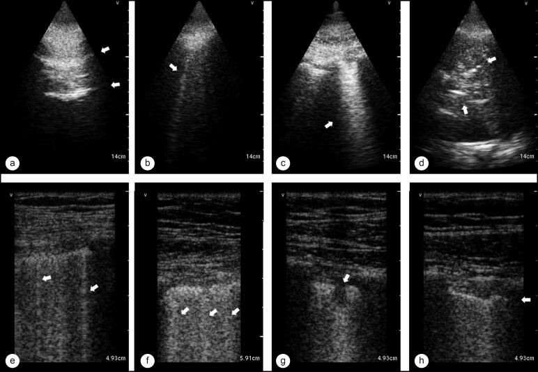
There is a scarcity of data on lung ultrasound (LUS) in SARS-CoV-2 pneumonia. As with many other pulmonary conditions, ultrasound may be a better diagnostic tool than routine chest radiography. In an era where computed tomography scanning is deferred because of the potential for cross-contamination, we evaluated the ability of LUS to detect a pattern of lung injury in SARS-CoV-2 pneumonia. A limited anterolateral LUS was performed to limit time spent in isolation rooms by ultrasound operators. We chose to use a hand-held ultrasound device due to portability and superior confidence in infection control. Both linear and phased array probes were used to obtain images of the pleura and lung. Of 69 patients who had lung ultrasound images saved and were included in the analysis, 36 were positive for SARS-CoV-2. Multifocal confluent B-lines, pleural irregularities, and the absence of moderate or large pleural effusions were the predominant pattern observed in most (86%) of SARS-CoV-2-positive patients. We evaluated the accuracy of the above criteria (LUS-CoV) and report a high sensitivity (91%) and specificity (86%) for SARS-CoV-2 pneumonia. In conclusion, a characteristic sonographic pattern of multifocal confluent B-lines with irregular pleural markings was seen on LUS in patients with SARS-CoV-2 pneumonia.
关于新型冠状病毒肺炎的肺部超声(LUS)数据匮乏。与许多其他肺部疾病一样,超声可能是比常规胸部X线摄影更好的诊断工具。在因存在交叉污染风险而推迟计算机断层扫描的时代,我们评估了LUS检测新型冠状病毒肺炎肺损伤模式的能力。进行有限的前外侧LUS检查,以减少超声检查人员在隔离病房的停留时间。由于便携性和对感染控制更有信心,我们选择使用手持式超声设备。使用线性和相控阵探头获取胸膜和肺部图像。在保存了肺部超声图像并纳入分析的69例患者中,36例新型冠状病毒检测呈阳性。多灶性融合B线、胸膜不规则以及无中、大量胸腔积液是大多数(86%)新型冠状病毒阳性患者中观察到的主要模式。我们评估了上述标准(LUS-CoV)的准确性,报告其对新型冠状病毒肺炎具有高敏感性(91%)和特异性(86%)。总之,新型冠状病毒肺炎患者的LUS检查可见多灶性融合B线伴胸膜标记不规则的特征性超声模式。