Departments of Pharmacology and Experimental Therapeutics, Boston University School of Medicine, Boston, MA, 02118, USA.
Department of Cell Biology, Harvard Medical School, Boston, MA, 02115, USA.
Mol Neurodegener. 2021 Mar 22;16(1):18. doi: 10.1186/s13024-021-00440-9.
Recent studies suggest that microglia contribute to tau pathology progression in Alzheimer's disease. Amyloid plaque accumulation transforms microglia, the primary innate immune cells in the brain, into neurodegenerative microglia (MGnD), which exhibit enhanced phagocytosis of plaques, apoptotic neurons and dystrophic neurites containing aggregated and phosphorylated tau (p-tau). It remains unclear how microglia promote disease progression while actively phagocytosing pathological proteins, therefore ameliorating pathology.
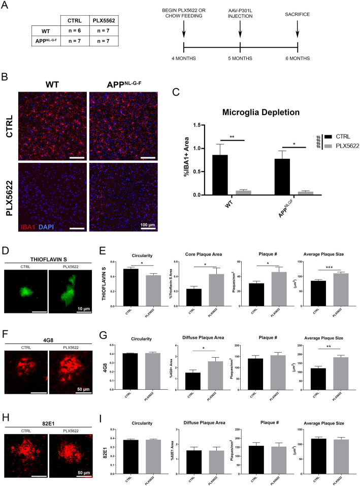
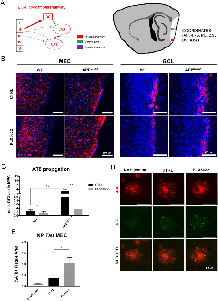
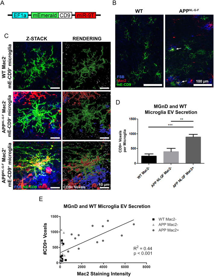
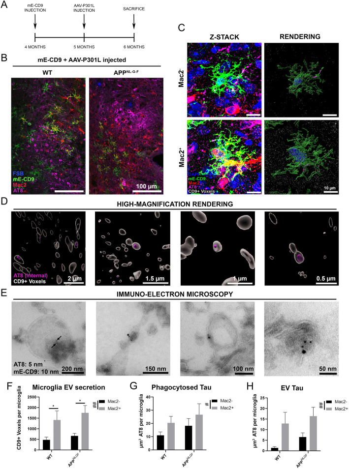
Adeno-associated virus expressing P301L tau mutant (AAV-P301L-tau) was stereotaxically injected into the medial entorhinal cortex (MEC) in C57BL/6 (WT) and humanized APP mutant knock-in homozygote (App) mice at 5 months of age. Mice were fed either chow containing a colony stimulating factor-1 receptor inhibitor (PLX5622) or control chow from 4 to 6 months of age to test the effect of microglia depletion. Animals were tested at 6 months of age for immunofluorescence, biochemistry, and FACS of microglia. In order to monitor microglial extracellular vesicle secretion in vivo, a novel lentiviral EV reporter system was engineered to express mEmerald-CD9 (mE-CD9) specifically in microglia, which was injected into the same region of MEC.
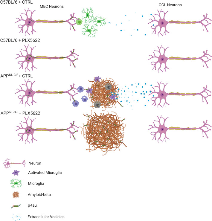
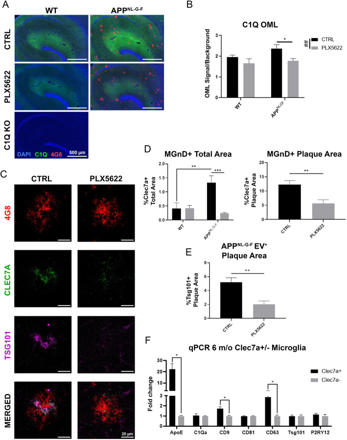
Expressing P301L tau mutant in the MEC induced tau propagation to the granule cell layer of the hippocampal dentate gyrus, which was significantly exacerbated in App mice compared to WT control mice. Administration of PLX5622 depleted nearly all microglia in mouse brains and dramatically reduced propagation of p-tau in WT and to a greater extent in App mice, although it increased plaque burden and plaque-associated p-tau dystrophic neurites. Plaque-associated MGnD microglia strongly expressed an EV marker, tumor susceptibility gene 101, indicative of heightened synthesis of EVs. Intracortical injection of mE-CD9 lentivirus successfully induced microglia-specific expression of mE-CD9 EV particles, which were significantly enhanced in Mac2 MGnD microglia compared to Mac2 homeostatic microglia. Finally, consecutive intracortical injection of mE-CD9 lentivirus and AAV-P301L-tau into App mice revealed encapsulation of p-tau in microglia-specific mE-CD9 EVs as determined by super-resolution microscopy and immuno-electron microscopy.
Our findings suggest that MGnD microglia hyper-secrete p-tau EVs while compacting Aβ plaques and clearing NP tau, which we propose as a novel mechanistic link between amyloid plaque deposition and exacerbation of tau propagation in App mice.
最近的研究表明,小胶质细胞有助于阿尔茨海默病中 tau 病理的进展。淀粉样斑块的积累使大脑中的主要先天免疫细胞小胶质细胞转化为神经退行性小胶质细胞(MGnD),其表现为增强对斑块、凋亡神经元和含有聚集和磷酸化 tau(p-tau)的变性神经突的吞噬作用。目前尚不清楚小胶质细胞如何在积极吞噬病理蛋白的同时促进疾病进展,从而改善病理。
腺相关病毒表达 P301L tau 突变体(AAV-P301L-tau)在 5 月龄时通过立体定向注射到 C57BL/6(WT)和人源化 APP 突变体敲入纯合子(App)小鼠的内侧隔核(MEC)中。从 4 到 6 月龄,用含有集落刺激因子 1 受体抑制剂(PLX5622)的饮食或对照饮食喂养小鼠,以测试小胶质细胞耗竭的效果。在 6 月龄时对动物进行免疫荧光、生物化学和小胶质细胞的流式细胞术检测。为了监测体内小胶质细胞细胞外囊泡的分泌,设计了一种新型慢病毒 EV 报告系统,该系统专门在小胶质细胞中表达 mEmerald-CD9(mE-CD9),并将其注射到 MEC 的同一区域。
在 MEC 中表达 P301L tau 突变体诱导 tau 向海马齿状回颗粒细胞层传播,与 WT 对照小鼠相比,在 App 小鼠中这种传播明显加剧。PLX5622 的给药耗尽了小鼠大脑中的几乎所有小胶质细胞,并显著减少了 WT 和 App 小鼠中 p-tau 的传播,但增加了斑块负担和斑块相关的 p-tau 神经突变性。斑块相关的 MGnD 小胶质细胞强烈表达 EV 标志物肿瘤易感性基因 101,表明 EV 的合成增强。皮层内注射 mE-CD9 慢病毒成功诱导了 mE-CD9 EV 颗粒在小胶质细胞中的特异性表达,与 Mac2 静息小胶质细胞相比,Mac2 MGnD 小胶质细胞中 mE-CD9 EV 颗粒显著增强。最后,连续在 App 小鼠的皮层内注射 mE-CD9 慢病毒和 AAV-P301L-tau 揭示了 p-tau 在小胶质细胞特异性 mE-CD9 EV 中的封装,这通过超分辨率显微镜和免疫电子显微镜确定。
我们的发现表明,MGnD 小胶质细胞过度分泌 p-tau EVs,同时使 Aβ 斑块致密化并清除 NP tau,我们认为这是 APP 小鼠中淀粉样斑块沉积和 tau 传播加剧之间的一种新的机制联系。