Lin Ling, Chen Shuyi, Wang Hua, Gao Bin, Kallakury Bhaskar, Bhuvaneshwar Krithika, Cahn Katherine, Gusev Yuriy, Wang Xue, Wu Yunan, Marshall John L, Zhi Xiuling, He Aiwu Ruth
Department of Medicine and Oncology, Lombardi Comprehensive Cancer Center, Georgetown University, Washington, DC, USA.
Department of Physiology and Pathophysiology, School of Basic Medical Sciences, Fudan University, Shanghai, China.
Theranostics. 2021 Feb 20;11(9):4232-4250. doi: 10.7150/thno.49819. eCollection 2021.
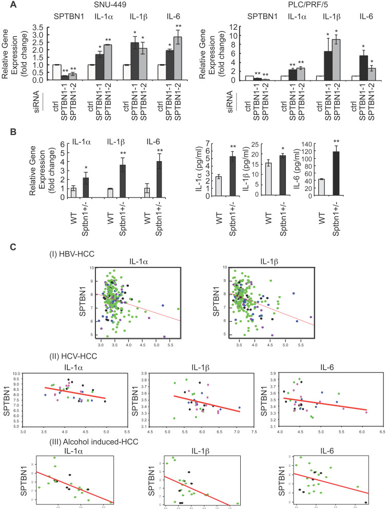
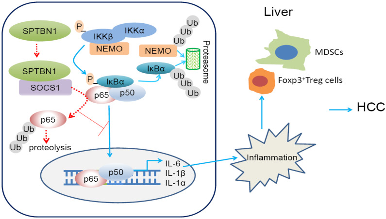
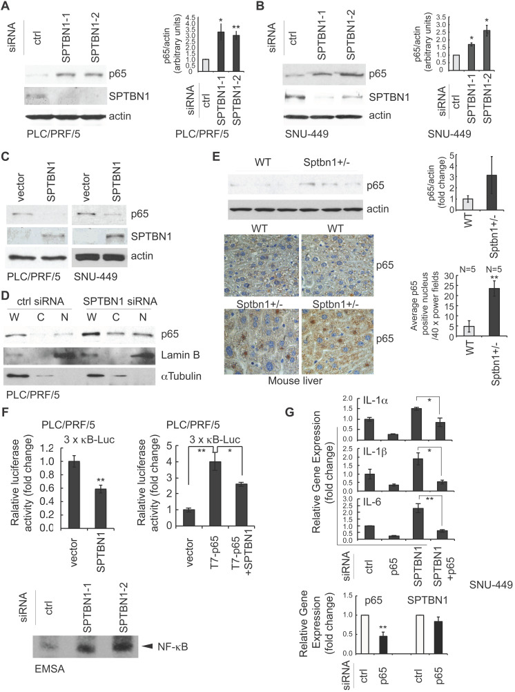
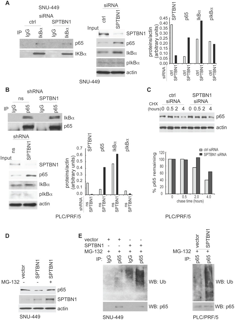
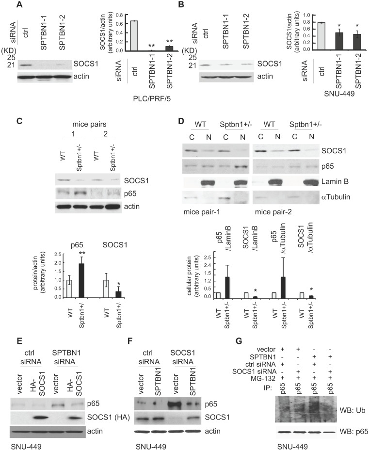
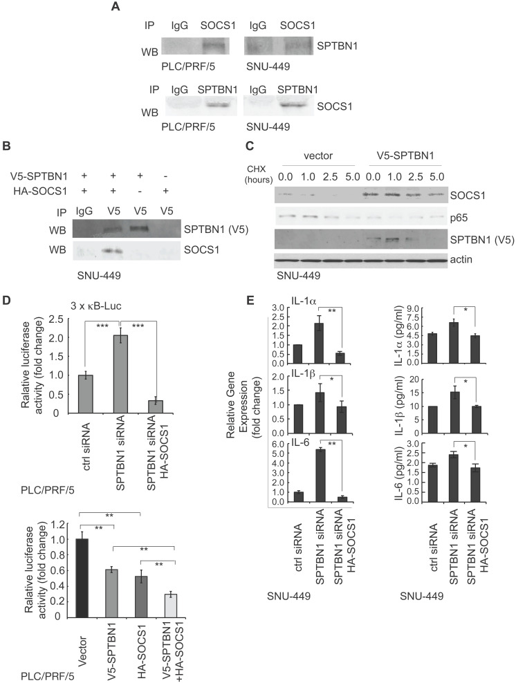
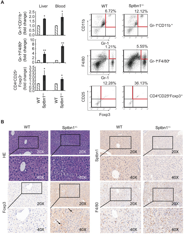
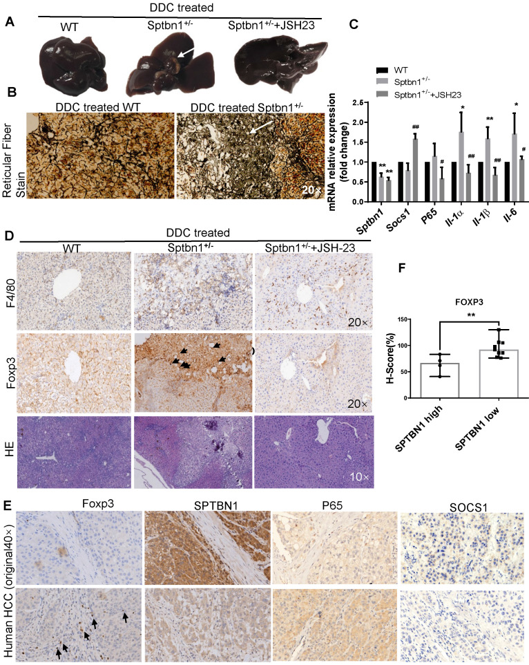
Spectrin, beta, non-erythrocytic 1 (SPTBN1), an adapter protein for transforming growth factor beta (TGF-β) signaling, is recognized as a tumor suppressor in the development of hepatocellular carcinoma (HCC); however, the underlying molecular mechanisms of this tumor suppression remain obscure. The effects on expression of pro-inflammatory cytokines upon the inhibition or impairment of SPTBN1 in HCC cell lines and liver tissues of and wild-type (WT) mice were assessed by analyses of quantitative real-time reverse-transcription polymerase chain reaction (QRT-PCR), enzyme linked immunosorbent assay (ELISA), Western blotting and gene array databases from HCC patients. We investigated the detailed molecular mechanisms underlying the inflammatory responses by immunoprecipitation-Western blotting, luciferase reporter assay, chromatin immunoprecipitation quantitative real time PCR (ChIP-qPCR), immunohistochemistry (IHC) and electrophoretic mobility shift assay (EMSA). The proportion of myeloid-derived suppressor cells in liver, spleen, bone marrow and peripheral blood samples from WT and mice were measured by fluorescence-activated cell sorting (FACS) analysis. Further, the hepatocacinogenesis and its correlation with inflammatory microenvironment by loss of SPTBN1/SOCS1 and induction of p65 were analyzed by treating WT and mice with 3,5-diethoxycarbonyl-1,4-dihydrocollidine (DDC). Loss of SPTBN1 in HCC cells upregulated the expression of pro-inflammatory cytokines including interleukin-1α (IL-1α), IL-1β, and IL-6, and enhanced NF-κB transcriptional activation. Mechanistic analyses revealed that knockdown of SPTBN1 by siRNA downregulated the expression of suppressor of cytokine signaling 1 (SOCS1), an E3 ligase of p65, and subsequently upregulated p65 accumulation in the nucleus of HCC cells. Restoration of SOCS1 abrogated this SPTBN1 loss-associated elevation of p65 in HCC cells. In human HCC tissues, SPTBN1 gene expression was inversely correlated with gene expression of IL-1α, IL-1β and IL-6. Furthermore, a decrease in the levels of SPTBN1 gene, as well as an increase in the gene expression of IL-1β or IL-6 predicted shorter relapse free survival in HCC patients, and that HCC patients with low expression of SPTBN1 or SOCS1 protein is associated with poor survival. Heterozygous loss of SPTBN1 ( ) in mice markedly upregulated hepatic expression of IL-1α, IL-1β and IL-6, and elevated the proportion of myeloid-derived suppressor cells (MDSCs) and CD4CD25Foxp3 regulatory T cells (Foxp3Treg) cells in the liver, promoting hepatocarcinogenesis of mouse fed by DDC. : Our findings provided evidence that loss of SPTBN1 in HCC cells increases p65 protein stability via the inhibition of SOCS1 and enhances NF-κB activation, stimulating the release of inflammatory cytokines, which are critical molecular mechanisms for the loss of SPTBN1-induced liver cancer formation. Reduced SPTBN1 and SOCS1 predict poor outcome in HCC patients.
血影蛋白β非红细胞型1(SPTBN1)是转化生长因子β(TGF-β)信号传导的衔接蛋白,被认为是肝细胞癌(HCC)发生发展过程中的一种肿瘤抑制因子;然而,这种肿瘤抑制作用的潜在分子机制仍不清楚。通过定量实时逆转录聚合酶链反应(QRT-PCR)分析、酶联免疫吸附测定(ELISA)、蛋白质免疫印迹法以及来自HCC患者的基因芯片数据库,评估了在HCC细胞系以及野生型(WT)小鼠的肝脏组织中抑制或损伤SPTBN1后对促炎细胞因子表达的影响。我们通过免疫沉淀-蛋白质免疫印迹法、荧光素酶报告基因检测、染色质免疫沉淀定量实时PCR(ChIP-qPCR)、免疫组织化学(IHC)和电泳迁移率变动分析(EMSA),研究了炎症反应背后的详细分子机制。通过荧光激活细胞分选(FACS)分析测量WT和特定基因敲除小鼠肝脏、脾脏、骨髓和外周血样本中髓源性抑制细胞的比例。此外,通过用3,5-二乙氧基羰基-1,4-二氢可力丁(DDC)处理WT和特定基因敲除小鼠,分析了SPTBN1/SOCS1缺失和p65诱导对肝癌发生及其与炎症微环境的相关性。HCC细胞中SPTBN1的缺失上调了包括白细胞介素-1α(IL-1α)、IL-1β和IL-6在内的促炎细胞因子的表达,并增强了NF-κB转录激活。机制分析表明,小干扰RNA(siRNA)敲低SPTBN1下调了细胞因子信号传导抑制因子1(SOCS1)的表达,SOCS1是p65的E3连接酶,随后上调了HCC细胞核中p65的积累。恢复SOCS1可消除HCC细胞中这种与SPTBN1缺失相关的p65升高。在人类HCC组织中,SPTBN1基因表达与IL-1α、IL-1β和IL-6的基因表达呈负相关。此外,SPTBN1基因水平的降低以及IL-1β或IL-6基因表达的增加预示着HCC患者无复发生存期较短,并且SPTBN1或SOCS1蛋白低表达的HCC患者生存情况较差。小鼠中SPTBN1的杂合缺失(特定基因敲除)显著上调了肝脏中IL-1α、IL-1β和IL-6的表达,并提高了肝脏中髓源性抑制细胞(MDSCs)和CD4CD25Foxp3调节性T细胞(Foxp3Treg)的比例,促进了DDC喂养小鼠的肝癌发生。我们的研究结果表明,HCC细胞中SPTBN1的缺失通过抑制SOCS1增加了p65蛋白的稳定性并增强了NF-κB激活,刺激了炎症细胞因子的释放,这是SPTBN1缺失诱导肝癌形成的关键分子机制。SPTBN1和SOCS1降低预示着HCC患者预后不良。