Tsuchiya Junichi, Yokoyama Kota, Yamagiwa Ken, Watanabe Ryosuke, Kimura Koichiro, Kishino Mitsuhiro, Chan Chung, Asma Evren, Tateishi Ukihide
Department of Diagnostic Radiology and Nuclear Medicine, Tokyo Medical and Dental University, 1-5-45 Yushima, Bunkyo-ku, Tokyo, 113-8510, Japan.
Canon Medical Research USA, Inc., 706 N. Deerpath Drive, Vernon Hills, IL, 60061, USA.
EJNMMI Phys. 2021 Mar 25;8(1):31. doi: 10.1186/s40658-021-00377-4.
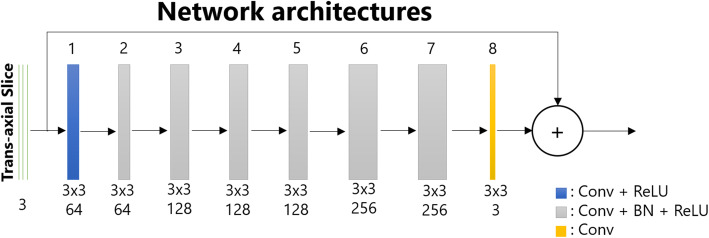
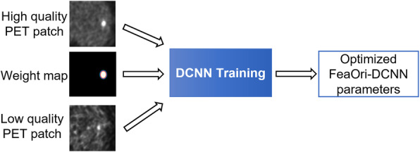
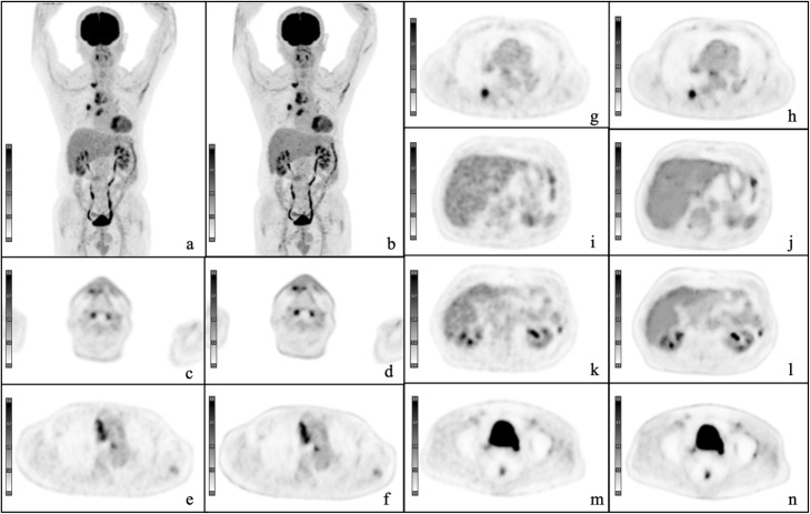
Deep learning (DL)-based image quality improvement is a novel technique based on convolutional neural networks. The aim of this study was to compare the clinical value of F-fluorodeoxyglucose positron emission tomography (F-FDG PET) images obtained with the DL method with those obtained using a Gaussian filter.
Fifty patients with a mean age of 64.4 (range, 19-88) years who underwent F-FDG PET/CT between April 2019 and May 2019 were included in the study. PET images were obtained with the DL method in addition to conventional images reconstructed with three-dimensional time of flight-ordered subset expectation maximization and filtered with a Gaussian filter as a baseline for comparison. The reconstructed images were reviewed by two nuclear medicine physicians and scored from 1 (poor) to 5 (excellent) for tumor delineation, overall image quality, and image noise. For the semi-quantitative analysis, standardized uptake values in tumors and healthy tissues were compared between images obtained using the DL method and those obtained with a Gaussian filter.
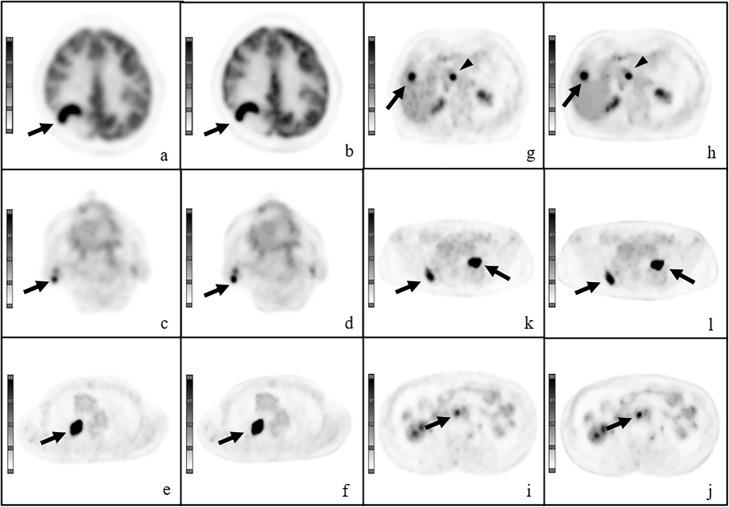
Images acquired using the DL method scored significantly higher for tumor delineation, overall image quality, and image noise compared to baseline (P < 0.001). The Fleiss' kappa value for overall inter-reader agreement was 0.78. The standardized uptake values in tumor obtained by DL were significantly higher than those acquired using a Gaussian filter (P < 0.001).
Deep learning method improves the quality of PET images.
基于深度学习(DL)的图像质量改善是一种基于卷积神经网络的新技术。本研究的目的是比较使用DL方法获得的F-氟脱氧葡萄糖正电子发射断层扫描(F-FDG PET)图像与使用高斯滤波器获得的图像的临床价值。
纳入2019年4月至2019年5月期间接受F-FDG PET/CT检查的50例患者,平均年龄64.4岁(范围19 - 88岁)。除了用三维飞行时间有序子集期望最大化重建并用高斯滤波器滤波作为比较基线的传统图像外,还用DL方法获得PET图像。由两名核医学医师对重建图像进行评估,在肿瘤轮廓、整体图像质量和图像噪声方面从1分(差)到5分(优)进行评分。对于半定量分析,比较使用DL方法获得的图像与使用高斯滤波器获得的图像中肿瘤和健康组织的标准化摄取值。
与基线相比,使用DL方法获得的图像在肿瘤轮廓、整体图像质量和图像噪声方面得分显著更高(P < 0.001)。读者间总体一致性的Fleiss' kappa值为0.78。DL获得的肿瘤标准化摄取值显著高于使用高斯滤波器获得的值(P < 0.001)。
深度学习方法可提高PET图像质量。