Department of Kidney Transplantation, The Third Affiliated Hospital of Sun Yat-sen University, Guangzhou, China.
State Key Laboratory of Membrane Biology, Institute of Zoology, Chinese Academy of Sciences, Beijing, China.
Front Immunol. 2021 Mar 9;12:637335. doi: 10.3389/fimmu.2021.637335. eCollection 2021.
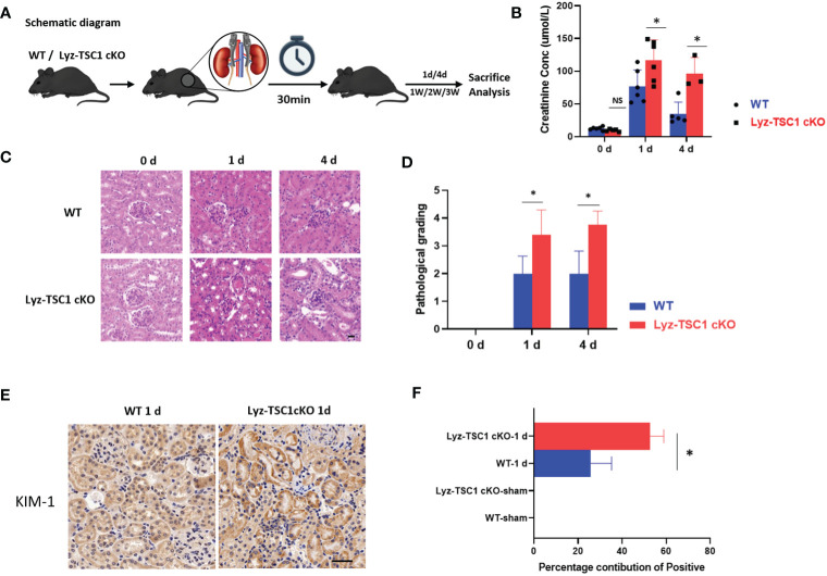
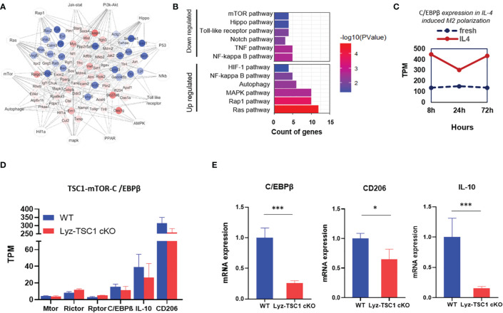
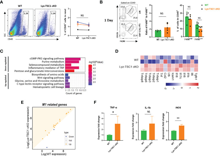
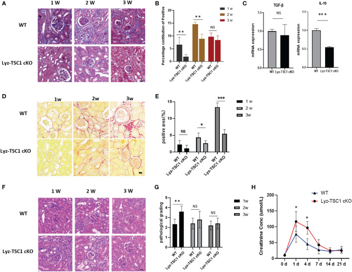
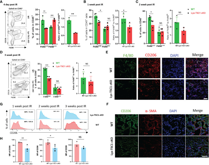
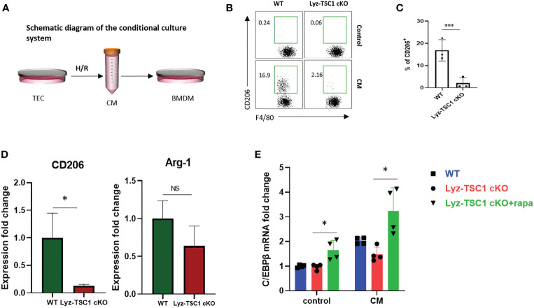
Renal ischemia-reperfusion injury (IRI) contributes to acute kidney injury (AKI), increases morbidity and mortality, and is a significant risk factor for chronic kidney disease (CKD). Macrophage infiltration is a common feature after renal IRI, and infiltrating macrophages can be polarized into the following two distinct types: M1 macrophages, i.e., classically activated macrophages, which can not only inhibit infection but also accelerate renal injury, and M2 macrophages, i.e., alternatively activated macrophages, which have a repair phenotype that can promote wound healing and subsequent fibrosis. The role of TSC1, which is a negative regulator of mTOR signaling that regulates macrophage polarization in inflammation-linked diseases, has been well documented, but whether TSC1 contributes to macrophage polarization in the process of IRI is still unknown. Here, by using a mouse model of renal ischemia-reperfusion, we found that myeloid cell-specific TSC1 knockout mice (termed Lyz-TSC1 cKO mice) had higher serum creatinine levels, more severe histological damage, and greater proinflammatory cytokine production than wild-type (WT) mice during the early phase after renal ischemia-reperfusion. Furthermore, the Lyz-TSC1 cKO mice showed attenuated renal fibrosis during the repair phase of IRI with decreased levels of M2 markers on macrophages in the operated kidneys, which was further confirmed in a cell model of hypoxia-reoxygenation (H/R) . Mechanistically, by using RNA sequencing of sorted renal macrophages, we found that the expression of most M1-related genes was upregulated in the Lyz-TSC1 cKO group (Supplemental Table 1) during the early phase. However, C/EBPβ and CD206 expression was decreased during the repair phase compared to in the WT group. Overall, our findings demonstrate that the expression of TSC1 in macrophages contributes to the whole process of IRI but serves as an inflammation suppressor during the early phase and a fibrosis promoter during the repair phase.
肾缺血再灌注损伤 (IRI) 导致急性肾损伤 (AKI),增加发病率和死亡率,是慢性肾脏病 (CKD) 的重要危险因素。巨噬细胞浸润是肾 IRI 后的一个常见特征,浸润的巨噬细胞可以极化为以下两种截然不同的类型:M1 巨噬细胞,即经典激活的巨噬细胞,不仅能抑制感染,还能加速肾损伤,以及 M2 巨噬细胞,即替代激活的巨噬细胞,具有修复表型,能促进伤口愈合和随后的纤维化。TSC1 是 mTOR 信号的负调节剂,它在炎症相关疾病中调节巨噬细胞极化的作用已得到充分证实,但 TSC1 是否有助于 IRI 过程中的巨噬细胞极化仍不清楚。在这里,我们通过使用肾缺血再灌注的小鼠模型,发现骨髓细胞特异性 TSC1 敲除小鼠 (称为 Lyz-TSC1 cKO 小鼠) 在肾缺血再灌注后早期的血清肌酐水平更高,组织学损伤更严重,促炎细胞因子产生更多,而野生型 (WT) 小鼠则没有。此外,Lyz-TSC1 cKO 小鼠在 IRI 的修复阶段表现出肾纤维化减轻,在手术肾脏的巨噬细胞上 M2 标志物水平降低,这在缺氧再复氧 (H/R) 的细胞模型中得到了进一步证实。从机制上讲,我们通过对分选的肾巨噬细胞进行 RNA 测序,发现 Lyz-TSC1 cKO 组在早期阶段大多数 M1 相关基因的表达上调 (补充表 1)。然而,与 WT 组相比,修复阶段 C/EBPβ 和 CD206 的表达减少。总之,我们的研究结果表明,巨噬细胞中 TSC1 的表达有助于 IRI 的全过程,但在早期阶段作为炎症抑制因子,在修复阶段作为纤维化促进因子。