Department of Nephrology, Affiliated Hangzhou First People's Hospital, Zhejiang University School of Medicine, No. 261, Huansha Road, Hangzhou, 310006, Zhejiang, China.
Department of Nephrology, School of Medicine, The Second Affiliated Hospital, Zhejiang University, Hangzhou, China.
Stem Cell Res Ther. 2022 Jul 28;13(1):367. doi: 10.1186/s13287-022-03075-9.
Ischemia-reperfusion injury (IRI)-induced acute kidney injury (AKI) can repair itself completely. However, most moderate and severe patients undergoing IRI-AKI progress to chronic kidney disease due to incomplete repair. The present study is aimed to investigate the role of bone marrow mesenchymal stem cell-derived exosomes (MSC-Exo) with indoleamine 2,3-dioxygenase (IDO) overexpression on incomplete repair in mice after IRI.
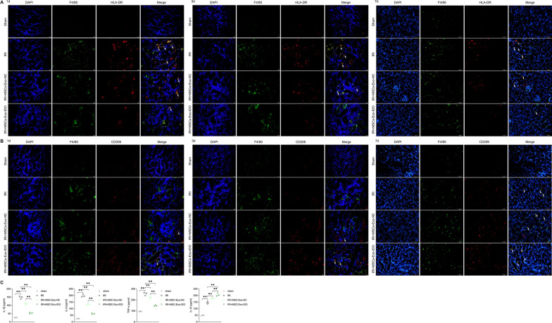
IRI mice was established by clamping the unilateral renal pedicles and challenged with MSC-Exo. Blood biochemical indexes and inflammation factors contents were measured by ELISA assay. Histopathological examinations were monitored by HE, Masson, Immunohistochemical and TUNEL staining. Immunofluorescence, flow cytometry and immunoblotting were used to detect the polarization of macrophages, respectively.
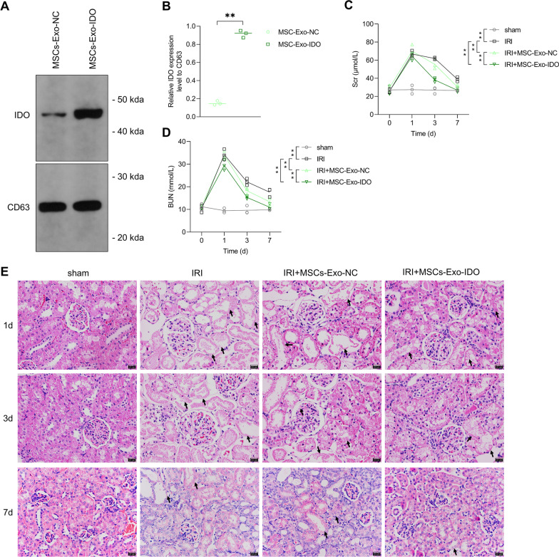
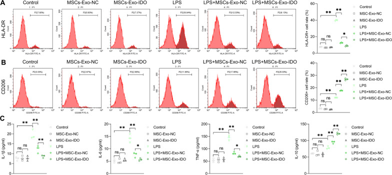
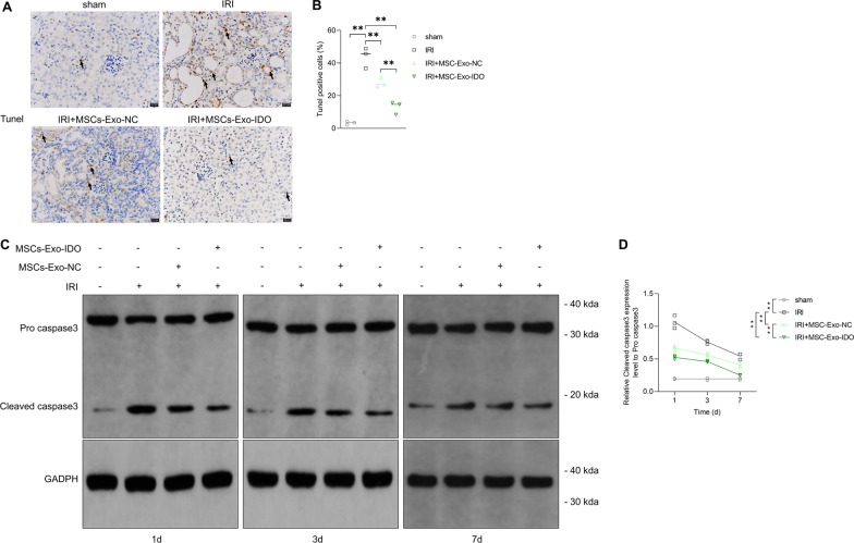
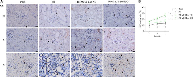
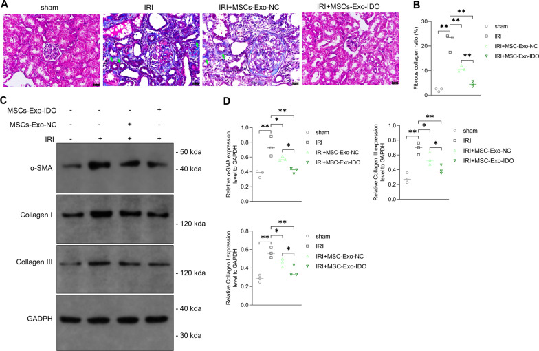
As compared to sham operation mice, IRI mice showed high contents of serum BUN and Scr, and more severe damaged kidney tissues on days 1 and 3, which all gradually declined over time, showing the lowest level on day 7 after injury. Once treated with MSCs-Exo that could directly transfer to kidney tubular cells, the restoration of kidney functions significantly accelerated by contrast to IRI mice, and the promotive effects were more obvious in IDO-overexpressed MSCs-Exo (MSCs-Exo-IDO)-treated IRI mice. Furthermore, MSCs-Exo-IDO administration also accelerated renal tubular cells proliferation, restrained tubular cells apoptosis, fibrosis and inflammation factor secretions during self-repair process compared to IRI mice, whose effects were higher than MSCs-Exo-NC-challenged IRI mice and IDO overexpressing plasmid-injected IRI mice. Mechanistically, MSCs-Exo-NC and MSCs-Exo-IDO exposure promoted the polarization from M1 macrophage to M2 macrophage, leading to more anti-inflammatory factors production, and subsequently altered the inflammatory microenvironment of renal tubular cells, which facilitated the self-repair process in mice after IRI.
MSCs-derived exosome accelerated renal self-repair in IRI mice by activating M2 macrophages polarization, which effects were amplified by IDO overexpression in MSCs. Potentially, genetically modified MSCs-Exo is an effective approach to improve renal self-repair in IRI-AKI mice.
缺血再灌注损伤(IRI)引起的急性肾损伤(AKI)可完全自行修复。然而,大多数经历 IRI-AKI 的中度和重度患者由于修复不完全而进展为慢性肾脏病。本研究旨在探讨过表达吲哚胺 2,3-双加氧酶(IDO)的骨髓间充质干细胞衍生外泌体(MSC-Exo)在 IRI 后小鼠不完全修复中的作用。
通过夹闭单侧肾蒂建立 IRI 小鼠模型,并给予 MSC-Exo 处理。通过 ELISA 测定血液生化指标和炎症因子含量。通过 HE、Masson、免疫组化和 TUNEL 染色监测组织病理学检查。免疫荧光、流式细胞术和免疫印迹法分别用于检测巨噬细胞的极化。
与假手术组小鼠相比,IRI 组小鼠在第 1 天和第 3 天血清 BUN 和 Scr 含量较高,肾脏组织损伤较严重,随着时间的推移逐渐下降,损伤后第 7 天降至最低水平。给予可直接转染至肾小管细胞的 MSC-Exo 处理后,与 IRI 组小鼠相比,肾功能恢复明显加快,而过表达 IDO 的 MSC-Exo(MSC-Exo-IDO)处理的 IRI 组小鼠的促进作用更为明显。此外,与 IRI 组小鼠相比,MSC-Exo-IDO 给药还可在自我修复过程中加速肾小管细胞增殖,抑制肾小管细胞凋亡、纤维化和炎症因子分泌,其作用高于 MSC-Exo-NC 处理的 IRI 组小鼠和 IDO 过表达质粒注射的 IRI 组小鼠。机制上,MSC-Exo-NC 和 MSC-Exo-IDO 暴露促进了 M1 巨噬细胞向 M2 巨噬细胞的极化,导致更多抗炎因子的产生,进而改变了肾小管细胞的炎症微环境,促进了 IRI 后小鼠的自我修复过程。
MSC 衍生的外泌体通过激活 M2 巨噬细胞极化加速 IRI 小鼠的肾自我修复,而 MSC 中的 IDO 过表达则放大了这一效应。潜在地,基因修饰的 MSC-Exo 是改善 IRI-AKI 小鼠肾自我修复的有效方法。