• 文献检索
  • 文档翻译
  • 深度研究
  • 学术资讯
  • Suppr Zotero 插件Zotero 插件
  • 邀请有礼
  • 套餐&价格
  • 历史记录
应用&插件
Suppr Zotero 插件Zotero 插件浏览器插件Mac 客户端Windows 客户端微信小程序
定价
高级版会员购买积分包购买API积分包
服务
文献检索文档翻译深度研究API 文档MCP 服务
关于我们
关于 Suppr公司介绍联系我们用户协议隐私条款
关注我们

Suppr 超能文献

核心技术专利:CN118964589B侵权必究
粤ICP备2023148730 号-1Suppr @ 2026

文献检索

告别复杂PubMed语法,用中文像聊天一样搜索,搜遍4000万医学文献。AI智能推荐,让科研检索更轻松。

立即免费搜索

文件翻译

保留排版,准确专业,支持PDF/Word/PPT等文件格式,支持 12+语言互译。

免费翻译文档

深度研究

AI帮你快速写综述,25分钟生成高质量综述,智能提取关键信息,辅助科研写作。

立即免费体验

利用后巩膜断层抬高进行病理性近视的机器学习预测。

Machine learning prediction of pathologic myopia using tomographic elevation of the posterior sclera.

机构信息

Department of Ophthalmology, Incheon St. Mary's Hospital, College of Medicine, The Catholic University of Korea, Seoul, Republic of Korea.

Department of Medical Informatics, College of Medicine, The Catholic University of Korea, Seoul, Republic of Korea.

出版信息

Sci Rep. 2021 Mar 26;11(1):6950. doi: 10.1038/s41598-021-85699-0.

DOI:10.1038/s41598-021-85699-0
PMID:33772040
原文链接:https://pmc.ncbi.nlm.nih.gov/articles/PMC7997908/
Abstract

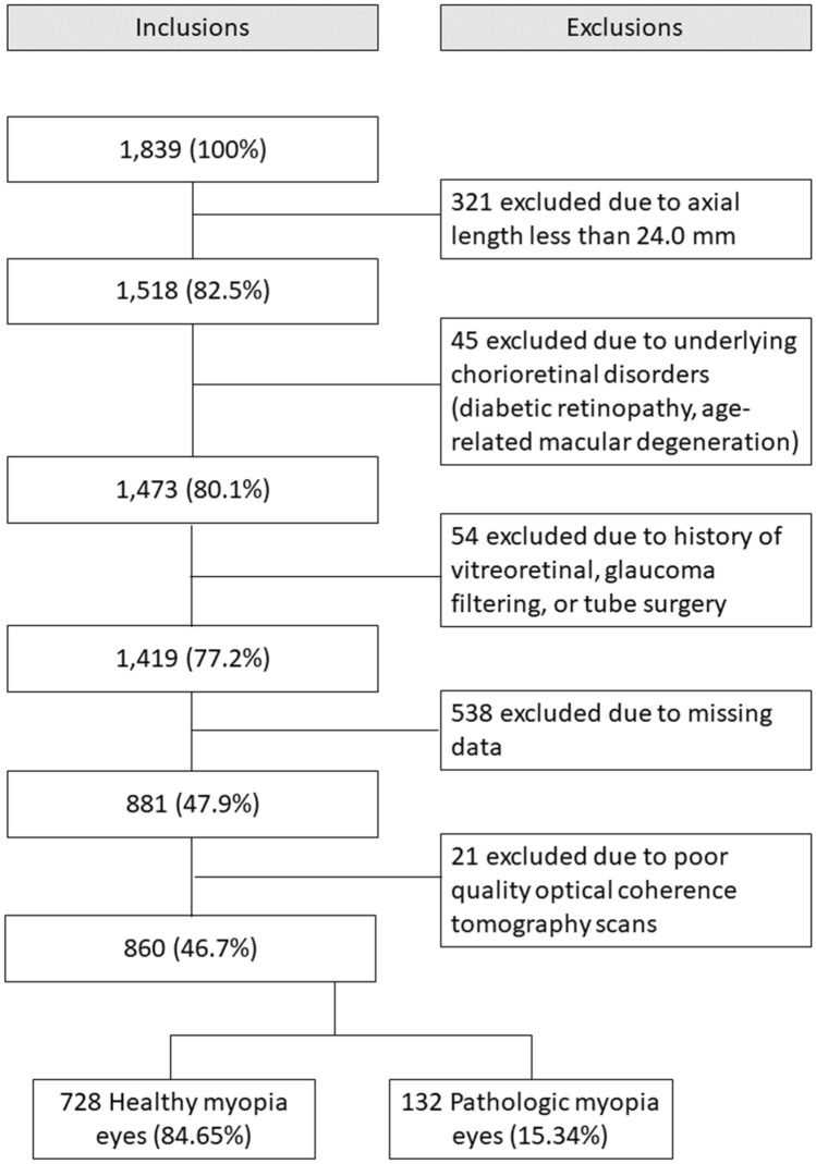
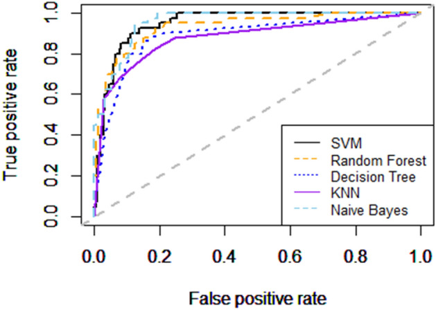
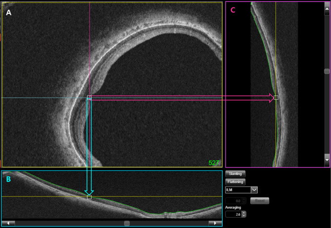
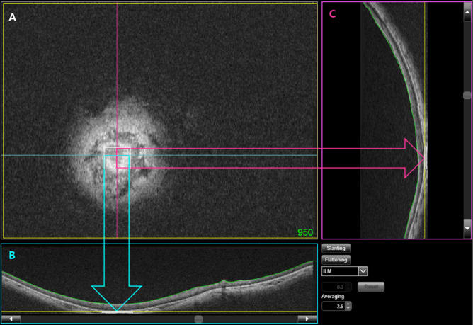
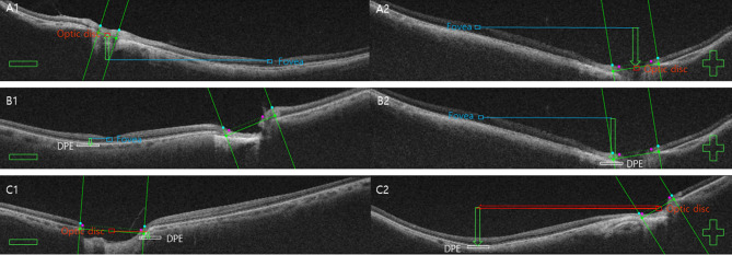
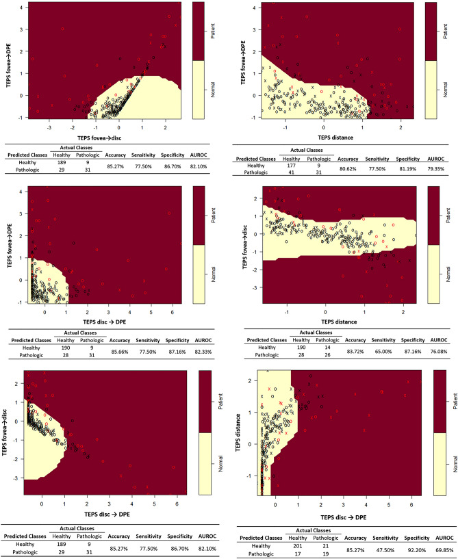
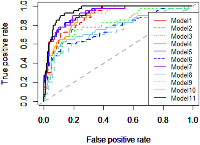
Qualitative analysis of fundus photographs enables straightforward pattern recognition of advanced pathologic myopia. However, it has limitations in defining the classification of the degree or extent of early disease, such that it may be biased by subjective interpretation. In this study, we used the fovea, optic disc, and deepest point of the eye (DPE) as the three major markers (i.e., key indicators) of the posterior globe to quantify the relative tomographic elevation of the posterior sclera (TEPS). Using this quantitative index from eyes of 860 myopic patients, support vector machine based machine learning classifier predicted pathologic myopia an AUROC of 0.828, with 77.5% sensitivity and 88.07% specificity. Axial length and choroidal thickness, the existing quantitative indicator of pathologic myopia only reached an AUROC of 0.758, with 75.0% sensitivity and 76.61% specificity. When all six indices were applied (four TEPS, AxL, and SCT), the discriminative ability of the SVM model was excellent, demonstrating an AUROC of 0.868, with 80.0% sensitivity and 93.58% specificity. Our model provides an accurate modality for identification of patients with pathologic myopia and may help prioritize these patients for further treatment.

摘要

眼底照相的定性分析能够直接识别高度近视的病理性改变。但是,它在定义疾病程度或早期病变范围方面存在局限性,可能会受到主观解释的影响。在这项研究中,我们使用黄斑、视盘和眼球最深处(DPE)作为后极部的三个主要标志物(即关键指标)来量化后巩膜的相对断层抬高(TEPS)。使用 860 名近视患者的眼部定量指标,基于支持向量机的机器学习分类器预测病理性近视的 AUC 为 0.828,敏感性为 77.5%,特异性为 88.07%。眼轴长度和脉络膜厚度是病理性近视的现有定量指标,其 AUC 仅为 0.758,敏感性为 75.0%,特异性为 76.61%。当应用所有六个指标(四个 TEPS、AxL 和 SCT)时,SVM 模型的判别能力非常出色,AUC 为 0.868,敏感性为 80.0%,特异性为 93.58%。我们的模型为病理性近视患者的识别提供了一种准确的方法,可能有助于为这些患者的进一步治疗提供优先级。

https://cdn.ncbi.nlm.nih.gov/pmc/blobs/f4c7/7997908/ad9f319daa16/41598_2021_85699_Fig7_HTML.jpg
https://cdn.ncbi.nlm.nih.gov/pmc/blobs/f4c7/7997908/ec24ebf18242/41598_2021_85699_Fig1_HTML.jpg
https://cdn.ncbi.nlm.nih.gov/pmc/blobs/f4c7/7997908/3bf3dabfa1e0/41598_2021_85699_Fig2_HTML.jpg
https://cdn.ncbi.nlm.nih.gov/pmc/blobs/f4c7/7997908/9494972b0dd3/41598_2021_85699_Fig3_HTML.jpg
https://cdn.ncbi.nlm.nih.gov/pmc/blobs/f4c7/7997908/d4d458fc8105/41598_2021_85699_Fig4_HTML.jpg
https://cdn.ncbi.nlm.nih.gov/pmc/blobs/f4c7/7997908/da5f6b3c75e3/41598_2021_85699_Fig5_HTML.jpg
https://cdn.ncbi.nlm.nih.gov/pmc/blobs/f4c7/7997908/910e39d64f79/41598_2021_85699_Fig6_HTML.jpg
https://cdn.ncbi.nlm.nih.gov/pmc/blobs/f4c7/7997908/ad9f319daa16/41598_2021_85699_Fig7_HTML.jpg

相似文献

1
Machine learning prediction of pathologic myopia using tomographic elevation of the posterior sclera.利用后巩膜断层抬高进行病理性近视的机器学习预测。
Sci Rep. 2021 Mar 26;11(1):6950. doi: 10.1038/s41598-021-85699-0.
2
Optic Disc-Fovea Distance, Axial Length and Parapapillary Zones. The Beijing Eye Study 2011.视盘-黄斑中心凹距离、眼轴长度及视乳头旁区域。2011年北京眼病研究
PLoS One. 2015 Sep 21;10(9):e0138701. doi: 10.1371/journal.pone.0138701. eCollection 2015.
3
Histologic differences between primary high myopia and secondary high myopia due to congenital glaucoma.原发性高度近视与先天性青光眼所致继发性高度近视之间的组织学差异。
Acta Ophthalmol. 2016 Mar;94(2):147-53. doi: 10.1111/aos.12937. Epub 2015 Dec 23.
4
Comparison of Diagnostic Power of Optic Nerve Head and Posterior Sclera Configuration Parameters on Myopic Normal Tension Glaucoma.视神经头和后巩膜形态参数对近视性正常眼压青光眼诊断效能的比较。
J Glaucoma. 2019 Sep;28(9):834-842. doi: 10.1097/IJG.0000000000001328.
5
Peripapillary Diffuse Chorioretinal Atrophy in Children as a Sign of Eventual Pathologic Myopia in Adults.儿童周边弥漫性脉络膜视网膜萎缩是成人病理性近视的潜在标志。
Ophthalmology. 2016 Aug;123(8):1783-1787. doi: 10.1016/j.ophtha.2016.04.029. Epub 2016 May 22.
6
The height of the posterior staphyloma and corneal hysteresis is associated with the scleral thickness at the staphyloma region in highly myopic normal-tension glaucoma eyes.在高度近视性正常眼压性青光眼眼中,后巩膜葡萄肿的高度和角膜滞后与葡萄肿区域的巩膜厚度相关。
Br J Ophthalmol. 2016 Sep;100(9):1251-6. doi: 10.1136/bjophthalmol-2015-307292. Epub 2015 Dec 11.
7
Association between shape of sclera and myopic retinochoroidal lesions in patients with pathologic myopia.病理性近视患者巩膜形态与近视性脉络膜视网膜病变的关系。
Invest Ophthalmol Vis Sci. 2012 Sep 7;53(10):6046-61. doi: 10.1167/iovs.12-10161.
8
Bruch's membrane thickness in high myopia.高度近视的脉络膜厚度。
Acta Ophthalmol. 2014 Sep;92(6):e470-4. doi: 10.1111/aos.12372. Epub 2014 Mar 11.
9
TEMPORAL VASCULAR ARCADE WIDTH AND ANGLE IN HIGH AXIAL MYOPIA.高度近视的眼轴血管丛宽度和角度。
Retina. 2018 Sep;38(9):1839-1847. doi: 10.1097/IAE.0000000000001786.
10
Parapapillary Gamma Zone and Axial Elongation-Associated Optic Disc Rotation: The Beijing Eye Study.视乳头旁γ区与眼轴长度相关的视盘旋转:北京眼病研究
Invest Ophthalmol Vis Sci. 2016 Feb;57(2):396-402. doi: 10.1167/iovs.15-18263.

引用本文的文献

1
Prediction of Myopia Among Undergraduate Students Using Ensemble Machine Learning Techniques.使用集成机器学习技术预测本科生近视情况。
Health Sci Rep. 2025 May 26;8(5):e70874. doi: 10.1002/hsr2.70874. eCollection 2025 May.
2
Artificial intelligence in pathologic myopia: a review of clinical research studies.病理性近视中的人工智能:临床研究综述
Front Med (Lausanne). 2025 Apr 23;12:1572750. doi: 10.3389/fmed.2025.1572750. eCollection 2025.
3
Artificial Intelligence in Optometry: Current and Future Perspectives.验光领域的人工智能:现状与未来展望

本文引用的文献

1
Influence of the foveal curvature on myopic macular complications.黄斑曲率对近视性黄斑病变的影响。
Sci Rep. 2019 Nov 15;9(1):16936. doi: 10.1038/s41598-019-53443-4.
2
Impact of Posterior Sclera on Glaucoma Progression in Treated Myopic Normal-Tension Glaucoma Using Reconstructed Optical Coherence Tomographic Images.基于重建光学相干断层扫描图像探讨后巩膜对治疗性近视性正常眼压性青光眼进展的影响。
Invest Ophthalmol Vis Sci. 2019 May 1;60(6):2198-2207. doi: 10.1167/iovs.19-26794.
3
Visual Acuity in Pathological Myopia Is Correlated With the Photoreceptor Myoid and Ellipsoid Zone Thickness and Affected by Choroid Thickness.
Clin Optom (Auckl). 2025 Mar 12;17:83-114. doi: 10.2147/OPTO.S494911. eCollection 2025.
4
Machine Learning Approaches in High Myopia: Systematic Review and Meta-Analysis.高度近视的机器学习方法:系统评价与荟萃分析
J Med Internet Res. 2025 Jan 3;27:e57644. doi: 10.2196/57644.
5
Artificial intelligence-aided diagnosis and treatment in the field of optometry.验光领域的人工智能辅助诊断与治疗。
Int J Ophthalmol. 2023 Sep 18;16(9):1406-1416. doi: 10.18240/ijo.2023.09.06. eCollection 2023.
6
Performances of artificial intelligence in detecting pathologic myopia: a systematic review and meta-analysis.人工智能在病理性近视检测中的表现:系统评价和荟萃分析。
Eye (Lond). 2023 Dec;37(17):3565-3573. doi: 10.1038/s41433-023-02551-7. Epub 2023 Apr 28.
7
Identification of ocular refraction based on deep learning algorithm as a novel retinoscopy method.基于深度学习算法的眼屈光识别:一种新型检影法。
Biomed Eng Online. 2022 Dec 17;21(1):87. doi: 10.1186/s12938-022-01057-9.
8
3D Reconstruction of a Unitary Posterior Eye by Converging Optically Corrected Optical Coherence and Magnetic Resonance Tomography Images via 3D CAD.通过 3D CAD 将经光学矫正的相干光断层扫描和磁共振断层扫描图像汇聚进行整体后眼部的 3D 重建。
Transl Vis Sci Technol. 2022 Jul 8;11(7):24. doi: 10.1167/tvst.11.7.24.
9
Predicting Axial Length From Choroidal Thickness on Optical Coherence Tomography Images With Machine Learning Based Algorithms.基于机器学习算法,通过光学相干断层扫描图像上的脉络膜厚度预测眼轴长度。
Front Med (Lausanne). 2022 Jun 28;9:850284. doi: 10.3389/fmed.2022.850284. eCollection 2022.
10
Deep Learning Model Based on 3D Optical Coherence Tomography Images for the Automated Detection of Pathologic Myopia.基于三维光学相干断层扫描图像的深度学习模型用于病理性近视的自动检测
Diagnostics (Basel). 2022 Mar 18;12(3):742. doi: 10.3390/diagnostics12030742.
病理性近视的视力与光感受器的髓鞘和椭圆体带厚度相关,并受脉络膜厚度的影响。
Invest Ophthalmol Vis Sci. 2019 Apr 1;60(5):1714-1723. doi: 10.1167/iovs.18-26086.
4
IMI - Defining and Classifying Myopia: A Proposed Set of Standards for Clinical and Epidemiologic Studies.IMI - 定义和分类近视:临床和流行病学研究的一套标准建议。
Invest Ophthalmol Vis Sci. 2019 Feb 28;60(3):M20-M30. doi: 10.1167/iovs.18-25957.
5
OCT-Based Diagnostic Criteria for Different Stages of Myopic Maculopathy.基于 OCT 的不同阶段近视性黄斑病变的诊断标准。
Ophthalmology. 2019 Jul;126(7):1018-1032. doi: 10.1016/j.ophtha.2019.01.012. Epub 2019 Jan 29.
6
Posterior staphyloma in pathologic myopia.病理性近视的后葡萄肿。
Prog Retin Eye Res. 2019 May;70:99-109. doi: 10.1016/j.preteyeres.2018.12.001. Epub 2018 Dec 8.
7
Myopic maculopathy: Current status and proposal for a new classification and grading system (ATN).近视性黄斑病变:现状与新分类分级系统(ATN)的建议
Prog Retin Eye Res. 2019 Mar;69:80-115. doi: 10.1016/j.preteyeres.2018.10.005. Epub 2018 Nov 1.
8
Applications of Support Vector Machine (SVM) Learning in Cancer Genomics.支持向量机(SVM)学习在癌症基因组学中的应用。
Cancer Genomics Proteomics. 2018 Jan-Feb;15(1):41-51. doi: 10.21873/cgp.20063.
9
The Location of the Deepest Point of the Eyeball Determines the Optic Disc Configuration.眼球最深处的位置决定了视盘的形态。
Sci Rep. 2017 Jul 19;7(1):5881. doi: 10.1038/s41598-017-06072-8.
10
Three-Dimensional Eye Shape, Myopic Maculopathy, and Visual Acuity: The Zhongshan Ophthalmic Center-Brien Holden Vision Institute High Myopia Cohort Study.三维眼形、近视性黄斑病变和视力:中山眼科中心-布莱恩·霍顿视觉研究所高度近视队列研究。
Ophthalmology. 2017 May;124(5):679-687. doi: 10.1016/j.ophtha.2017.01.009. Epub 2017 Feb 23.