Kapogianni Eleni, Alkildani Said, Radenkovic Milena, Xiong Xin, Krastev Rumen, Stöwe Ignacio, Bielenstein James, Jung Ole, Najman Stevo, Barbeck Mike, Rothamel Daniel
Private Practice, 10623 Berlin, Germany.
BerlinAnalytix GmbH, 12109 Berlin, Germany.
Membranes (Basel). 2021 Mar 9;11(3):185. doi: 10.3390/membranes11030185.
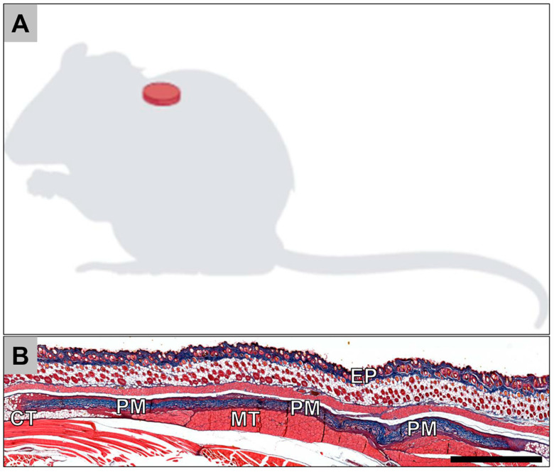
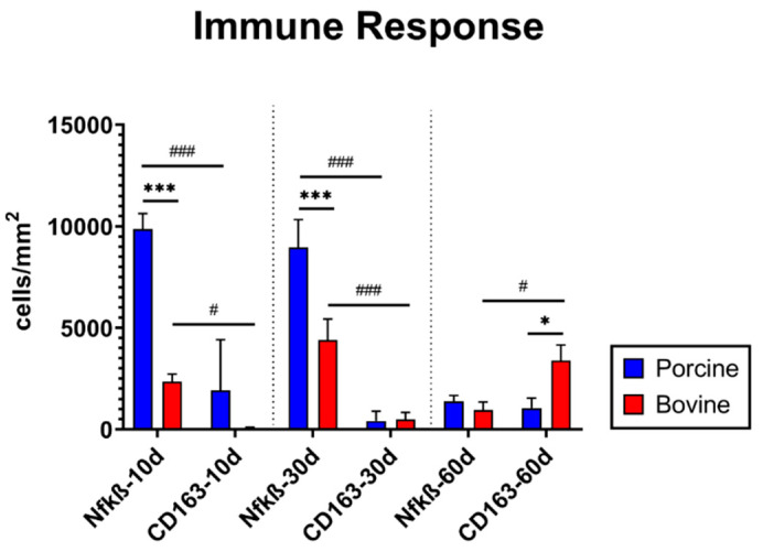
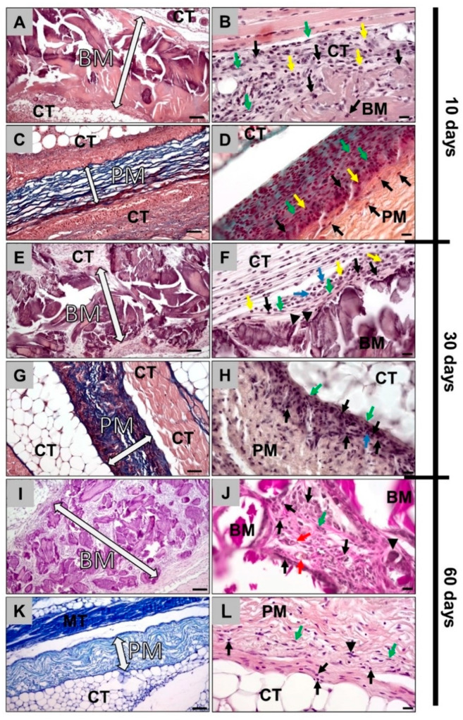
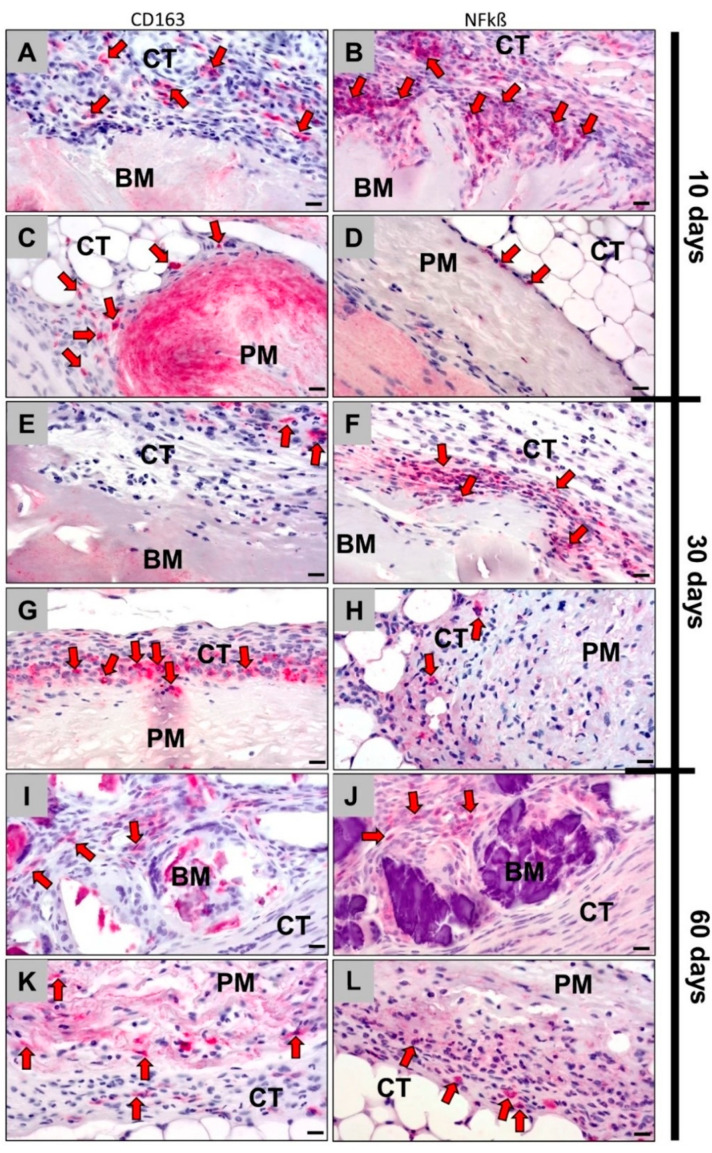
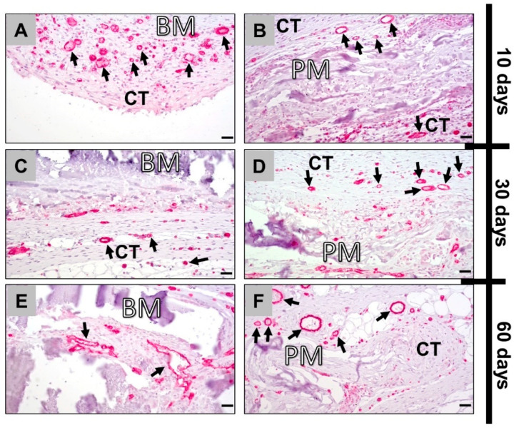
Collagen-based barrier membranes are an essential component in Guided Bone Regeneration (GBR) procedures. They act as cell-occlusive devices that should maintain a micromilieu where bone tissue can grow, which in turn provides a stable bed for prosthetic implantation. However, the standing time of collagen membranes has been a challenging area, as native membranes are often prematurely resorbed. Therefore, consolidation techniques, such as chemical cross-linking, have been used to enhance the structural integrity of the membranes, and by consequence, their standing time. However, these techniques have cytotoxic tendencies and can cause exaggerated inflammation and in turn, premature resorption, and material failures. However, tissues from different extraction sites and animals are variably cross-linked. For the present in vivo study, a new collagen membrane based on bovine dermis was extracted and compared to a commercially available porcine-sourced collagen membrane extracted from the pericardium. The membranes were implanted in Wistar rats for up to 60 days. The analyses included well-established histopathological and histomorphometrical methods, including histochemical and immunohistochemical staining procedures, to detect M1- and M2-macrophages as well as blood vessels. Initially, the results showed that both membranes remained intact up to day 30, while the bovine membrane was fragmented at day 60 with granulation tissue infiltrating the implantation beds. In contrast, the porcine membrane remained stable without signs of material-dependent inflammatory processes. Therefore, the bovine membrane showed a special integration pattern as the fragments were found to be overlapping, providing secondary porosity in combination with a transmembraneous vascularization. Altogether, the bovine membrane showed comparable results to the porcine control group in terms of biocompatibility and standing time. Moreover, blood vessels were found within the bovine membranes, which can potentially serve as an additional functionality of barrier membranes that conventional barrier membranes do not provide.
基于胶原蛋白的屏障膜是引导骨再生(GBR)手术中的重要组成部分。它们作为细胞封闭装置,应维持一个骨组织能够生长的微环境,进而为假体植入提供稳定的床。然而,胶原蛋白膜的留存时间一直是个具有挑战性的领域,因为天然膜常常过早被吸收。因此,诸如化学交联等加固技术已被用于增强膜的结构完整性,进而延长其留存时间。然而,这些技术具有细胞毒性倾向,会导致过度炎症,进而引发过早吸收和材料失效。此外,来自不同提取部位和动物的组织交联程度各不相同。在本体内研究中,提取了一种基于牛真皮的新型胶原蛋白膜,并与一种从心包提取的市售猪源胶原蛋白膜进行比较。将这些膜植入Wistar大鼠体内长达60天。分析采用了成熟的组织病理学和组织形态计量学方法,包括组织化学和免疫组织化学染色程序,以检测M1和M2巨噬细胞以及血管。最初,结果显示两种膜在第30天前都保持完整,而牛膜在第60天出现碎片化,肉芽组织侵入植入床。相比之下,猪膜保持稳定,没有材料依赖性炎症过程的迹象。因此,牛膜呈现出一种特殊的整合模式,发现碎片相互重叠,结合跨膜血管化提供了二次孔隙率。总体而言,牛膜在生物相容性和留存时间方面与猪对照组的结果相当。此外,在牛膜内发现了血管,这可能是传统屏障膜所不具备的屏障膜的额外功能。