• 文献检索
  • 文档翻译
  • 深度研究
  • 学术资讯
  • Suppr Zotero 插件Zotero 插件
  • 邀请有礼
  • 套餐&价格
  • 历史记录
应用&插件
Suppr Zotero 插件Zotero 插件浏览器插件Mac 客户端Windows 客户端微信小程序
定价
高级版会员购买积分包购买API积分包
服务
文献检索文档翻译深度研究API 文档MCP 服务
关于我们
关于 Suppr公司介绍联系我们用户协议隐私条款
关注我们

Suppr 超能文献

核心技术专利:CN118964589B侵权必究
粤ICP备2023148730 号-1Suppr @ 2026

文献检索

告别复杂PubMed语法,用中文像聊天一样搜索,搜遍4000万医学文献。AI智能推荐,让科研检索更轻松。

立即免费搜索

文件翻译

保留排版,准确专业,支持PDF/Word/PPT等文件格式,支持 12+语言互译。

免费翻译文档

深度研究

AI帮你快速写综述,25分钟生成高质量综述,智能提取关键信息,辅助科研写作。

立即免费体验

小鼠皮下移植后mPEG-聚(丙氨酸)水凝胶包埋的MIN6细胞的无创追踪

Noninvasive Tracking of mPEG-poly(Ala) Hydrogel-Embedded MIN6 Cells after Subcutaneous Transplantation in Mice.

作者信息

Juang Jyuhn-Huarng, Lin Hsiu-Chao, Chen Chen-Yi, Kao Chen-Wei, Chen Chen-Ling, Wu Shu-Ting, Lin Sung-Han, Shen Chia-Rui, Wang Jiun-Jie, Tsai Zei-Tsan, Chu I-Ming

机构信息

Division of Endocrinology and Metabolism, Department of Internal Medicine and Center for Tissue Engineering, Chang Gung Memorial Hospital, Taoyuan 33305, Taiwan.

Department of Medicine, College of Medicine, Chang Gung University, Taoyuan 33302, Taiwan.

出版信息

Polymers (Basel). 2021 Mar 13;13(6):885. doi: 10.3390/polym13060885.

DOI:10.3390/polym13060885
PMID:33805723
原文链接:https://pmc.ncbi.nlm.nih.gov/articles/PMC7998640/
Abstract

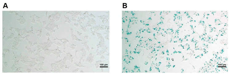
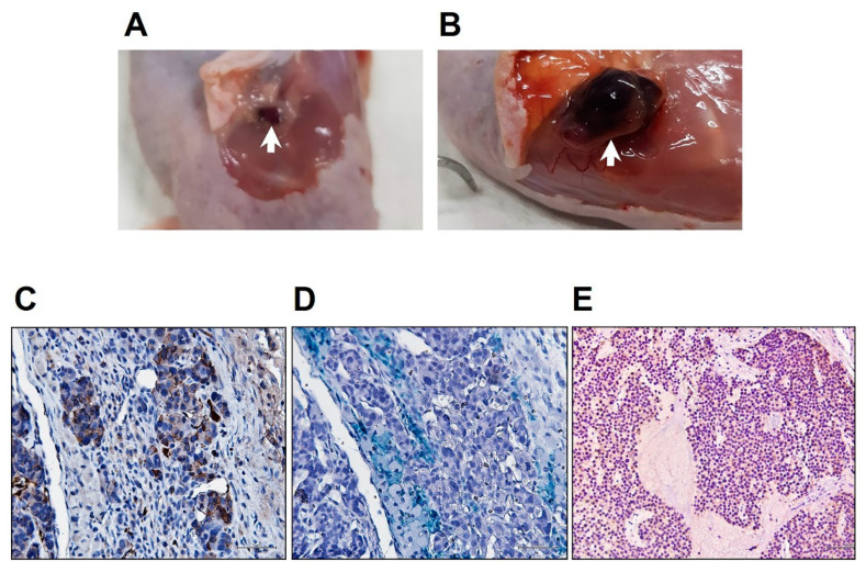
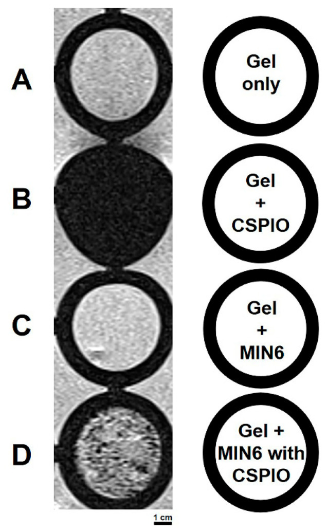
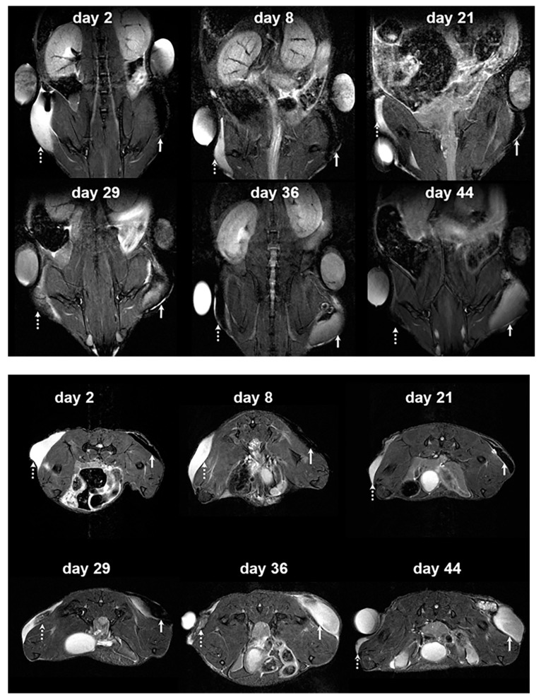
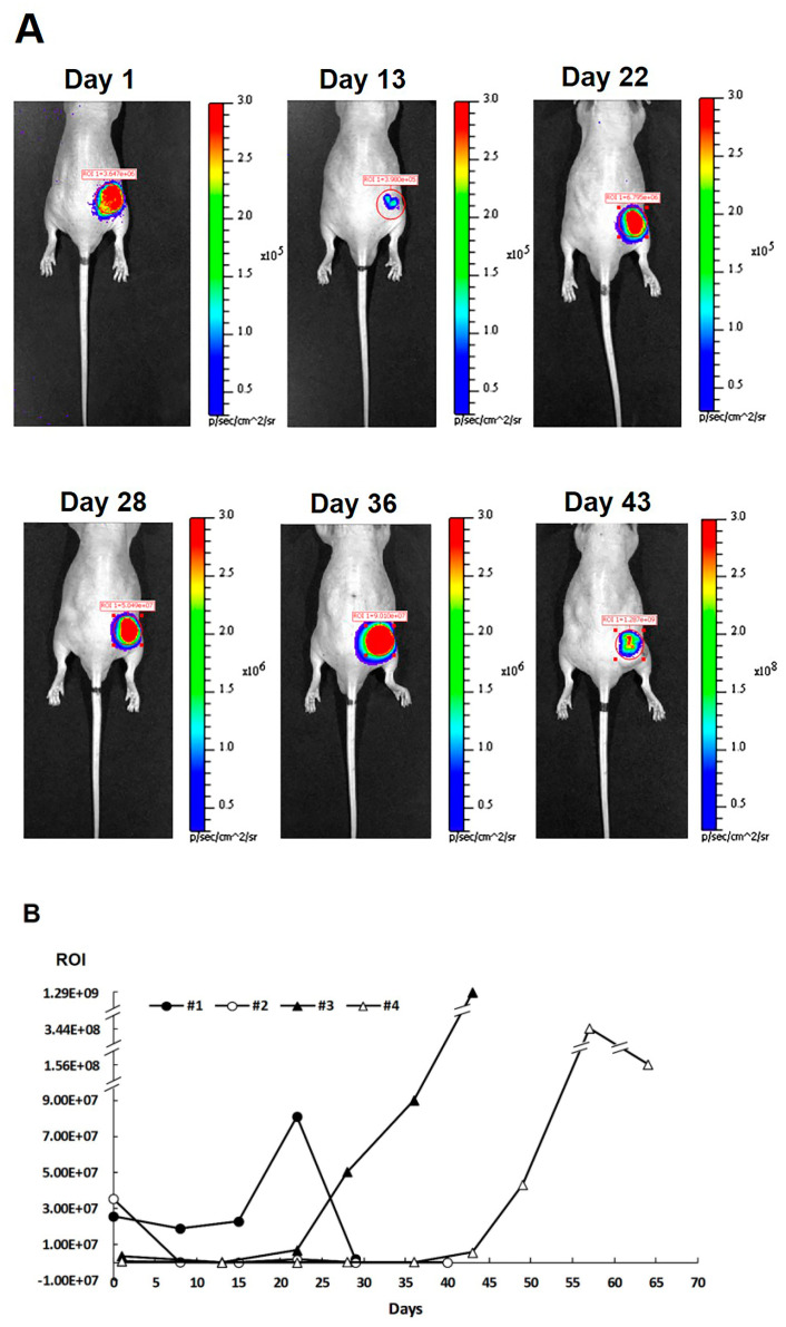
Recently, we demonstrated the feasibility of subcutaneous transplantation of MIN6 cells embedded in a scaffold with poly(ethylene glycol) methyl ether (mPEG)-poly(Ala) hydrogels. In this study, we further tracked these grafts using magnetic resonance (MR) and bioluminescence imaging. After being incubated overnight with chitosan-coated superparamagnetic iron oxide (CSPIO) nanoparticles and then mixed with mPEG-poly(Ala) hydrogels, MIN6 cells appeared as dark spots on MR scans. For in vivo experiments, we transfected MIN6 cells with luciferase and/or incubated them overnight with CSPIO overnight; 5 × 10 MIN6 cells embedded in mPEG-poly(Ala) hydrogels were transplanted into the subcutaneous space of each nude mouse. The graft of CSPIO-labeled MIN6 cells was visualized as a distinct hypointense area on MR images located at the implantation site before day 21. However, this area became hyperintense on MR scans for up to 64 days. In addition, positive bioluminescence images were also observed for up to 64 days after transplantation. The histology of removed grafts showed positive insulin and iron staining. These results indicate mPEG-poly(Ala) is a suitable scaffold for β-cell encapsulation and transplantation. Moreover, MR and bioluminescence imaging are useful noninvasive tools for detecting and monitoring mPEG-poly(Ala) hydrogel-embedded MIN6 cells at a subcutaneous site.

摘要

最近,我们证明了将MIN6细胞嵌入聚(乙二醇)甲醚(mPEG)-聚(丙氨酸)水凝胶支架中进行皮下移植的可行性。在本研究中,我们进一步使用磁共振(MR)和生物发光成像对这些移植物进行追踪。在用壳聚糖包被的超顺磁性氧化铁(CSPIO)纳米颗粒孵育过夜后,将MIN6细胞与mPEG-聚(丙氨酸)水凝胶混合,MIN6细胞在MR扫描中表现为黑点。对于体内实验,我们用荧光素酶转染MIN6细胞和/或使其与CSPIO孵育过夜;将5×10个嵌入mPEG-聚(丙氨酸)水凝胶的MIN6细胞移植到每只裸鼠的皮下空间。在第21天之前,CSPIO标记的MIN6细胞移植物在MR图像上显示为植入部位的一个明显的低信号区。然而,在长达64天的MR扫描中,该区域变为高信号。此外,移植后长达64天也观察到了阳性生物发光图像。取出的移植物的组织学检查显示胰岛素和铁染色呈阳性。这些结果表明mPEG-聚(丙氨酸)是用于β细胞包封和移植的合适支架。此外,MR和生物发光成像对于检测和监测皮下部位的mPEG-聚(丙氨酸)水凝胶包埋的MIN6细胞是有用的非侵入性工具。

https://cdn.ncbi.nlm.nih.gov/pmc/blobs/9d2e/7998640/f0bcebbd6203/polymers-13-00885-g005.jpg
https://cdn.ncbi.nlm.nih.gov/pmc/blobs/9d2e/7998640/412406672676/polymers-13-00885-g001.jpg
https://cdn.ncbi.nlm.nih.gov/pmc/blobs/9d2e/7998640/7a636b26245c/polymers-13-00885-g002.jpg
https://cdn.ncbi.nlm.nih.gov/pmc/blobs/9d2e/7998640/4715b4b67ef5/polymers-13-00885-g003.jpg
https://cdn.ncbi.nlm.nih.gov/pmc/blobs/9d2e/7998640/b16e69df8fb2/polymers-13-00885-g004.jpg
https://cdn.ncbi.nlm.nih.gov/pmc/blobs/9d2e/7998640/f0bcebbd6203/polymers-13-00885-g005.jpg

相似文献

1
Noninvasive Tracking of mPEG-poly(Ala) Hydrogel-Embedded MIN6 Cells after Subcutaneous Transplantation in Mice.小鼠皮下移植后mPEG-聚(丙氨酸)水凝胶包埋的MIN6细胞的无创追踪
Polymers (Basel). 2021 Mar 13;13(6):885. doi: 10.3390/polym13060885.
2
The Image-Histology Correlation of Subcutaneous mPEG-poly(Ala) Hydrogel-Embedded MIN6 Cell Grafts in Nude Mice.裸鼠皮下mPEG-聚(丙氨酸)水凝胶包埋的MIN6细胞移植瘤的图像与组织学相关性
Polymers (Basel). 2023 Jun 6;15(12):2584. doi: 10.3390/polym15122584.
3
Magnetic Resonance Imaging of Transplanted Porcine Neonatal Pancreatic Cell Clusters Labeled with Chitosan-Coated Superparamagnetic Iron Oxide Nanoparticles in Mice.壳聚糖包被的超顺磁性氧化铁纳米颗粒标记的猪新生胰岛细胞簇移植小鼠的磁共振成像
Polymers (Basel). 2021 Apr 11;13(8):1238. doi: 10.3390/polym13081238.
4
Magnetic resonance imaging of mouse islet grafts labeled with novel chitosan-coated superparamagnetic iron oxide nanoparticles.新型壳聚糖包裹超顺磁性氧化铁纳米粒子标记的小鼠胰岛移植的磁共振成像。
PLoS One. 2013 Apr 29;8(4):e62626. doi: 10.1371/journal.pone.0062626. Print 2013.
5
Magnetic resonance imaging of transplanted mouse islets labeled with chitosan-coated superparamagnetic iron oxide nanoparticles.用壳聚糖包被的超顺磁性氧化铁纳米颗粒标记的移植小鼠胰岛的磁共振成像
Transplant Proc. 2010 Jul-Aug;42(6):2104-8. doi: 10.1016/j.transproceed.2010.05.103.
6
In Vivo Imaging of Immune Rejection of MIN6 Cells Transplanted in C3H Mice.MIN6 细胞在 C3H 小鼠中移植后的免疫排斥的体内成像。
Cells. 2024 Jun 17;13(12):1044. doi: 10.3390/cells13121044.
7
Magnetic resonance imaging study of mouse islet allotransplantation.小鼠胰岛同种异体移植的磁共振成像研究
Transplant Proc. 2010 Dec;42(10):4217-20. doi: 10.1016/j.transproceed.2010.09.089.
8
Exendin-4-Conjugated Manganese Magnetism-Engineered Iron Oxide Nanoparticles as a Potential Magnetic Resonance Imaging Contrast Agent for Tracking Transplanted β-Cells.艾塞那肽-4缀合的锰磁性工程化氧化铁纳米颗粒作为用于追踪移植β细胞的潜在磁共振成像造影剂
Nanomaterials (Basel). 2021 Nov 21;11(11):3145. doi: 10.3390/nano11113145.
9
Evaluation of a mPEG-polyester-based hydrogel as cell carrier for chondrocytes.评价一种基于 mPEG-聚酯的水凝胶作为软骨细胞载体。
J Biomed Mater Res A. 2013 Nov;101(11):3311-9. doi: 10.1002/jbm.a.34632. Epub 2013 Aug 27.
10
Imaging specificity of MR-optical imaging agents following the masking of surface charge by poly(ethylene glycol).通过聚乙二醇掩蔽表面电荷后,磁共振-光学成像剂的成像特异性。
Biomaterials. 2013 May;34(16):4118-4127. doi: 10.1016/j.biomaterials.2013.02.025. Epub 2013 Mar 1.

引用本文的文献

1
In Vivo Imaging of Immune Rejection of MIN6 Cells Transplanted in C3H Mice.MIN6 细胞在 C3H 小鼠中移植后的免疫排斥的体内成像。
Cells. 2024 Jun 17;13(12):1044. doi: 10.3390/cells13121044.
2
The Image-Histology Correlation of Subcutaneous mPEG-poly(Ala) Hydrogel-Embedded MIN6 Cell Grafts in Nude Mice.裸鼠皮下mPEG-聚(丙氨酸)水凝胶包埋的MIN6细胞移植瘤的图像与组织学相关性
Polymers (Basel). 2023 Jun 6;15(12):2584. doi: 10.3390/polym15122584.
3
Magnetic Resonance Imaging of Transplanted Porcine Neonatal Pancreatic Cell Clusters Labeled with Exendin-4-Conjugated Manganese Magnetism-Engineered Iron Oxide Nanoparticles.

本文引用的文献

1
Islet Transplantation Imaging in vivo.体内胰岛移植成像
Diabetes Metab Syndr Obes. 2020 Sep 23;13:3301-3311. doi: 10.2147/DMSO.S263253. eCollection 2020.
2
Characterization of a mouse model of islet transplantation using MIN-6 cells.利用 MIN-6 细胞建立胰岛移植小鼠模型的研究。
Islets. 2020 Jul 3;12(4):71-86. doi: 10.1080/19382014.2020.1763719. Epub 2020 Jun 22.
3
Enhancement of Subcutaneously Transplanted β Cell Survival Using 3D Stem Cell Spheroids with Proangiogenic and Prosurvival Potential.利用具有促血管生成和促生存潜力的 3D 干细胞球体增强皮下移植的β细胞存活。
用艾塞那肽-4偶联的锰磁性工程氧化铁纳米颗粒标记的移植猪新生胰岛细胞团的磁共振成像
Nanomaterials (Basel). 2022 Apr 5;12(7):1222. doi: 10.3390/nano12071222.
4
Exendin-4-Conjugated Manganese Magnetism-Engineered Iron Oxide Nanoparticles as a Potential Magnetic Resonance Imaging Contrast Agent for Tracking Transplanted β-Cells.艾塞那肽-4缀合的锰磁性工程化氧化铁纳米颗粒作为用于追踪移植β细胞的潜在磁共振成像造影剂
Nanomaterials (Basel). 2021 Nov 21;11(11):3145. doi: 10.3390/nano11113145.
5
Two in One: Use of Divalent Manganese Ions as Both Cross-Linking and MRI Contrast Agent for Intrathecal Injection of Hydrogel-Embedded Stem Cells.二合一:使用二价锰离子作为交联剂和磁共振成像造影剂用于鞘内注射水凝胶包埋的干细胞
Pharmaceutics. 2021 Jul 13;13(7):1076. doi: 10.3390/pharmaceutics13071076.
Adv Biosyst. 2020 Mar;4(3):e1900254. doi: 10.1002/adbi.201900254. Epub 2020 Feb 10.
4
Injectable Macroporous Hydrogel Formed by Enzymatic Cross-Linking of Gelatin Microgels.通过明胶微凝胶的酶促交联形成的可注射大孔水凝胶。
ACS Appl Bio Mater. 2018 Nov 19;1(5):1430-1439. doi: 10.1021/acsabm.8b00380. Epub 2018 Oct 15.
5
Improved delivery of PLGA microparticles and microparticle-cell scaffolds in clinical needle gauges using modified viscosity formulations.采用改良黏度配方,提高临床用针规中 PLGA 微球和微球-细胞支架的递释效率。
Int J Pharm. 2018 Jul 30;546(1-2):272-278. doi: 10.1016/j.ijpharm.2018.05.025. Epub 2018 May 16.
6
Antimicrobial hydrogels: promising materials for medical application.抗菌水凝胶:有应用前景的医用材料。
Int J Nanomedicine. 2018 Apr 12;13:2217-2263. doi: 10.2147/IJN.S154748. eCollection 2018.
7
Discovering Cell-Adhesion Peptides in Tissue Engineering: Beyond RGD.在组织工程中发现细胞黏附肽:超越 RGD。
Trends Biotechnol. 2018 Apr;36(4):372-383. doi: 10.1016/j.tibtech.2018.01.008. Epub 2018 Feb 6.
8
Layer-by-layer cell coating technique using extracellular matrix facilitates rapid fabrication and function of pancreatic β-cell spheroids.采用细胞外基质的层层细胞包被技术可促进胰岛β细胞球体的快速构建和功能。
Biomaterials. 2018 Apr;160:82-91. doi: 10.1016/j.biomaterials.2018.01.020. Epub 2018 Jan 16.
9
Imaging of Transplanted Pancreatic Islets.移植胰岛的成像
Front Endocrinol (Lausanne). 2018 Jan 22;8:382. doi: 10.3389/fendo.2017.00382. eCollection 2017.
10
The journey of islet cell transplantation and future development.胰岛细胞移植的历程与未来发展。
Islets. 2018 Mar 4;10(2):80-94. doi: 10.1080/19382014.2018.1428511. Epub 2018 Feb 5.