Juang Jyuhn-Huarng, Chen Chen-Ling, Kao Chen-Wei, Chen Chen-Yi, Shen Chia-Rui, Wang Jiun-Jie, Tsai Zei-Tsan, Chu I-Ming
Division of Endocrinology and Metabolism, Department of Internal Medicine and Center for Tissue Engineering, Chang Gung Memorial Hospital, Taoyuan 33305, Taiwan.
Department of Medicine, College of Medicine, Chang Gung University, Taoyuan 33305, Taiwan.
Polymers (Basel). 2023 Jun 6;15(12):2584. doi: 10.3390/polym15122584.
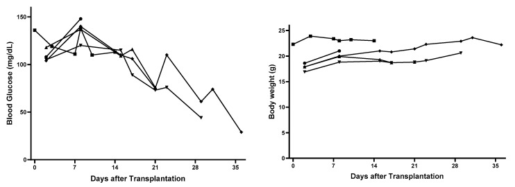
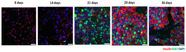
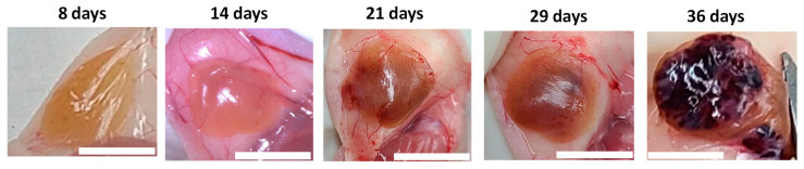
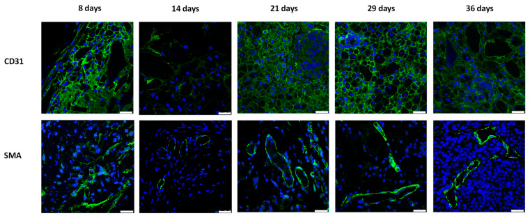
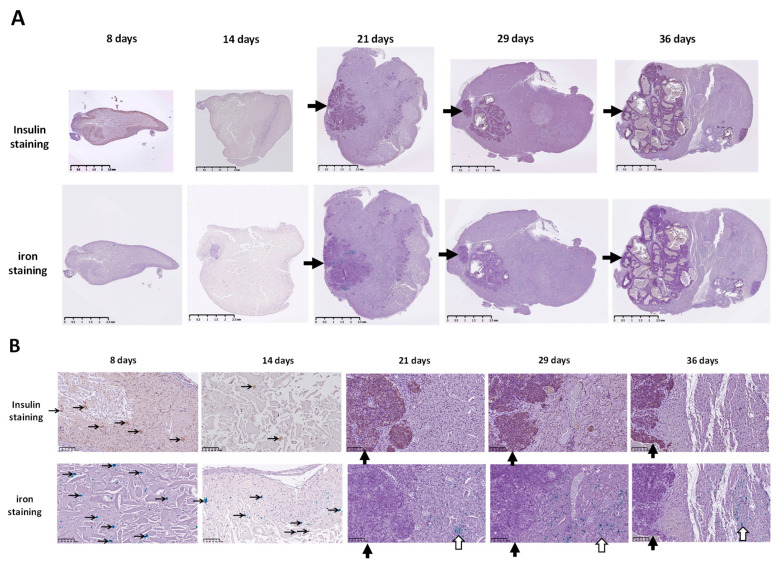
Previously, we have successfully used noninvasive magnetic resonance (MR) and bioluminescence imaging to detect and monitor mPEG-poly(Ala) hydrogel-embedded MIN6 cells at the subcutaneous space for up to 64 days. In this study, we further explored the histological evolution of MIN6 cell grafts and correlated it with image findings. MIN6 cells were incubated overnight with chitosan-coated superparamagnetic iron oxide (CSPIO) and then 5 × 10 cells in the 100 μL hydrogel solution were injected subcutaneously into each nude mouse. Grafts were removed and examined the vascularization, cell growth and proliferation with anti-CD31, SMA, insulin and ki67 antibodies, respectively, at 8, 14, 21, 29 and 36 days after transplantation. All grafts were well-vascularized with prominent CD31 and SMA staining at all time points. Interestingly, insulin-positive cells and iron-positive cells were scattered in the graft at 8 and 14 days; while clusters of insulin-positive cells without iron-positive cells appeared in the grafts at 21 days and persisted thereafter, indicating neogrowth of MIN6 cells. Moreover, proliferating MIN6 cells with strong ki67 staining was observed in 21-, 29- and 36-day grafts. Our results indicate that the originally transplanted MIN6 cells proliferated from 21 days that presented distinctive bioluminescence and MR images.
此前,我们已成功运用无创磁共振(MR)和生物发光成像技术,在皮下空间对包裹于甲氧基聚乙二醇-聚丙氨酸(mPEG-poly(Ala))水凝胶中的MIN6细胞进行了长达64天的检测与监测。在本研究中,我们进一步探究了MIN6细胞移植体的组织学演变,并将其与图像结果相关联。将MIN6细胞与壳聚糖包被的超顺磁性氧化铁(CSPIO)孵育过夜,然后将100μL水凝胶溶液中的5×10个细胞皮下注射到每只裸鼠体内。在移植后第8、14、21、29和36天分别取出移植体,并用抗CD31、平滑肌肌动蛋白(SMA)、胰岛素和ki67抗体检测血管生成、细胞生长及增殖情况。在所有时间点,所有移植体均有良好的血管生成,CD31和SMA染色显著。有趣的是,在第8天和第14天,胰岛素阳性细胞和铁阳性细胞散在于移植体中;而在第21天,移植体中出现了无铁阳性细胞的胰岛素阳性细胞簇,且此后一直存在,这表明MIN6细胞出现了新生长。此外,在第21天、29天和36天的移植体中观察到具有强烈ki67染色的增殖MIN6细胞。我们的结果表明,最初移植的MIN6细胞从第21天开始增殖,呈现出独特的生物发光和MR图像。