• 文献检索
  • 文档翻译
  • 深度研究
  • 学术资讯
  • Suppr Zotero 插件Zotero 插件
  • 邀请有礼
  • 套餐&价格
  • 历史记录
应用&插件
Suppr Zotero 插件Zotero 插件浏览器插件Mac 客户端Windows 客户端微信小程序
定价
高级版会员购买积分包购买API积分包
服务
文献检索文档翻译深度研究API 文档MCP 服务
关于我们
关于 Suppr公司介绍联系我们用户协议隐私条款
关注我们

Suppr 超能文献

核心技术专利:CN118964589B侵权必究
粤ICP备2023148730 号-1Suppr @ 2026

文献检索

告别复杂PubMed语法,用中文像聊天一样搜索,搜遍4000万医学文献。AI智能推荐,让科研检索更轻松。

立即免费搜索

文件翻译

保留排版,准确专业,支持PDF/Word/PPT等文件格式,支持 12+语言互译。

免费翻译文档

深度研究

AI帮你快速写综述,25分钟生成高质量综述,智能提取关键信息,辅助科研写作。

立即免费体验

通过结构光照明显微镜和图像互相关光谱学(SIM-ICCS)测量纳米级距离。

Measuring Nanoscale Distances by Structured Illumination Microscopy and Image Cross-Correlation Spectroscopy (SIM-ICCS).

机构信息

Nanoscopy and NIC@IIT, CHT Erzelli, Istituto Italiano di Tecnologia, Via Enrico Melen 83, Building B, 16152 Genoa, Italy.

DIFILAB, Department of Physics, University of Genoa, Via Dodecaneso 33, 16143 Genoa, Italy.

出版信息

Sensors (Basel). 2021 Mar 12;21(6):2010. doi: 10.3390/s21062010.

DOI:10.3390/s21062010
PMID:33809144
原文链接:https://pmc.ncbi.nlm.nih.gov/articles/PMC8001887/
Abstract

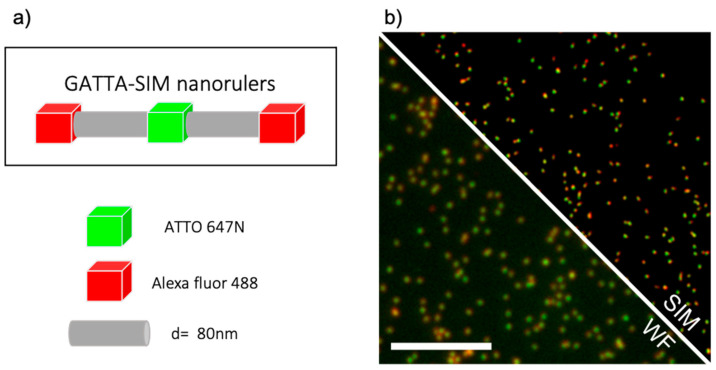
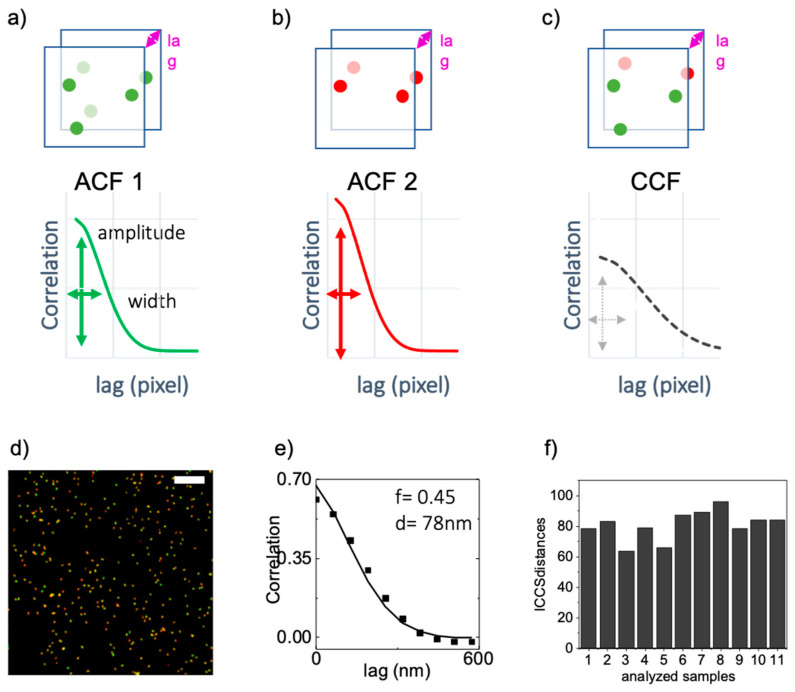
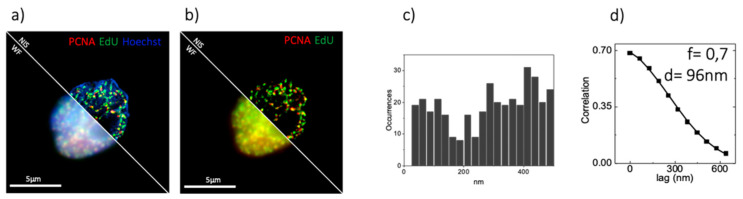
Since the introduction of super-resolution microscopy, there has been growing interest in quantifying the nanoscale spatial distributions of fluorescent probes to better understand cellular processes and their interactions. One way to check if distributions are correlated or not is to perform colocalization analysis of multi-color acquisitions. Among all the possible methods available to study and quantify the colocalization between multicolor images, there is image cross-correlation spectroscopy (ICCS). The main advantage of ICCS, in comparison with other co-localization techniques, is that it does not require pre-segmentation of the sample into single objects. Here we show that the combination of structured illumination microscopy (SIM) with ICCS (SIM-ICCS) is a simple approach to quantify colocalization and measure nanoscale distances from multi-color SIM images. We validate the SIM-ICCS analysis on SIM images of optical nanorulers, DNA-origami-based model samples containing fluorophores of different colors at a distance of 80 nm. The SIM-ICCS analysis is compared with an object-based analysis performed on the same samples. Finally, we show that SIM-ICCS can be used to quantify the nanoscale spatial distribution of functional nuclear sites in fixed cells.

摘要

自超分辨率显微镜问世以来,人们越来越感兴趣于定量分析荧光探针的纳米级空间分布,以更好地了解细胞过程及其相互作用。检查分布是否相关的一种方法是对多色采集进行共定位分析。在研究和量化多色图像之间共定位的所有可能方法中,有一种方法是图像互相关光谱学 (ICCS)。与其他共定位技术相比,ICCS 的主要优势在于它不需要将样品预先分割成单个物体。在这里,我们展示了将结构光照明显微镜 (SIM) 与 ICCS(SIM-ICCS)相结合,是一种从多色 SIM 图像定量共定位和测量纳米级距离的简单方法。我们在距离为 80nm 的包含不同颜色荧光团的 DNA 折纸模型样品的光学纳米标尺的 SIM 图像上验证了 SIM-ICCS 分析。将 SIM-ICCS 分析与对同一样品进行的基于对象的分析进行了比较。最后,我们证明了 SIM-ICCS 可用于定量固定细胞中功能性核位点的纳米级空间分布。

https://cdn.ncbi.nlm.nih.gov/pmc/blobs/2100/8001887/68cd43c21ce1/sensors-21-02010-g006.jpg
https://cdn.ncbi.nlm.nih.gov/pmc/blobs/2100/8001887/ed463de46685/sensors-21-02010-g001.jpg
https://cdn.ncbi.nlm.nih.gov/pmc/blobs/2100/8001887/56f6e7bedb8b/sensors-21-02010-g002.jpg
https://cdn.ncbi.nlm.nih.gov/pmc/blobs/2100/8001887/c1f01fd0c49e/sensors-21-02010-g003.jpg
https://cdn.ncbi.nlm.nih.gov/pmc/blobs/2100/8001887/26bc2673288a/sensors-21-02010-g004.jpg
https://cdn.ncbi.nlm.nih.gov/pmc/blobs/2100/8001887/20673af12209/sensors-21-02010-g005.jpg
https://cdn.ncbi.nlm.nih.gov/pmc/blobs/2100/8001887/68cd43c21ce1/sensors-21-02010-g006.jpg

相似文献

1
Measuring Nanoscale Distances by Structured Illumination Microscopy and Image Cross-Correlation Spectroscopy (SIM-ICCS).通过结构光照明显微镜和图像互相关光谱学(SIM-ICCS)测量纳米级距离。
Sensors (Basel). 2021 Mar 12;21(6):2010. doi: 10.3390/s21062010.
2
Nanoscale Distribution of Nuclear Sites by Super-Resolved Image Cross-Correlation Spectroscopy.纳米级核位点的超分辨图像互相关光谱分布。
Biophys J. 2019 Dec 3;117(11):2054-2065. doi: 10.1016/j.bpj.2019.10.036. Epub 2019 Nov 2.
3
Novel Tools to Measure Single Molecules Colocalization in Fluorescence Nanoscopy by Image Cross Correlation Spectroscopy.通过图像互相关光谱法测量荧光纳米显微镜中单个分子共定位的新型工具。
Nanomaterials (Basel). 2022 Feb 18;12(4):686. doi: 10.3390/nano12040686.
4
Imaging tissues and cells beyond the diffraction limit with structured illumination microscopy and Bayesian image reconstruction.利用结构光照明显微镜和贝叶斯图像重建技术对组织和细胞进行超衍射极限成像。
Gigascience. 2019 Jan 1;8(1):giy126. doi: 10.1093/gigascience/giy126.
5
Augmented Super-Resolution Radial Fluctuations (aSRRF) Pushing the Limits of Structured Illumination Microscopy.增强型超分辨率径向波动(aSRRF):突破结构光照明显微镜的极限
bioRxiv. 2023 Jul 6:2023.07.05.547885. doi: 10.1101/2023.07.05.547885.
6
Open-source image reconstruction of super-resolution structured illumination microscopy data in ImageJ.在ImageJ中对超分辨率结构光照明显微镜数据进行开源图像重建。
Nat Commun. 2016 Mar 21;7:10980. doi: 10.1038/ncomms10980.
7
Rapid Image Reconstruction of Structured Illumination Microscopy Directly in the Spatial Domain.直接在空间域中进行结构照明显微镜的快速图像重建。
IEEE Photonics J. 2021 Feb;13(1). doi: 10.1109/JPHOT.2021.3053110. Epub 2021 Jan 20.
8
High speed structured illumination microscopy in optically thick samples.光学厚样品中的高速结构照明显微镜术
Methods. 2015 Oct 15;88:11-9. doi: 10.1016/j.ymeth.2015.03.020. Epub 2015 Apr 1.
9
Simultaneous multicolour imaging using quantum dot structured illumination microscopy.使用量子点结构光照明显微镜进行的同时多色成像。
J Microsc. 2020 Jan;277(1):32-41. doi: 10.1111/jmi.12862. Epub 2020 Jan 23.
10
Easily scalable multi-color DMD-based structured illumination microscopy.基于数字微镜器件(DMD)的易于扩展的多色结构光照明显微术。
Opt Lett. 2024 Jan 1;49(1):77-80. doi: 10.1364/OL.507599.

引用本文的文献

1
Spectral phasor imaging on a commercial confocal microscope without a spectral detector.在没有光谱探测器的商用共聚焦显微镜上进行光谱相量成像。
Sci Rep. 2025 Aug 26;15(1):31431. doi: 10.1038/s41598-025-15637-x.
2
Location of oncogene-induced DNA damage sites revealed by quantitative analysis of a DNA counterstain.通过DNA复染剂的定量分析揭示癌基因诱导的DNA损伤位点的位置。
Eur Biophys J. 2025 May 7. doi: 10.1007/s00249-025-01755-x.
3
Super-resolved analysis of colocalization between replication and transcription along the cell cycle in a model of oncogene activation.

本文引用的文献

1
Measurement of co-localization of objects in dual-colour confocal images.双色共聚焦图像中物体共定位的测量。
J Microsc. 1993 Mar;169(3):375-382. doi: 10.1111/j.1365-2818.1993.tb03313.x.
2
Nanoscale Distribution of Nuclear Sites by Super-Resolved Image Cross-Correlation Spectroscopy.纳米级核位点的超分辨图像互相关光谱分布。
Biophys J. 2019 Dec 3;117(11):2054-2065. doi: 10.1016/j.bpj.2019.10.036. Epub 2019 Nov 2.
3
Chromatin nanoscale compaction in live cells visualized by acceptor-to-donor ratio corrected Förster resonance energy transfer between DNA dyes.
细胞周期中癌基因激活模型中复制和转录共定位的超分辨分析。
Commun Biol. 2024 Oct 4;7(1):1260. doi: 10.1038/s42003-024-06972-2.
4
MultiMatch: geometry-informed colocalization in multi-color super-resolution microscopy.多匹配:多色超分辨率显微镜中的几何信息共定位。
Commun Biol. 2024 Sep 13;7(1):1139. doi: 10.1038/s42003-024-06772-8.
5
Colocalization by cross-correlation, a new method of colocalization suited for super-resolution microscopy.通过互相关实现共定位,一种适用于超分辨率显微镜的共定位新方法。
BMC Bioinformatics. 2024 Feb 2;25(1):55. doi: 10.1186/s12859-024-05675-z.
6
Imaging-based study demonstrates how the DEK nanoscale distribution differentially correlates with epigenetic marks in a breast cancer model.基于成像的研究表明,DEK 的纳米级分布如何与乳腺癌模型中的表观遗传标记差异相关。
Sci Rep. 2023 Aug 7;13(1):12749. doi: 10.1038/s41598-023-38685-7.
7
SPLIT-PIN software enabling confocal and super-resolution imaging with a virtually closed pinhole.SPLIT-PIN 软件可实现带有虚拟封闭针孔的共聚焦和超分辨率成像。
Sci Rep. 2023 Feb 15;13(1):2741. doi: 10.1038/s41598-023-29951-9.
8
Alterations induced by the PML-RARα oncogene revealed by image cross correlation spectroscopy.PML-RARα 癌基因诱导的改变通过像关联光谱学揭示。
Biophys J. 2022 Nov 15;121(22):4358-4367. doi: 10.1016/j.bpj.2022.10.003. Epub 2022 Oct 4.
9
Novel Tools to Measure Single Molecules Colocalization in Fluorescence Nanoscopy by Image Cross Correlation Spectroscopy.通过图像互相关光谱法测量荧光纳米显微镜中单个分子共定位的新型工具。
Nanomaterials (Basel). 2022 Feb 18;12(4):686. doi: 10.3390/nano12040686.
10
Evaluation of sted super-resolution image quality by image correlation spectroscopy (QuICS).基于图像相关光谱学(QuICS)评估 sted 超分辨率图像质量。
Sci Rep. 2021 Oct 21;11(1):20782. doi: 10.1038/s41598-021-00301-x.
利用 DNA 染料之间的受体质子-给体质子比校正的福斯特共振能量转移,可视化活细胞中的染色质纳米级压缩。
J Biophotonics. 2019 Dec;12(12):e201900164. doi: 10.1002/jbio.201900164. Epub 2019 Aug 21.
4
Measuring Mobility in Chromatin by Intensity-Sorted FCS.通过强度分选 FCS 测量染色质的流动性。
Biophys J. 2019 Mar 19;116(6):987-999. doi: 10.1016/j.bpj.2019.02.003. Epub 2019 Feb 14.
5
SMoLR: visualization and analysis of single-molecule localization microscopy data in R.SMoLR:R 中的单分子定位显微镜数据的可视化和分析。
BMC Bioinformatics. 2019 Jan 15;20(1):30. doi: 10.1186/s12859-018-2578-3.
6
Super-resolution microscopy demystified.超分辨率显微镜解析。
Nat Cell Biol. 2019 Jan;21(1):72-84. doi: 10.1038/s41556-018-0251-8. Epub 2019 Jan 2.
7
Super-resolution chromatin tracing reveals domains and cooperative interactions in single cells.超分辨率染色质追踪揭示了单细胞中的域和协同相互作用。
Science. 2018 Oct 26;362(6413). doi: 10.1126/science.aau1783.
8
Local raster image correlation spectroscopy generates high-resolution intracellular diffusion maps.局部光栅图像相关光谱法可生成高分辨率的细胞内扩散图谱。
Commun Biol. 2018 Feb 8;1:10. doi: 10.1038/s42003-017-0010-6. eCollection 2018.
9
STED super-resolved microscopy.受激发射损耗超分辨显微镜。
Nat Methods. 2018 Mar;15(3):173-182. doi: 10.1038/nmeth.4593. Epub 2018 Jan 29.
10
Live-Cell Super-resolution Reveals F-Actin and Plasma Membrane Dynamics at the T Cell Synapse.活细胞超分辨率成像揭示T细胞突触处的F-肌动蛋白和质膜动力学
Biophys J. 2017 Apr 25;112(8):1703-1713. doi: 10.1016/j.bpj.2017.01.038.