Genah Shirley, Cialdai Francesca, Ciccone Valerio, Sereni Elettra, Morbidelli Lucia, Monici Monica
Department of Life Sciences, University of Siena, I-53100 Siena, Italy.
ASA Campus Joint Laboratory, ASA Research Division & Department of Experimental and Clinical Biomedical Sciences "Mario Serio", University of Florence, I-50139 Florence, Italy.
Biomedicines. 2021 Mar 16;9(3):307. doi: 10.3390/biomedicines9030307.
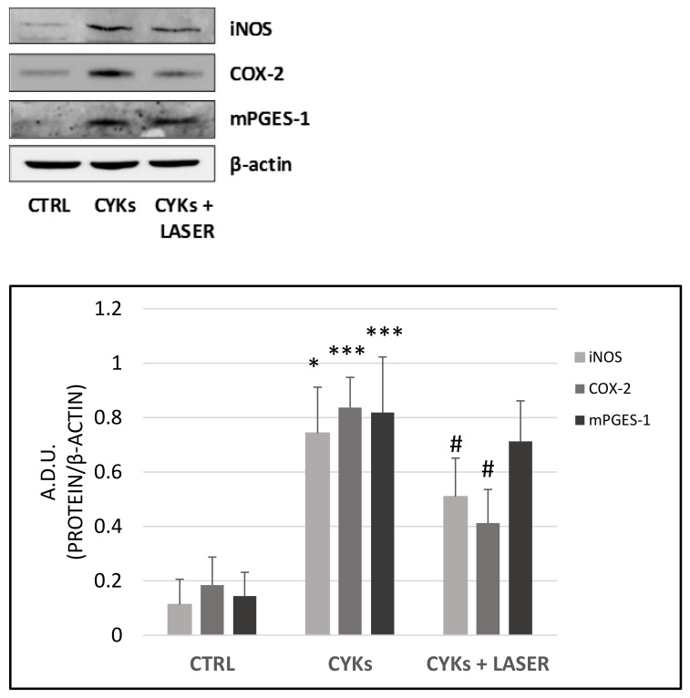
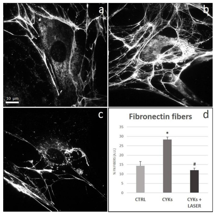
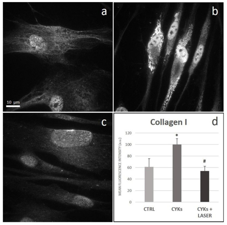
The fine control of inflammation following injury avoids fibrotic scars or impaired wounds. Due to side effects by anti-inflammatory drugs, the research is continuously active to define alternative therapies. Among them, physical countermeasures such as photobiomodulation therapy (PBMT) are considered effective and safe. To study the cellular and molecular events associated with the anti-inflammatory activity of PBMT by a dual-wavelength NIR laser source, human dermal fibroblasts were exposed to a mix of inflammatory cytokines (IL-1β and TNF-α) followed by laser treatment once a day for three days. Inducible inflammatory key enzymatic pathways, as iNOS and COX-2/mPGES-1/PGE2, were upregulated by the cytokine mix while PBMT reverted their levels and activities. The same behavior was observed with the proangiogenic factor vascular endothelial growth factor (VEGF), involved in neovascularization of granulation tissue. From a molecular point of view, PBMT retained NF-kB cytoplasmatic localization. According to a change in cell morphology, differences in expression and distribution of fundamental cytoskeletal proteins were observed following treatments. Tubulin, F-actin, and α-SMA changed their organization upon cytokine stimulation, while PBMT reestablished the basal localization. Cytoskeletal rearrangements occurring after inflammatory stimuli were correlated with reorganization of membrane α5β1 and fibronectin network as well as with their upregulation, while PBMT induced significant downregulation. Similar changes were observed for collagen I and the gelatinolytic enzyme MMP-1. In conclusion, the present study demonstrates that the proposed NIR laser therapy is effective in controlling fibroblast activation induced by IL-1β and TNF-α, likely responsible for a deleterious effect of persistent inflammation.
损伤后炎症的精细调控可避免形成纤维化瘢痕或伤口愈合受损。由于抗炎药物存在副作用,因此不断有研究致力于确定替代疗法。其中,诸如光生物调节疗法(PBMT)等物理对策被认为是有效且安全的。为了通过双波长近红外激光源研究与PBMT抗炎活性相关的细胞和分子事件,将人皮肤成纤维细胞暴露于炎性细胞因子(IL-1β和TNF-α)混合物中,随后每天进行一次激光治疗,持续三天。炎性关键酶途径如诱导型一氧化氮合酶(iNOS)和环氧化酶-2/微粒体前列腺素E合酶-1/前列腺素E2(COX-2/mPGES-1/PGE2)被细胞因子混合物上调,而PBMT使其水平和活性恢复正常。促血管生成因子血管内皮生长因子(VEGF)也观察到同样的情况,VEGF参与肉芽组织的新血管形成。从分子角度来看,PBMT使核因子κB(NF-κB)保持在细胞质定位。根据细胞形态的变化,处理后观察到基本细胞骨架蛋白的表达和分布存在差异。微管蛋白、F-肌动蛋白和α-平滑肌肌动蛋白(α-SMA)在细胞因子刺激后改变了它们的组织形式,而PBMT重新建立了基础定位。炎症刺激后发生的细胞骨架重排与膜α5β1和纤连蛋白网络的重组及其上调相关,而PBMT则导致显著下调。胶原蛋白I和明胶溶解酶基质金属蛋白酶-1(MMP-1)也观察到类似变化。总之,本研究表明,所提出的近红外激光疗法可有效控制由IL-1β和TNF-α诱导的成纤维细胞活化,而这种活化可能是持续性炎症产生有害作用的原因。