• 文献检索
  • 文档翻译
  • 深度研究
  • 学术资讯
  • Suppr Zotero 插件Zotero 插件
  • 邀请有礼
  • 套餐&价格
  • 历史记录
应用&插件
Suppr Zotero 插件Zotero 插件浏览器插件Mac 客户端Windows 客户端微信小程序
定价
高级版会员购买积分包购买API积分包
服务
文献检索文档翻译深度研究API 文档MCP 服务
关于我们
关于 Suppr公司介绍联系我们用户协议隐私条款
关注我们

Suppr 超能文献

核心技术专利:CN118964589B侵权必究
粤ICP备2023148730 号-1Suppr @ 2026

文献检索

告别复杂PubMed语法,用中文像聊天一样搜索,搜遍4000万医学文献。AI智能推荐,让科研检索更轻松。

立即免费搜索

文件翻译

保留排版,准确专业,支持PDF/Word/PPT等文件格式,支持 12+语言互译。

免费翻译文档

深度研究

AI帮你快速写综述,25分钟生成高质量综述,智能提取关键信息,辅助科研写作。

立即免费体验

SAFE:一种用于乳腺癌筛查和早期检测的新型微波成像系统设计——临床评估

SAFE: A Novel Microwave Imaging System Design for Breast Cancer Screening and Early Detection-Clinical Evaluation.

作者信息

Janjic Aleksandar, Cayoren Mehmet, Akduman Ibrahim, Yilmaz Tuba, Onemli Emre, Bugdayci Onur, Aribal Mustafa Erkin

机构信息

Mitos Medical Technologies, ITU Ayazaga Ari Teknokent 2-B Block 2-2-E, Maslak, 34469 Istanbul, Turkey.

Electrical and Electronics Engineering Faculty, Istanbul Technical University, Maslak, 34469 Istanbul, Turkey.

出版信息

Diagnostics (Basel). 2021 Mar 16;11(3):533. doi: 10.3390/diagnostics11030533.

DOI:10.3390/diagnostics11030533
PMID:33809770
原文链接:https://pmc.ncbi.nlm.nih.gov/articles/PMC8002280/
Abstract

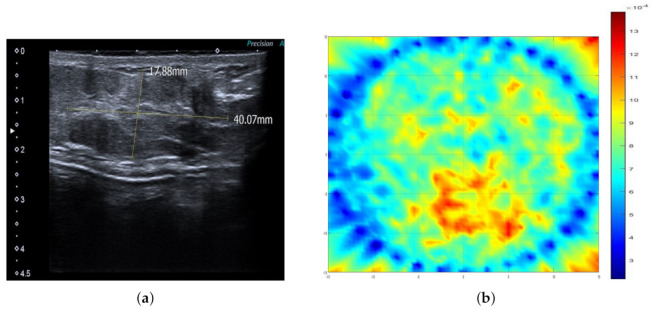
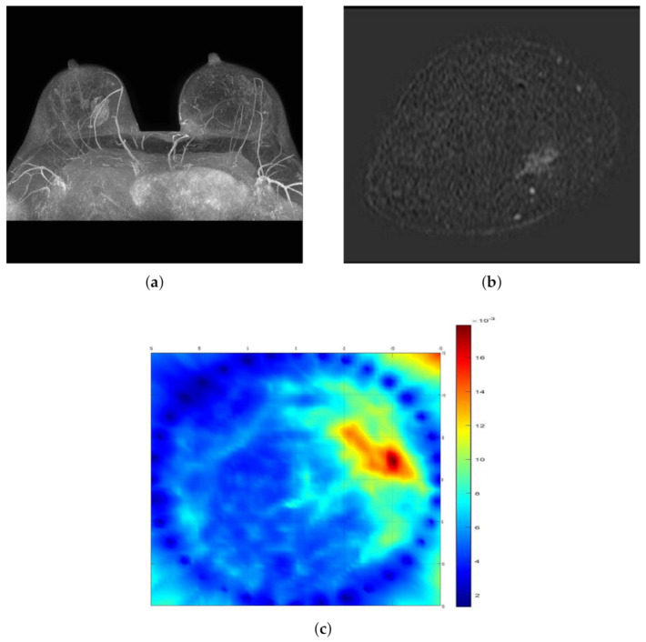
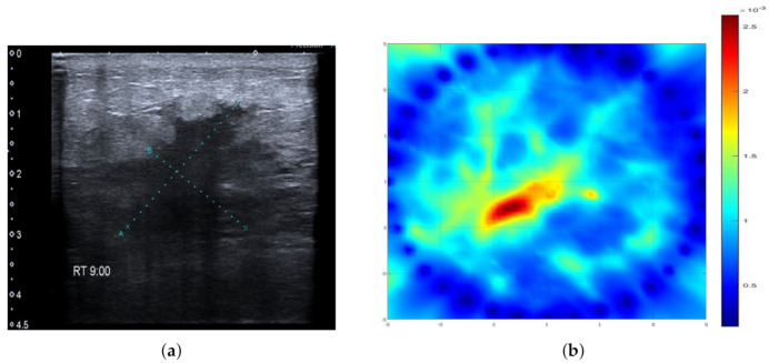
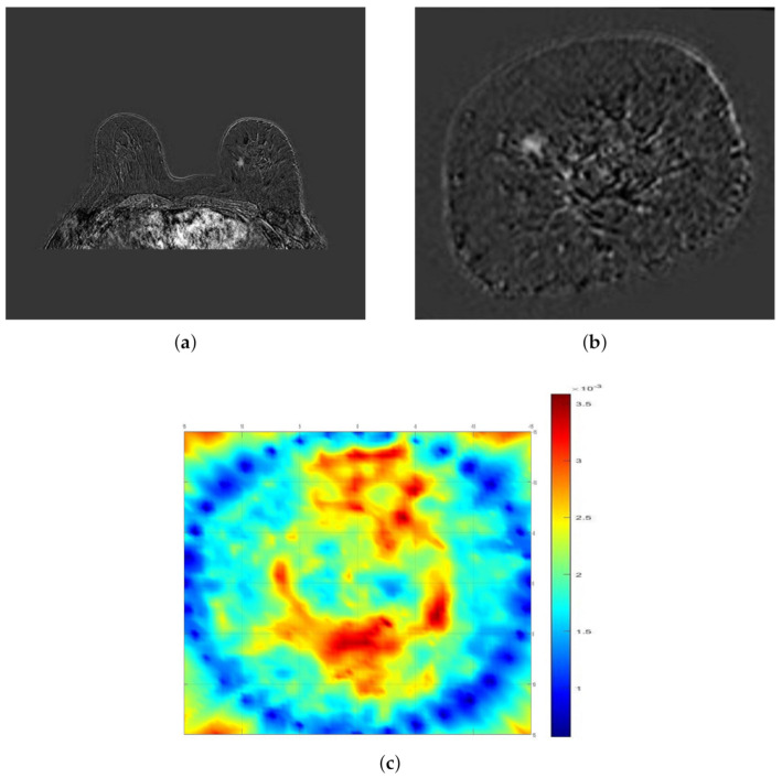
SAFE (Scan and Find Early) is a novel microwave imaging device intended for breast cancer screening and early detection. SAFE is based on the use of harmless electromagnetic waves and can provide relevant initial diagnostic information without resorting to X-rays. Because of SAFE's harmless effect on organic tissue, imaging can be performed repeatedly. In addition, the scanning process itself is not painful since breast compression is not required. Because of the absence of physical compression, SAFE can also detect tumors that are close to the thoracic wall. A total number of 115 patients underwent the SAFE scanning procedure, and the resultant images were compared with available magnetic resonance (MR), ultrasound, and mammography images in order to determine the correct detection rate. A sensitivity of 63% was achieved. Breast size influenced overall sensitivity, as sensitivity was lower in smaller breasts (51%) compared to larger ones (74%). Even though this is only a preliminary study, the results show promising concordance with clinical reports, thus encouraging further SAFE clinical studies.

摘要

SAFE(扫描并早期发现)是一种用于乳腺癌筛查和早期检测的新型微波成像设备。SAFE基于无害电磁波的使用,无需借助X射线就能提供相关的初步诊断信息。由于SAFE对有机组织无害,成像可反复进行。此外,由于无需乳房压迫,扫描过程本身不会产生疼痛。由于不存在物理压迫,SAFE还能检测靠近胸壁的肿瘤。共有115名患者接受了SAFE扫描程序,并将所得图像与现有的磁共振(MR)、超声和乳腺X线摄影图像进行比较,以确定正确的检测率。灵敏度达到了63%。乳房大小影响总体灵敏度,较小乳房(51%)的灵敏度低于较大乳房(74%)。尽管这只是一项初步研究,但结果显示与临床报告有良好的一致性,从而鼓励进一步开展SAFE临床研究。

https://cdn.ncbi.nlm.nih.gov/pmc/blobs/c3ec/8002280/487f2a948a84/diagnostics-11-00533-g005.jpg
https://cdn.ncbi.nlm.nih.gov/pmc/blobs/c3ec/8002280/521b22f2015f/diagnostics-11-00533-g001.jpg
https://cdn.ncbi.nlm.nih.gov/pmc/blobs/c3ec/8002280/8cf319f55fea/diagnostics-11-00533-g002.jpg
https://cdn.ncbi.nlm.nih.gov/pmc/blobs/c3ec/8002280/b9f537706e78/diagnostics-11-00533-g003.jpg
https://cdn.ncbi.nlm.nih.gov/pmc/blobs/c3ec/8002280/7157e7cc8736/diagnostics-11-00533-g004.jpg
https://cdn.ncbi.nlm.nih.gov/pmc/blobs/c3ec/8002280/487f2a948a84/diagnostics-11-00533-g005.jpg

相似文献

1
SAFE: A Novel Microwave Imaging System Design for Breast Cancer Screening and Early Detection-Clinical Evaluation.SAFE:一种用于乳腺癌筛查和早期检测的新型微波成像系统设计——临床评估
Diagnostics (Basel). 2021 Mar 16;11(3):533. doi: 10.3390/diagnostics11030533.
2
3
4
MR Imaging as an Additional Screening Modality for the Detection of Breast Cancer in Women Aged 50-75 Years with Extremely Dense Breasts: The DENSE Trial Study Design.磁共振成像作为一种额外的筛查手段,用于检测 50-75 岁致密型乳腺女性的乳腺癌:DENSE 试验研究设计。
Radiology. 2015 Nov;277(2):527-37. doi: 10.1148/radiol.2015141827. Epub 2015 Jun 23.
5
Emerging technologies in breast cancer detection.乳腺癌检测中的新兴技术。
Radiol Manage. 2004 Jul-Aug;26(4):16-24; quiz 25-7.
6
More advantages in detecting bone and soft tissue metastases from prostate cancer using F-PSMA PET/CT.使用F-PSMA PET/CT检测前列腺癌骨和软组织转移方面有更多优势。
Hell J Nucl Med. 2019 Jan-Apr;22(1):6-9. doi: 10.1967/s002449910952. Epub 2019 Mar 7.
7
Gradient-Boosting Algorithm for Microwave Breast Lesion Classification-SAFE Clinical Investigation.用于微波乳腺病变分类的梯度提升算法——SAFE临床研究。
Diagnostics (Basel). 2022 Dec 13;12(12):3151. doi: 10.3390/diagnostics12123151.
8
Feasibility of Portable Microwave Imaging Device for Breast Cancer Detection.用于乳腺癌检测的便携式微波成像设备的可行性
Diagnostics (Basel). 2021 Dec 23;12(1):27. doi: 10.3390/diagnostics12010027.
9
Retrospective and comparative analysis of (99m)Tc-Sestamibi breast specific gamma imaging versus mammography, ultrasound, and magnetic resonance imaging for the detection of breast cancer in Chinese women.(99m)锝-司他米比乳腺特异性γ成像与乳腺X线摄影、超声及磁共振成像在中国女性乳腺癌检测中的回顾性对比分析
BMC Cancer. 2016 Jul 11;16:450. doi: 10.1186/s12885-016-2537-1.
10
Responding to the challenges of breast cancer in egypt and other arab countries.应对埃及及其他阿拉伯国家乳腺癌带来的挑战。
J Egypt Natl Canc Inst. 2008 Dec;20(4):309-12.

引用本文的文献

1
Feasibility of tumor detection with a transmission-based microwave imaging system.基于透射的微波成像系统用于肿瘤检测的可行性。
Med Phys. 2025 Sep;52(9):e18080. doi: 10.1002/mp.18080.
2
Advances in breast cancer diagnosis: a comprehensive review of imaging, biosensors, and emerging wearable technologies.乳腺癌诊断的进展:影像学、生物传感器及新兴可穿戴技术的全面综述
Front Oncol. 2025 Jun 18;15:1587517. doi: 10.3389/fonc.2025.1587517. eCollection 2025.
3
Emerging paradigms in microwave imaging technology for biomedical applications: unleashing the power of artificial intelligence.

本文引用的文献

1
Overdiagnosis and Risks of Breast Cancer Screening.乳腺癌筛查的过度诊断与风险
Radiol Clin North Am. 2021 Jan;59(1):19-27. doi: 10.1016/j.rcl.2020.09.005.
2
Cancer statistics, 2020.癌症统计数据,2020 年。
CA Cancer J Clin. 2020 Jan;70(1):7-30. doi: 10.3322/caac.21590. Epub 2020 Jan 8.
3
European cancer mortality predictions for the year 2019 with focus on breast cancer.2019 年欧洲癌症死亡率预测,重点关注乳腺癌。
用于生物医学应用的微波成像技术的新兴范式:释放人工智能的力量。
Npj Imaging. 2024 Jun 3;2(1):13. doi: 10.1038/s44303-024-00012-8.
4
A Line-Source Approach for Simulating MammoWave Microwave Imaging Apparatus for Breast Lesion Detection.一种用于模拟乳腺波微波成像设备以检测乳腺病变的线源方法。
Sensors (Basel). 2025 Jun 10;25(12):3640. doi: 10.3390/s25123640.
5
Nano-formulations in disease therapy: designs, advances, challenges, and future directions.疾病治疗中的纳米制剂:设计、进展、挑战及未来方向。
J Nanobiotechnology. 2025 May 30;23(1):396. doi: 10.1186/s12951-025-03442-7.
6
XGBoost Enhances the Performance of SAFE: A Novel Microwave Imaging System for Early Detection of Malignant Breast Cancer.XGBoost提升SAFE的性能:一种用于早期检测恶性乳腺癌的新型微波成像系统。
Cancers (Basel). 2025 Jan 10;17(2):214. doi: 10.3390/cancers17020214.
7
Review of Microwave Near-Field Sensing and Imaging Devices in Medical Applications.医学应用中微波近场传感与成像设备的综述。
Sensors (Basel). 2024 Jul 12;24(14):4515. doi: 10.3390/s24144515.
8
Repository of MRI-derived models of the breast with single and multiple benign and malignant tumors for microwave imaging research.用于微波成象研究的具有单个和多个良性及恶性肿瘤的 MRI 模型的储存库。
PLoS One. 2024 May 17;19(5):e0302974. doi: 10.1371/journal.pone.0302974. eCollection 2024.
9
Microwave Imaging System Based on Signal Analysis in a Planar Environment for Detection of Abdominal Aortic Aneurysms.基于平面环境中信号分析的用于检测腹主动脉瘤的微波成象系统。
Biosensors (Basel). 2024 Mar 18;14(3):149. doi: 10.3390/bios14030149.
10
Impact of Imaging Biomarkers and AI on Breast Cancer Management: A Brief Review.影像生物标志物与人工智能对乳腺癌管理的影响:简要综述
Cancers (Basel). 2023 Oct 30;15(21):5216. doi: 10.3390/cancers15215216.
Ann Oncol. 2019 May 1;30(5):781-787. doi: 10.1093/annonc/mdz051.
4
Cancer statistics, 2019.癌症统计数据,2019 年。
CA Cancer J Clin. 2019 Jan;69(1):7-34. doi: 10.3322/caac.21551. Epub 2019 Jan 8.
5
Compact ultrasound-guided diffuse optical tomography system for breast cancer imaging.紧凑型超声引导漫射光学断层成像系统用于乳腺癌成像。
J Biomed Opt. 2018 Oct;24(2):1-9. doi: 10.1117/1.JBO.24.2.021203.
6
Estimating the global cancer incidence and mortality in 2018: GLOBOCAN sources and methods.估算 2018 年全球癌症发病率和死亡率:GLOBOCAN 来源和方法。
Int J Cancer. 2019 Apr 15;144(8):1941-1953. doi: 10.1002/ijc.31937. Epub 2018 Dec 6.
7
Microwave Breast Imaging: Clinical Advances and Remaining Challenges.微波乳腺成像:临床进展和遗留挑战。
IEEE Trans Biomed Eng. 2018 Nov;65(11):2580-2590. doi: 10.1109/TBME.2018.2809541. Epub 2018 Feb 26.
8
Review of breast screening: Toward clinical realization of microwave imaging.乳腺筛查综述:迈向微波成象技术的临床应用。
Med Phys. 2017 Dec;44(12):e446-e458. doi: 10.1002/mp.12611. Epub 2017 Nov 1.
9
MARIA M4: clinical evaluation of a prototype ultrawideband radar scanner for breast cancer detection.玛丽亚M4:用于乳腺癌检测的超宽带雷达扫描仪原型的临床评估。
J Med Imaging (Bellingham). 2016 Jul;3(3):033502. doi: 10.1117/1.JMI.3.3.033502. Epub 2016 Jul 20.
10
US-localized diffuse optical tomography in breast cancer: comparison with pharmacokinetic parameters of DCE-MRI and with pathologic biomarkers.美国局部乳腺癌扩散光学断层扫描:与动态对比增强磁共振成像的药代动力学参数及病理生物标志物的比较
BMC Cancer. 2016 Feb 1;16:50. doi: 10.1186/s12885-016-2086-7.