• 文献检索
  • 文档翻译
  • 深度研究
  • 学术资讯
  • Suppr Zotero 插件Zotero 插件
  • 邀请有礼
  • 套餐&价格
  • 历史记录
应用&插件
Suppr Zotero 插件Zotero 插件浏览器插件Mac 客户端Windows 客户端微信小程序
定价
高级版会员购买积分包购买API积分包
服务
文献检索文档翻译深度研究API 文档MCP 服务
关于我们
关于 Suppr公司介绍联系我们用户协议隐私条款
关注我们

Suppr 超能文献

核心技术专利:CN118964589B侵权必究
粤ICP备2023148730 号-1Suppr @ 2026

文献检索

告别复杂PubMed语法,用中文像聊天一样搜索,搜遍4000万医学文献。AI智能推荐,让科研检索更轻松。

立即免费搜索

文件翻译

保留排版,准确专业,支持PDF/Word/PPT等文件格式,支持 12+语言互译。

免费翻译文档

深度研究

AI帮你快速写综述,25分钟生成高质量综述,智能提取关键信息,辅助科研写作。

立即免费体验

单分子网络分析揭示了由于小窝蛋白-1支架结构域突变导致的小窝和支架的结构变化。

Single molecule network analysis identifies structural changes to caveolae and scaffolds due to mutation of the caveolin-1 scaffolding domain.

作者信息

Wong Timothy H, Khater Ismail M, Joshi Bharat, Shahsavari Mona, Hamarneh Ghassan, Nabi Ivan R

机构信息

Life Sciences Institute, Department of Cellular and Physiological Sciences, University of British Columbia, Vancouver, BC, V6T 1Z3, Canada.

School of Computing Science, Simon Fraser University, Burnaby, BC, V5A 1S6, Canada.

出版信息

Sci Rep. 2021 Apr 8;11(1):7810. doi: 10.1038/s41598-021-86770-6.

DOI:10.1038/s41598-021-86770-6
PMID:33833286
原文链接:https://pmc.ncbi.nlm.nih.gov/articles/PMC8032680/
Abstract

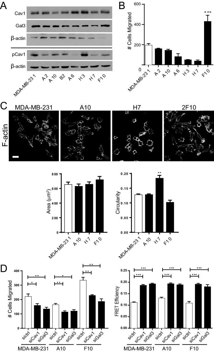
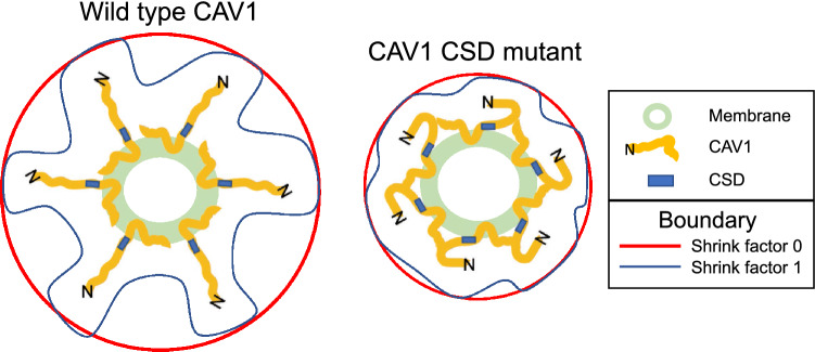
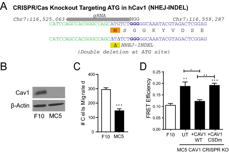
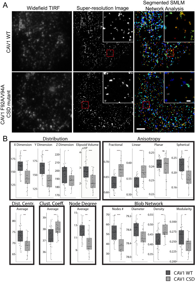
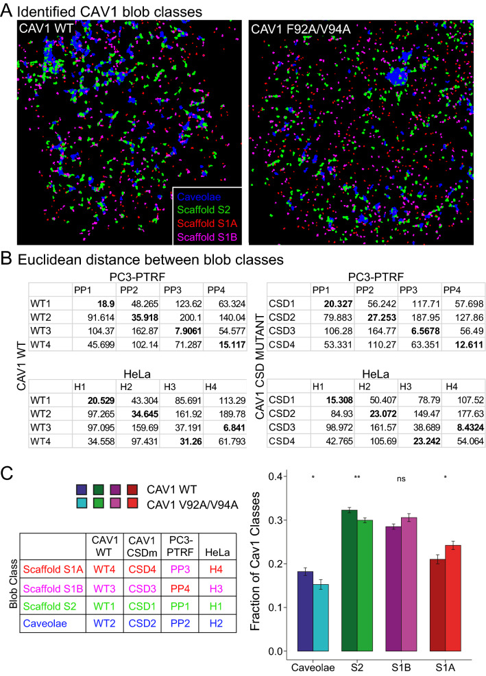
Caveolin-1 (CAV1), the caveolae coat protein, also associates with non-caveolar scaffold domains. Single molecule localization microscopy (SMLM) network analysis distinguishes caveolae and three scaffold domains, hemispherical S2 scaffolds and smaller S1B and S1A scaffolds. The caveolin scaffolding domain (CSD) is a highly conserved hydrophobic region that mediates interaction of CAV1 with multiple effector molecules. F92A/V94A mutation disrupts CSD function, however the structural impact of CSD mutation on caveolae or scaffolds remains unknown. Here, SMLM network analysis quantitatively shows that expression of the CAV1 CSD F92A/V94A mutant in CRISPR/Cas CAV1 knockout MDA-MB-231 breast cancer cells reduces the size and volume and enhances the elongation of caveolae and scaffold domains, with more pronounced effects on S2 and S1B scaffolds. Convex hull analysis of the outer surface of the CAV1 point clouds confirms the size reduction of CSD mutant CAV1 blobs and shows that CSD mutation reduces volume variation amongst S2 and S1B CAV1 blobs at increasing shrink values, that may reflect retraction of the CAV1 N-terminus towards the membrane, potentially preventing accessibility of the CSD. Detection of point mutation-induced changes to CAV1 domains highlights the utility of SMLM network analysis for mesoscale structural analysis of oligomers in their native environment.

摘要

小窝蛋白-1(CAV1)是小窝的包被蛋白,也与非小窝支架结构域相关联。单分子定位显微镜(SMLM)网络分析可区分小窝和三个支架结构域,即半球形S2支架以及较小的S1B和S1A支架。小窝蛋白支架结构域(CSD)是一个高度保守的疏水区域,介导CAV1与多种效应分子的相互作用。F92A/V94A突变会破坏CSD功能,然而CSD突变对小窝或支架的结构影响仍不清楚。在此,SMLM网络分析定量显示,在CRISPR/Cas CAV1基因敲除的MDA-MB-231乳腺癌细胞中表达CAV1 CSD F92A/V94A突变体,会减小小窝和支架结构域的尺寸和体积,并增加其伸长率,对S2和S1B支架的影响更为明显。对CAV1点云外表面的凸包分析证实了CSD突变体CAV1斑点的尺寸减小,并表明CSD突变在收缩值增加时减少了S2和S1B CAV1斑点之间的体积变化,这可能反映了CAV1 N端向膜的回缩,可能阻止了CSD的可及性。检测点突变引起的CAV1结构域变化,突出了SMLM网络分析在其天然环境中对寡聚体进行中尺度结构分析的实用性。

https://cdn.ncbi.nlm.nih.gov/pmc/blobs/5dbd/8032680/75e5876857b5/41598_2021_86770_Fig7_HTML.jpg
https://cdn.ncbi.nlm.nih.gov/pmc/blobs/5dbd/8032680/d4d0303ec8c3/41598_2021_86770_Fig1_HTML.jpg
https://cdn.ncbi.nlm.nih.gov/pmc/blobs/5dbd/8032680/081acfc04395/41598_2021_86770_Fig2_HTML.jpg
https://cdn.ncbi.nlm.nih.gov/pmc/blobs/5dbd/8032680/fcaa4b48e5a4/41598_2021_86770_Fig3_HTML.jpg
https://cdn.ncbi.nlm.nih.gov/pmc/blobs/5dbd/8032680/eb43ea76e667/41598_2021_86770_Fig4_HTML.jpg
https://cdn.ncbi.nlm.nih.gov/pmc/blobs/5dbd/8032680/291719ea1515/41598_2021_86770_Fig5_HTML.jpg
https://cdn.ncbi.nlm.nih.gov/pmc/blobs/5dbd/8032680/14c96c7958f5/41598_2021_86770_Fig6_HTML.jpg
https://cdn.ncbi.nlm.nih.gov/pmc/blobs/5dbd/8032680/75e5876857b5/41598_2021_86770_Fig7_HTML.jpg

相似文献

1
Single molecule network analysis identifies structural changes to caveolae and scaffolds due to mutation of the caveolin-1 scaffolding domain.单分子网络分析揭示了由于小窝蛋白-1支架结构域突变导致的小窝和支架的结构变化。
Sci Rep. 2021 Apr 8;11(1):7810. doi: 10.1038/s41598-021-86770-6.
2
Super-resolution modularity analysis shows polyhedral caveolin-1 oligomers combine to form scaffolds and caveolae.超分辨率模块化分析表明,多面体型窖蛋白-1 寡聚物结合形成支架和小窝。
Sci Rep. 2019 Jul 8;9(1):9888. doi: 10.1038/s41598-019-46174-z.
3
Scaffolds and the scaffolding domain: an alternative paradigm for caveolin-1 signaling.支架与支架结构域:小窝蛋白-1信号传导的另一种模式
Biochem Soc Trans. 2024 Apr 24;52(2):947-959. doi: 10.1042/BST20231570.
4
Identification of caveolin-1 domain signatures via machine learning and graphlet analysis of single-molecule super-resolution data.通过机器学习和单分子超分辨率数据的图元分析鉴定窖蛋白-1 结构域特征。
Bioinformatics. 2019 Sep 15;35(18):3468-3475. doi: 10.1093/bioinformatics/btz113.
5
Super Resolution Network Analysis Defines the Molecular Architecture of Caveolae and Caveolin-1 Scaffolds.超分辨率网络分析定义了质膜微囊泡和窖蛋白-1 支架的分子结构。
Sci Rep. 2018 Jun 13;8(1):9009. doi: 10.1038/s41598-018-27216-4.
6
The phospho-caveolin-1 scaffolding domain dampens force fluctuations in focal adhesions and promotes cancer cell migration.磷酸化的小窝蛋白-1支架结构域可抑制粘着斑中的力波动并促进癌细胞迁移。
Mol Biol Cell. 2017 Aug 1;28(16):2190-2201. doi: 10.1091/mbc.E17-05-0278. Epub 2017 Jun 7.
7
Molecular Characterization of Caveolin-induced Membrane Curvature.小窝蛋白诱导的膜曲率的分子特征
J Biol Chem. 2015 Oct 9;290(41):24875-90. doi: 10.1074/jbc.M115.644336. Epub 2015 Aug 24.
8
Caveolin-1 Scaffolding Domain Peptides Alleviate Liver Fibrosis by Inhibiting TGF-β1/Smad Signaling in Mice.窖蛋白-1 支架结构域肽通过抑制 TGF-β1/Smad 信号通路减轻小鼠肝纤维化。
Int J Mol Sci. 2018 Jun 11;19(6):1729. doi: 10.3390/ijms19061729.
9
Differential impact of caveolae and caveolin-1 scaffolds on the membrane raft proteome.网格蛋白小窝及其相关蛋白 1 支架对膜筏蛋白组的差异影响。
Mol Cell Proteomics. 2011 Oct;10(10):M110.007146. doi: 10.1074/mcp.M110.007146. Epub 2011 Jul 13.
10
Caveolin-1 interacts directly with dynamin-2.小窝蛋白-1与发动蛋白-2直接相互作用。
J Mol Biol. 2005 Apr 29;348(2):491-501. doi: 10.1016/j.jmb.2005.02.003.

引用本文的文献

1
SuperResNET: Model-Free Single-Molecule Network Analysis Software Achieves Molecular Resolution of Nup96.SuperResNET:无模型单分子网络分析软件实现了Nup96的分子分辨率。
Adv Intell Syst. 2025 Mar;7(3):2400521. doi: 10.1002/aisy.202400521. Epub 2024 Dec 25.
2
Scaffolds and the scaffolding domain: an alternative paradigm for caveolin-1 signaling.支架与支架结构域:小窝蛋白-1信号传导的另一种模式
Biochem Soc Trans. 2024 Apr 24;52(2):947-959. doi: 10.1042/BST20231570.
3
Are There Lipid Membrane-Domain Subtypes in Neurons with Different Roles in Calcium Signaling?

本文引用的文献

1
Structure and assembly of CAV1 8S complexes revealed by single particle electron microscopy.单颗粒电子显微镜揭示的CAV1 8S复合物的结构与组装
Sci Adv. 2020 Dec 2;6(49). doi: 10.1126/sciadv.abc6185. Print 2020 Dec.
2
A Review of Super-Resolution Single-Molecule Localization Microscopy Cluster Analysis and Quantification Methods.超分辨率单分子定位显微镜簇分析与量化方法综述
Patterns (N Y). 2020 Jun 12;1(3):100038. doi: 10.1016/j.patter.2020.100038.
3
Endothelial caveolin and its scaffolding domain in cancer.内皮细胞 caveolin 及其在癌症中的支架结构域。
神经元在钙信号转导中具有不同作用,是否存在脂质膜域亚型?
Molecules. 2023 Dec 2;28(23):7909. doi: 10.3390/molecules28237909.
4
Physiological functions of caveolae in endothelium.内皮细胞中质膜小窝的生理功能。
Curr Opin Physiol. 2023 Oct;35. doi: 10.1016/j.cophys.2023.100701. Epub 2023 Jun 28.
5
SPECHT: Self-tuning Plausibility based object detection Enables quantification of Conflict in Heterogeneous multi-scale microscopy.斯佩克特:基于自调谐似真性的目标检测可实现异质多尺度显微镜下冲突的量化。
PLoS One. 2022 Dec 29;17(12):e0276726. doi: 10.1371/journal.pone.0276726. eCollection 2022.
6
A deep learning-based tool for the automated detection and analysis of caveolae in transmission electron microscopy images.一种基于深度学习的工具,用于在透射电子显微镜图像中自动检测和分析小窝。
Comput Struct Biotechnol J. 2022 Dec 5;21:224-237. doi: 10.1016/j.csbj.2022.11.062. eCollection 2023.
7
Studying the Dynamics of Chromatin-Binding Proteins in Mammalian Cells Using Single-Molecule Localization Microscopy.使用单分子定位显微镜研究哺乳动物细胞中染色质结合蛋白的动力学。
Methods Mol Biol. 2022;2476:209-247. doi: 10.1007/978-1-0716-2221-6_16.
8
Caveolin-1 Regulates Cellular Metabolism: A Potential Therapeutic Target in Kidney Disease.小窝蛋白-1调节细胞代谢:肾脏疾病的潜在治疗靶点。
Front Pharmacol. 2021 Dec 10;12:768100. doi: 10.3389/fphar.2021.768100. eCollection 2021.
Cancer Metastasis Rev. 2020 Jun;39(2):471-483. doi: 10.1007/s10555-020-09895-6.
4
Tyrosine phosphorylation of tumor cell caveolin-1: impact on cancer progression.肿瘤细胞窖蛋白-1 的酪氨酸磷酸化:对癌症进展的影响。
Cancer Metastasis Rev. 2020 Jun;39(2):455-469. doi: 10.1007/s10555-020-09892-9.
5
Caveolae and scaffold detection from single molecule localization microscopy data using deep learning.使用深度学习从单分子定位显微镜数据中检测质膜微囊和支架
PLoS One. 2019 Aug 26;14(8):e0211659. doi: 10.1371/journal.pone.0211659. eCollection 2019.
6
Super-resolution modularity analysis shows polyhedral caveolin-1 oligomers combine to form scaffolds and caveolae.超分辨率模块化分析表明,多面体型窖蛋白-1 寡聚物结合形成支架和小窝。
Sci Rep. 2019 Jul 8;9(1):9888. doi: 10.1038/s41598-019-46174-z.
7
Deletion of caveolin scaffolding domain alters cancer cell migration.缺失窖蛋白 scaffolding 结构域改变癌细胞迁移。
Cell Cycle. 2019 Jun;18(11):1268-1280. doi: 10.1080/15384101.2019.1618118. Epub 2019 May 22.
8
Challenges and opportunities in cryo-EM single-particle analysis.冷冻电镜单颗粒分析的挑战与机遇。
J Biol Chem. 2019 Mar 29;294(13):5181-5197. doi: 10.1074/jbc.REV118.005602. Epub 2019 Feb 25.
9
Multi-omic measurements of heterogeneity in HeLa cells across laboratories.多组学测量 HeLa 细胞在不同实验室间的异质性。
Nat Biotechnol. 2019 Mar;37(3):314-322. doi: 10.1038/s41587-019-0037-y. Epub 2019 Feb 18.
10
Recent advances in proximity-based labeling methods for interactome mapping.用于相互作用组图谱绘制的基于邻近标记方法的最新进展。
F1000Res. 2019 Jan 31;8. doi: 10.12688/f1000research.16903.1. eCollection 2019.