Read Scott A, Gloss Brian S, Liddle Christopher, George Jacob, Ahlenstiel Golo
Blacktown Clinical School, Western Sydney University, Blacktown, NSW, 2148, Australia.
Blacktown Hospital, WSLHD, Blacktown, NSW, 2148, Australia.
J Inflamm Res. 2021 Apr 1;14:1257-1270. doi: 10.2147/JIR.S301476. eCollection 2021.
Interferon lambdas (IFN-λs) are antiviral cytokines that restrict pathogen infection and dissemination at barrier surfaces. Controlled expression of IFN-λs efficiently eliminates acute infections by activating a suite of interferon stimulated genes that inhibit viral propagation and activate local immune cells. Excessive or prolonged production of IFN-λs can however mediate tissue inflammation and disrupt epithelial barriers in both viral and non-viral disease. The mechanism by which IFN-λs drive this disease pathogenesis is poorly understood but may be caused by IFN-λ-mediated amplification of other innate immune signaling pathways.
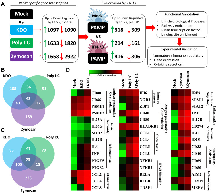
Monocyte-derived macrophages were differentiated ± IFN-λ3 and treated with KDO-lipid A, poly I:C or zymosan, representing bacterial, viral or fungal ligands, respectively. Transcriptome and protein expression were quantified by RNA sequencing/PCR and ELISA/bead array, respectively. Bioinformatic analysis was used to define transcription factor profiles and signaling pathways amplified by IFN-λ3. Finally, the SARS-CoV-2 dataset GSE152075 was queried to compare the effects of versus expression in relation to viral load and nasopharyngeal transcriptomes.
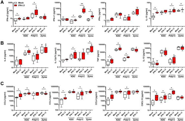
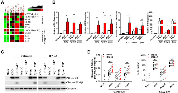
IFN-λ3 exacerbated inflammatory and chemotactic responses unique to each microbial ligand, as measured by RNA sequencing and by ELISA/bead array. Functional annotation identified pathways amplified by IFN-λ3, including inflammasome activation. Inflammasome amplification was confirmed in vitro, as measured by caspase 1 activity and IL-1β cleavage. Lastly, SARS-CoV-2 infected nasopharyngeal transcriptomes expressing IFN-λs but not IFN-αs were implicated in myeloid cell-driven pathogenesis including neutrophil degranulation, complement and coagulation cascades.
These data suggest that IFN-λs contribute to disease pathology by exacerbating innate immune responses during chronic or severe disease states. IFN-λs may contribute to SARS-CoV-2 disease severity, however further study is required to confirm true causation.
干扰素λ(IFN-λ)是一类抗病毒细胞因子,可在屏障表面限制病原体感染和传播。通过激活一系列抑制病毒繁殖并激活局部免疫细胞的干扰素刺激基因,IFN-λ的可控表达能有效消除急性感染。然而,在病毒性和非病毒性疾病中,IFN-λ的过量或长期产生都可能介导组织炎症并破坏上皮屏障。IFN-λ驱动这种疾病发病机制的原因尚不清楚,但可能是由IFN-λ介导的其他固有免疫信号通路的放大所致。
单核细胞衍生的巨噬细胞在有或无IFN-λ3的情况下分化,并用分别代表细菌、病毒或真菌配体的KDO-脂多糖、聚肌胞苷酸或酵母聚糖进行处理。转录组和蛋白质表达分别通过RNA测序/PCR和ELISA/微珠阵列进行定量。生物信息学分析用于定义转录因子谱和由IFN-λ3放大的信号通路。最后,查询了严重急性呼吸综合征冠状病毒2(SARS-CoV-2)数据集GSE152075,以比较IFN-λ与IFN-α表达在病毒载量和鼻咽转录组方面的影响。
通过RNA测序以及ELISA/微珠阵列检测发现,IFN-λ3加剧了每种微生物配体特有的炎症和趋化反应。功能注释确定了由IFN-λ3放大的信号通路,包括炎性小体激活。通过半胱天冬酶1活性和白细胞介素-1β裂解检测,在体外证实了炎性小体的放大。最后,感染SARS-CoV-2的鼻咽转录组中,表达IFN-λ而非IFN-α与髓样细胞驱动的发病机制有关,包括中性粒细胞脱颗粒、补体和凝血级联反应。
这些数据表明,在慢性或严重疾病状态下,IFN-λ通过加剧固有免疫反应而导致疾病病理变化。IFN-λ可能会导致SARS-CoV-2疾病的严重程度增加,然而,需要进一步研究来证实真正的因果关系。