Kang Jae Myeong, Cho Seo-Eun, Na Kyoung-Sae, Kang Seung-Gul
Sleep Medicine Center, Gil Medical Center, Gachon University College of Medicine, Incheon, Republic of Korea.
Department of Psychiatry, Gil Medical Center, Gachon University College of Medicine, Incheon, Republic of Korea.
Nat Sci Sleep. 2021 Apr 1;13:477-486. doi: 10.2147/NSS.S295742. eCollection 2021.
Previous spectral analysis studies on obstructive sleep apnea (OSA) involved small samples, and the results were inconsistent. We performed a spectral analysis of sleep EEG based on different severities of OSA using the Sleep Heart Health Study data. This study aimed to determine the difference in EEG spectral power during sleep in the non-OSA group and with different severities of OSA in the general population.
The participants (n = 5,804) underwent polysomnography, and they were classified into non-OSA, mild OSA, moderate OSA, and severe OSA groups. The fast Fourier transformation was used to compute the EEG power spectrum for total sleep duration within contiguous 30-second epochs of sleep. The EEG spectral powers of the groups were compared using 4,493 participants after adjusting potential confounding factors that could affect sleep EEG.
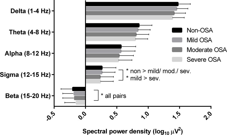
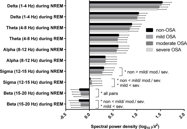
The power spectra differed significantly among the groups for all frequency bands ( corr < 0.001). We found that the quantitative EEG spectral powers in the beta and sigma bands of total sleep differed ( corr < 0.001) among the participants in the non-OSA group and with different severities of OSA, controlling for covariates. The beta power was higher and the sigma power was lower in the OSA groups than in the non-OSA group. The beta power decreased in the order of severe OSA, moderate OSA, mild OSA, and non-OSA.
This study suggests that there are differences between the microstructures of PSG-derived sleep EEG of non-OSA participants and those with different severities of OSA.
以往关于阻塞性睡眠呼吸暂停(OSA)的频谱分析研究样本量较小,结果不一致。我们使用睡眠心脏健康研究数据,基于OSA的不同严重程度对睡眠脑电图进行了频谱分析。本研究旨在确定非OSA组以及一般人群中不同严重程度OSA患者睡眠期间脑电图频谱功率的差异。
参与者(n = 5804)接受了多导睡眠图检查,并被分为非OSA组、轻度OSA组、中度OSA组和重度OSA组。使用快速傅里叶变换计算连续30秒睡眠时段内总睡眠时间的脑电图功率谱。在调整了可能影响睡眠脑电图的潜在混杂因素后,对4493名参与者的各小组脑电图频谱功率进行了比较。
所有频段各小组之间的功率谱存在显著差异(corr < 0.001)。我们发现,在控制协变量的情况下,非OSA组以及不同严重程度OSA患者总睡眠期间β和σ频段的定量脑电图频谱功率存在差异(corr < 0.001)。OSA组的β功率高于非OSA组,σ功率低于非OSA组。β功率按重度OSA、中度OSA、轻度OSA和非OSA的顺序递减。
本研究表明,非OSA参与者与不同严重程度OSA患者的多导睡眠图衍生睡眠脑电图微观结构存在差异。