Harmon Stephanie A, Gesztes William, Young Denise, Mehralivand Sherif, McKinney Yolanda, Sanford Thomas, Sackett Jonathan, Cullen Jennifer, Rosner Inger L, Srivastava Shiv, Merino Maria J, Wood Bradford J, Pinto Peter A, Choyke Peter L, Dobi Albert, Sesterhenn Isabell A, Turkbey Baris
From the Clinical Research Directorate, Frederick National Laboratory for Cancer Research sponsored by the National Cancer Institute (S.A.H.); Molecular Imaging Branch (S.A.H., S.M., Y.M., T.S., J.S., P.L.C., B.T.), Laboratory of Pathology (M.J.M.), Center for Interventional Oncology (B.J.W.), and Urologic Oncology Branch (S.M., P.A.P.), National Cancer Institute, National Institutes of Health, 9000 Rockville Pike, Building 10, Room B3B85, Bethesda, Md 20892; Center for Prostate Disease Research, John P. Murtha Cancer Center, Department of Surgery, Uniformed Services University of the Health Sciences (W.G., D.Y., J.C., I.L.R., S.S., A.D., I.A.S.) and Urology Service (I.L.R.), Walter Reed National Military Medical Center, Bethesda, Md; and Department of Genitourinary Pathology, Joint Pathology Center, Silver Spring, Md (I.A.S.).
Radiology. 2021 Jun;299(3):613-623. doi: 10.1148/radiol.2021202425. Epub 2021 Apr 13.
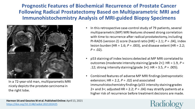
Background Although prostate MRI is routinely used for the detection and staging of localized prostate cancer, imaging-based assessment and targeted molecular sampling for risk stratification are an active area of research. Purpose To evaluate features of preoperative MRI and MRI-guided biopsy immunohistochemistry (IHC) findings associated with biochemical recurrence (BCR) of prostate cancer after surgery. Materials and Methods In this retrospective case-control study, patients underwent multiparametric MRI before MRI-guided biopsy followed by radical prostatectomy between 2008 and 2016. Lesions were retrospectively scored with the Prostate Imaging Reporting and Data System (PI-RADS) (version 2) by radiologists who were blinded to the clinical-pathologic results. The IHC staining, including stains for the ETS-related gene, phosphatase and tensin homolog, androgen receptor, prostate specific antigen, and p53, was performed with targeted biopsy specimens of the index lesion (highest suspicion at MRI and pathologic grade) and scored by pathologists who were blinded to clinical-pathologic outcomes. Cox proportional hazards regression analysis was used to evaluate associations with recurrence-free survival (RFS). Results The median RFS was 31.7 months (range, 1-101 months) for 39 patients (median age, 62 years; age range, 47-76 years) without BCR and 14.6 months (range, 1-61 months) for 40 patients (median age, 59 years; age range, 47-73 years) with BCR. MRI features that showed a significant relationship with the RFS interval included an index lesion with a PI-RADS score of 5 (hazard ratio [HR], 2.10; 95% CI: 1.05, 4.21; = .04); index lesion burden, defined as ratio of index lesion volume to prostate volume (HR, 1.55; 95% CI: 1.2, 2.1; = .003); and suspicion of extraprostatic extension (EPE) (HR, 2.18; 95% CI: 1.1, 4.2; = .02). Presurgical multivariable analysis indicated that suspicion of EPE at MRI (adjusted HR, 2.19; 95% CI: 1.1, 4.3; = .02) and p53 stain intensity (adjusted HR, 2.22; 95% CI: 1.0, 4.7; = .04) were significantly associated with RFS. Conclusion MRI features, including Prostate Imaging Reporting and Data System score, index lesion burden, extraprostatic extension, and preoperative guided biopsy p53 immunohistochemistry stain intensity are associated with biochemical relapse of prostate cancer after surgery. © RSNA, 2021 . See also the editorial by Costa in this issue.
尽管前列腺MRI常用于局限性前列腺癌的检测和分期,但基于影像学的评估和用于风险分层的靶向分子采样仍是一个活跃的研究领域。目的:评估术前MRI特征以及MRI引导下活检免疫组化(IHC)结果与前列腺癌术后生化复发(BCR)的相关性。材料与方法:在这项回顾性病例对照研究中,2008年至2016年间,患者在接受MRI引导下活检前行多参数MRI检查,随后接受根治性前列腺切除术。放射科医生对病变进行回顾性的前列腺影像报告和数据系统(PI-RADS)(第2版)评分,这些医生对临床病理结果不知情。对索引病变(MRI上怀疑度最高且病理分级最高)的靶向活检标本进行IHC染色,包括对ETS相关基因、磷酸酶和张力蛋白同源物、雄激素受体、前列腺特异性抗原和p53的染色,由对临床病理结果不知情的病理科医生进行评分。采用Cox比例风险回归分析评估与无复发生存期(RFS)的相关性。结果:39例无BCR患者(中位年龄62岁;年龄范围47 - 76岁)的中位RFS为31.7个月(范围1 - 101个月),40例有BCR患者(中位年龄59岁;年龄范围47 - 73岁)的中位RFS为14.6个月(范围1 - 61个月)。与RFS间隔显示出显著关系的MRI特征包括PI-RADS评分为5的索引病变(风险比[HR],2.10;95%可信区间:1.05,4.21;P = .04);索引病变负荷,定义为索引病变体积与前列腺体积之比(HR,1.55;95%可信区间:1.2,2.1;P = .003);以及怀疑前列腺外侵犯(EPE)(HR,2.18;95%可信区间:1.1,4.2;P = .02)。术前多变量分析表明,MRI上怀疑EPE(调整后HR,2.19;95%可信区间:1.1,4.3;P = .02)和p53染色强度(调整后HR,2.22;95%可信区间:1.0,4.7;P = .04)与RFS显著相关。结论:MRI特征,包括前列腺影像报告和数据系统评分、索引病变负荷、前列腺外侵犯以及术前引导活检p53免疫组化染色强度与前列腺癌术后生化复发相关。©RSNA,2021 。另见本期Costa的社论。