Deinsberger Julia, Brugger Jonas, Tschandl Philipp, Meier-Schiesser Barbara, Anzengruber Florian, Bossart Simon, Tzaneva Stanislava, Petzelbauer Peter, Böhler Kornelia, Beltraminelli Helmut, Hafner Jürg, Weber Benedikt
Skin and Endothelium Research Division (SERD), Department of Dermatology, Medical University of Vienna, AT-1090 Vienna, Austria.
Acta Derm Venereol. 2021 May 4;101(5):adv00449. doi: 10.2340/00015555-3804.
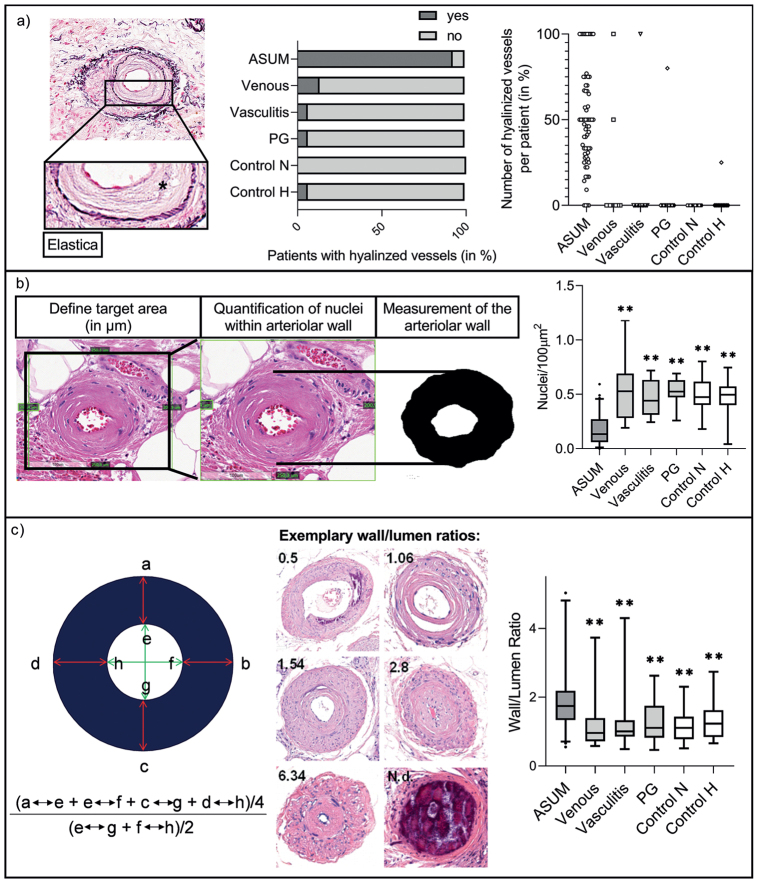
Clinical differential diagnosis of arteriolosclerotic ulcers of Martorell is challenging due to the lack of clearly affirmative instrument-based diagnostic criteria. The aim of this study was to develop vascular histomorphological diagnostic criteria differentiating Martorell ulcers from other types of leg ulcers. The histomorphology of patients diagnosed with arteriolosclerotic ulcers of Martorell (n = 67) was compared with that of patients with venous leg ulcers, necrotizing leukocytoclastic vasculitis, pyoderma gangrenosum, and non-ulcerative controls (n = 15 each). In a multivariable logistic regression model, the rates of arteriolar calcification (odds ratio (OR) 42.71, 95% confidence interval (CI) 7.43-443.96, p < 0.001) and subendothelial hyalinosis (OR 29.28, 95% CI 4.88-278.21, p <0.001) were significantly higher in arteriolosclerotic ulcers of Martorell. Arteriolar cellularity was significantly lower in Martorell ulcers than in controls (OR 0.003, 95 CI < 0.001-0.97, p = 0.05). However, the wall-to-lumen ratio was similar in all ulcers (OR 0.975, 95% CI 0.598-2.04, p =0.929). Based on the Youden index, a wall cellularity of < 0.24 cells/100 μm2 was determined as the optimum cut-off point (sensitivity 0.955, specificity 0.944). Thus, arteriolar calcification, subendothelial hyalinosis, and arteriolar cellularity revealed high discriminatory power for arteriolosclerotic ulcers of Martorell.
由于缺乏明确肯定的基于仪器的诊断标准,马托雷尔小动脉硬化性溃疡的临床鉴别诊断具有挑战性。本研究的目的是制定血管组织形态学诊断标准,以区分马托雷尔溃疡与其他类型的腿部溃疡。将诊断为马托雷尔小动脉硬化性溃疡的患者(n = 67)的组织形态学与腿部静脉溃疡、坏死性白细胞破碎性血管炎、坏疽性脓皮病患者以及非溃疡对照组(每组n = 15)的组织形态学进行比较。在多变量逻辑回归模型中,马托雷尔小动脉硬化性溃疡的小动脉钙化率(比值比(OR)42.71,95%置信区间(CI)7.43 - 443.96,p < 0.001)和内皮下玻璃样变性率(OR 29.28,95% CI 4.88 - 278.21,p < 0.001)显著更高。马托雷尔溃疡的小动脉细胞密度显著低于对照组(OR 0.003,95% CI < 0.001 - 0.97,p = 0.05)。然而,所有溃疡的壁腔比相似(OR 0.975,95% CI 0.598 - 2.04,p = 0.929)。基于约登指数,确定壁细胞密度< 0.24个细胞/100μm²为最佳切点(敏感性0.955,特异性0.944)。因此,小动脉钙化、内皮下玻璃样变性和小动脉细胞密度对马托雷尔小动脉硬化性溃疡具有很高的鉴别能力。