• 文献检索
  • 文档翻译
  • 深度研究
  • 学术资讯
  • Suppr Zotero 插件Zotero 插件
  • 邀请有礼
  • 套餐&价格
  • 历史记录
应用&插件
Suppr Zotero 插件Zotero 插件浏览器插件Mac 客户端Windows 客户端微信小程序
定价
高级版会员购买积分包购买API积分包
服务
文献检索文档翻译深度研究API 文档MCP 服务
关于我们
关于 Suppr公司介绍联系我们用户协议隐私条款
关注我们

Suppr 超能文献

核心技术专利:CN118964589B侵权必究
粤ICP备2023148730 号-1Suppr @ 2026

文献检索

告别复杂PubMed语法,用中文像聊天一样搜索,搜遍4000万医学文献。AI智能推荐,让科研检索更轻松。

立即免费搜索

文件翻译

保留排版,准确专业,支持PDF/Word/PPT等文件格式,支持 12+语言互译。

免费翻译文档

深度研究

AI帮你快速写综述,25分钟生成高质量综述,智能提取关键信息,辅助科研写作。

立即免费体验

从基因表达差异探讨心外膜脂肪组织在冠状动脉疾病中的作用

Exploring the Role of Epicardial Adipose Tissue in Coronary Artery Disease From the Difference of Gene Expression.

作者信息

Wang Qian-Chen, Wang Zhen-Yu, Xu Qian, Li Ruo-Bing, Zhang Guo-Gang, Shi Rui-Zheng

机构信息

Department of Cardiovascular Medicine, Xiangya Hospital, Central South University, Changsha, China.

Department of Cardiovascular Medicine, The Second Xiangya Hospital, Central South University, Changsha, China.

出版信息

Front Physiol. 2021 Mar 30;12:605811. doi: 10.3389/fphys.2021.605811. eCollection 2021.

DOI:10.3389/fphys.2021.605811
PMID:33859569
原文链接:https://pmc.ncbi.nlm.nih.gov/articles/PMC8042318/
Abstract

OBJECTIVES

Epicardial adipose tissue (EAT) is closely adjacent to the coronary arteries and myocardium, its role as an endocrine organ to affect the pathophysiological processes of the coronary arteries and myocardium has been increasingly recognized. However, the specific gene expression profiles of EAT in coronary artery disease (CAD) has not been well characterized. Our aim was to investigate the role of EAT in CAD at the gene level.

METHODS

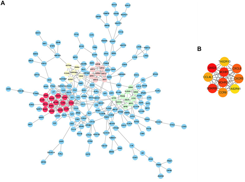
Here, we compared the histological and gene expression difference of EAT between CAD and non-CAD. We investigated the gene expression profiles in the EAT of patients with CAD through the high-throughput RNA sequencing. We performed bioinformatics analysis such as functional enrichment analysis and protein-protein interaction network construction to obtain and verify the hub differentially expressed genes (DEGs) in the EAT of CAD.

RESULTS

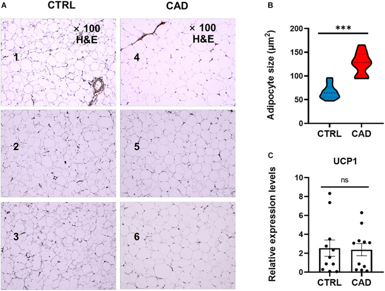
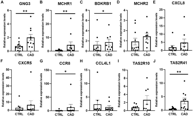
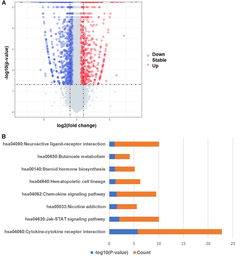
Our results showed that the size of epicardial adipocytes in the CAD group was larger than in the control group. Our findings on the EAT gene expression profiles of CAD showed a total of 747 DEGs (fold change >2, value <0.05). The enrichment analysis of DEGs showed that more pro-inflammatory and immunological genes and pathways were involved in CAD. Ten hub DEGs (, , , , , , , , , and ) were identified.

CONCLUSION

Epicardial adipose tissue in CAD shows unique gene expression profiles and may act as key regulators in the CAD pathological process.

摘要

目的

心外膜脂肪组织(EAT)紧邻冠状动脉和心肌,其作为内分泌器官影响冠状动脉和心肌病理生理过程的作用已日益得到认可。然而,冠状动脉疾病(CAD)中EAT的特定基因表达谱尚未得到充分表征。我们的目的是在基因水平上研究EAT在CAD中的作用。

方法

在此,我们比较了CAD患者与非CAD患者EAT的组织学和基因表达差异。我们通过高通量RNA测序研究了CAD患者EAT中的基因表达谱。我们进行了功能富集分析和蛋白质-蛋白质相互作用网络构建等生物信息学分析,以获取并验证CAD患者EAT中的核心差异表达基因(DEG)。

结果

我们的结果表明,CAD组的心外膜脂肪细胞大小大于对照组。我们对CAD患者EAT基因表达谱的研究结果显示共有747个DEG(倍数变化>2,P值<0.05)。DEG的富集分析表明,CAD涉及更多的促炎和免疫基因及通路。确定了10个核心DEG([此处原文缺失具体基因名称])。

结论

CAD中的心外膜脂肪组织表现出独特的基因表达谱,可能在CAD病理过程中充当关键调节因子。

https://cdn.ncbi.nlm.nih.gov/pmc/blobs/a759/8042318/94c00c0e8266/fphys-12-605811-g004.jpg
https://cdn.ncbi.nlm.nih.gov/pmc/blobs/a759/8042318/4a44eeb932ac/fphys-12-605811-g001.jpg
https://cdn.ncbi.nlm.nih.gov/pmc/blobs/a759/8042318/4310886e5d1c/fphys-12-605811-g002.jpg
https://cdn.ncbi.nlm.nih.gov/pmc/blobs/a759/8042318/05f20171ce9a/fphys-12-605811-g003.jpg
https://cdn.ncbi.nlm.nih.gov/pmc/blobs/a759/8042318/94c00c0e8266/fphys-12-605811-g004.jpg

相似文献

1
Exploring the Role of Epicardial Adipose Tissue in Coronary Artery Disease From the Difference of Gene Expression.从基因表达差异探讨心外膜脂肪组织在冠状动脉疾病中的作用
Front Physiol. 2021 Mar 30;12:605811. doi: 10.3389/fphys.2021.605811. eCollection 2021.
2
Identification of key genes and pathways affected in epicardial adipose tissue from patients with coronary artery disease by integrated bioinformatics analysis.通过综合生物信息学分析鉴定冠心病患者心外膜脂肪组织中受影响的关键基因和通路。
PeerJ. 2020 Mar 25;8:e8763. doi: 10.7717/peerj.8763. eCollection 2020.
3
lncRNA expression profiles and associated ceRNA network analyses in epicardial adipose tissue of patients with coronary artery disease.lncRNA 表达谱及冠心病患者心外膜脂肪组织中相关 ceRNA 网络分析。
Sci Rep. 2021 Jan 15;11(1):1567. doi: 10.1038/s41598-021-81038-5.
4
Extension of coronary artery disease is associated with increased IL-6 and decreased adiponectin gene expression in epicardial adipose tissue.冠状动脉疾病的扩展与心外膜脂肪组织中白细胞介素-6增加和脂联素基因表达降低有关。
Cytokine. 2008 Aug;43(2):174-80. doi: 10.1016/j.cyto.2008.05.006. Epub 2008 Jun 17.
5
ANGPTL4 Expression Is Increased in Epicardial Adipose Tissue of Patients with Coronary Artery Disease.冠状动脉疾病患者心外膜脂肪组织中血管生成素样蛋白4(ANGPTL4)表达增加。
J Clin Med. 2022 Apr 27;11(9):2449. doi: 10.3390/jcm11092449.
6
Expression Profiles of Long Noncoding and Messenger RNAs in Epicardial Adipose Tissue-Derived from Patients with Coronary Atherosclerosis.冠状动脉粥样硬化患者心外膜脂肪组织中长链非编码RNA和信使RNA的表达谱
Curr Vasc Pharmacol. 2022;20(2):189-200. doi: 10.2174/1570161120666220114095320.
7
Examining the effects of coronary artery disease- and mitochondrial biogenesis-related genes' and microRNAs' expression levels on metabolic disorders in epicardial adipose tissue.探讨冠状动脉疾病和线粒体生物发生相关基因及 microRNAs 表达水平对心外膜脂肪组织代谢紊乱的影响。
Gene. 2024 Feb 15;895:147988. doi: 10.1016/j.gene.2023.147988. Epub 2023 Nov 17.
8
Association between omentin-1 expression in human epicardial adipose tissue and coronary atherosclerosis.人的心外膜脂肪组织中网膜素-1表达与冠状动脉粥样硬化之间的关联。
Cardiovasc Diabetol. 2016 Jun 28;15:90. doi: 10.1186/s12933-016-0406-5.
9
Relationship between Epicardial and Coronary Adipose Tissue and the Expression of Adiponectin, Leptin, and Interleukin 6 in Patients with Coronary Artery Disease.冠心病患者心外膜脂肪组织与冠状动脉脂肪组织的关系以及脂联素、瘦素和白细胞介素6的表达
J Pers Med. 2022 Jan 19;12(2):129. doi: 10.3390/jpm12020129.
10
Relationships between visceral/subcutaneous adipose tissue FABP4 expression and coronary atherosclerosis in patients with metabolic syndrome.代谢综合征患者内脏/皮下脂肪组织脂肪酸结合蛋白4(FABP4)表达与冠状动脉粥样硬化的关系。
Cardiovasc Pathol. 2020 May-Jun;46:107192. doi: 10.1016/j.carpath.2019.107192. Epub 2019 Dec 6.

引用本文的文献

1
Mechanisms and novel therapeutic roles of bitter taste receptors in diseases.疾病中苦味受体的机制及新的治疗作用
Theranostics. 2025 Mar 3;15(9):3961-3978. doi: 10.7150/thno.107406. eCollection 2025.
2
Single-Nucleus Transcriptomic Atlas of Human Pericoronary Epicardial Adipose Tissue in Normal and Pathological Conditions.人类正常和病理条件下冠状动脉心外膜脂肪组织的单核转录组图谱。
Arterioscler Thromb Vasc Biol. 2024 Jul;44(7):1628-1645. doi: 10.1161/ATVBAHA.124.320923. Epub 2024 May 30.
3
Single-nucleus transcriptomics of epicardial adipose tissue from female pigs reveals effects of exercise training on resident innate and adaptive immune cells.

本文引用的文献

1
White adipose tissue browning in critical illness: A review of the evidence, mechanisms and future perspectives.危重病患者白色脂肪组织棕色化:证据、机制和未来展望的综述。
Obes Rev. 2020 Dec;21(12):e13085. doi: 10.1111/obr.13085. Epub 2020 Jul 1.
2
Regulation of thermogenic adipocytes during fasting and cold.禁食和寒冷时生热脂肪细胞的调节。
Mol Cell Endocrinol. 2020 Jul 15;512:110869. doi: 10.1016/j.mce.2020.110869. Epub 2020 May 18.
3
Validity of epicardial fat volume as biomarker of coronary artery disease in symptomatic individuals: Results from the ALTER-BIO registry.
雌性猪心外膜脂肪组织的单核转录组学揭示了运动训练对驻留固有免疫细胞和适应性免疫细胞的影响。
Cell Commun Signal. 2024 Apr 26;22(1):243. doi: 10.1186/s12964-024-01587-w.
4
Single-nucleus transcriptomics of epicardial adipose tissue from females reveals exercise control of innate and adaptive immune cells.对女性心外膜脂肪组织进行的单核转录组学研究揭示了运动对先天性和适应性免疫细胞的调控作用。
bioRxiv. 2023 Nov 5:2023.11.02.565385. doi: 10.1101/2023.11.02.565385.
5
Association of Epicardial Adipose Tissue Adipocytes Hypertrophy with Biomarkers of Low-Grade Inflammation and Extracellular Matrix Remodeling in Patients with Coronary Artery Disease.冠心病患者心外膜脂肪组织脂肪细胞肥大与低度炎症和细胞外基质重塑生物标志物的关联
Biomedicines. 2023 Jan 17;11(2):241. doi: 10.3390/biomedicines11020241.
6
Obesity and diabetes are major risk factors for epicardial adipose tissue inflammation.肥胖和糖尿病是心外膜脂肪组织炎症的主要危险因素。
JCI Insight. 2021 Aug 23;6(16):e145495. doi: 10.1172/jci.insight.145495.
有症状个体中心外膜脂肪体积作为冠状动脉疾病生物标志物的有效性:来自ALTER-BIO注册研究的结果。
Int J Cardiol. 2020 Sep 1;314:20-24. doi: 10.1016/j.ijcard.2020.04.031. Epub 2020 Apr 13.
4
Association between perivascular inflammation and downstream myocardial perfusion in patients with suspected coronary artery disease.血管周围炎症与疑似冠心病患者下游心肌灌注的关系。
Eur Heart J Cardiovasc Imaging. 2020 Jun 1;21(6):599-605. doi: 10.1093/ehjci/jeaa023.
5
Adipocytes Directly Affect Coronary Artery Disease Pathogenesis via Induction of Adipokine and Cytokine Imbalances.脂肪细胞通过诱导脂联素和细胞因子失衡直接影响冠状动脉疾病的发病机制。
Front Immunol. 2019 Sep 19;10:2163. doi: 10.3389/fimmu.2019.02163. eCollection 2019.
6
Relationship between epicardial adipose tissue and coronary vascular function in patients with suspected coronary artery disease and normal myocardial perfusion imaging.疑似冠心病且心肌灌注成像正常患者的心外膜脂肪组织与冠状动脉血管功能之间的关系
Eur Heart J Cardiovasc Imaging. 2019 Dec 1;20(12):1379-1387. doi: 10.1093/ehjci/jez182.
7
Radiopharmaceutical Evidence for MCHR1 Binding Sites in Murine Brown Adipocytes.小鼠棕色脂肪细胞中MCHR1结合位点的放射性药物证据。
Front Endocrinol (Lausanne). 2019 Jun 11;10:324. doi: 10.3389/fendo.2019.00324. eCollection 2019.
8
Dendritic Cells in Subcutaneous and Epicardial Adipose Tissue of Subjects with Type 2 Diabetes, Obesity, and Coronary Artery Disease.2 型糖尿病、肥胖症和冠状动脉疾病患者皮下和心外膜脂肪组织中的树突状细胞。
Mediators Inflamm. 2019 May 9;2019:5481725. doi: 10.1155/2019/5481725. eCollection 2019.
9
Statins Reduce Epicardial Adipose Tissue Attenuation Independent of Lipid Lowering: A Potential Pleiotropic Effect.他汀类药物降低心外膜脂肪组织衰减而不降低血脂:一种潜在的多效性效应。
J Am Heart Assoc. 2019 Jun 18;8(12):e013104. doi: 10.1161/JAHA.119.013104. Epub 2019 Jun 13.
10
Risk Factor Variability and Cardiovascular Outcome: JACC Review Topic of the Week.风险因素变异性与心血管结局:JACC 本周综述专题
J Am Coll Cardiol. 2019 May 28;73(20):2596-2603. doi: 10.1016/j.jacc.2019.02.063.