Yoshikawa Masato, Morine Yuji, Yamada Shinichiro, Miyazaki Katsuki, Tokuda Kazunori, Saito Yu, Arakawa Yusuke, Ikemoto Tetsuya, Imura Satoru, Shimada Mitsuo
Department of Surgery Institute of Biomedical Sciences University of Tokushima Tokushima Japan.
Ann Gastroenterol Surg. 2020 Oct 5;5(2):252-258. doi: 10.1002/ags3.12404. eCollection 2021 Mar.
Diffusion-weighted magnetic resonance imaging (DWI-MRI) is used to predict tumor malignancy. Here we explored the role of apparent diffusion coefficient (ADC) values in the treatment of patients with resectable colorectal liver metastasis (CRLM).
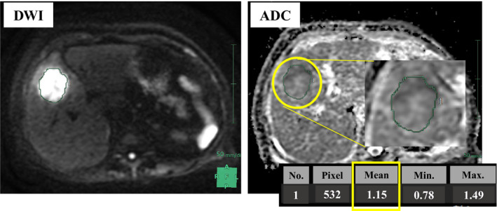
Magnetic resonance imaging (MRI) scans were conducted using a Signa HDe or Signa Explorer 1.5-T scanner (GE Healthcare). ADC maps were calculated using DWI with values of 0, 20, and 800 s/mm. We enrolled 60 patients who underwent upfront hepatic resection for CRLM and divided them into ADC-high (n = 30) and ADC-low (n = 30) groups. Clinicopathological variables of the groups were compared. Immunohistochemical analysis of HIF-1α expression in tumor tissues was performed, and the relationship between the ADC value and HIF-1α expression was evaluated.
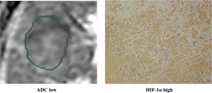
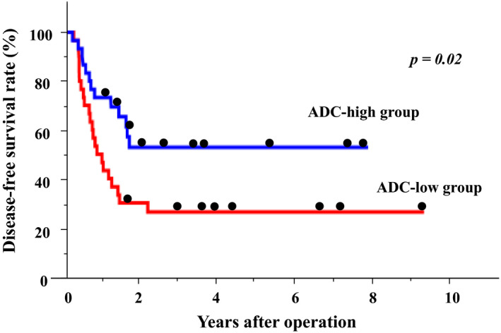
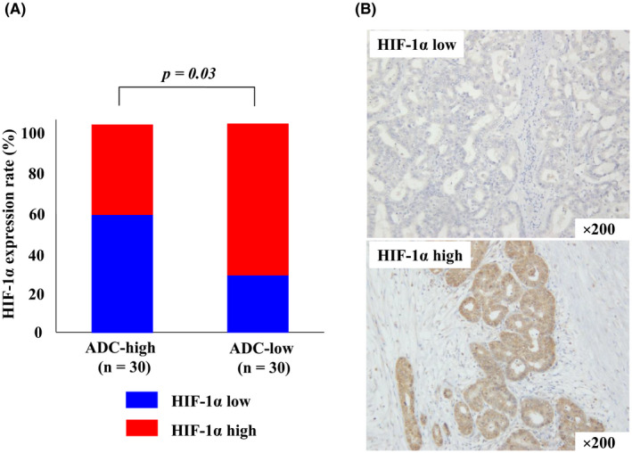
The disease-free survival rate of the ADC-low group was significantly lower than that of the ADC-high group ( < .05). Univariate analysis revealed that tumor number (more than five), synchronous metastasis, and low ADC were prognostic factors. Multivariate analysis identified low ADC as an independent prognostic factor. Furthermore, the ADC-low group more frequently expressed high levels of HIF-1α than the ADC-high group.
Low ADC values were an independent prognostic factor of resectable CRLM and correlated with HIF-1α expression.
弥散加权磁共振成像(DWI-MRI)用于预测肿瘤恶性程度。在此,我们探讨了表观扩散系数(ADC)值在可切除性结直肠癌肝转移(CRLM)患者治疗中的作用。
使用Signa HDe或Signa Explorer 1.5-T扫描仪(GE医疗)进行磁共振成像(MRI)扫描。使用DWI并设置值为0、20和800 s/mm来计算ADC图。我们纳入了60例行CRLM初次肝切除的患者,并将他们分为ADC高组(n = 30)和ADC低组(n = 30)。比较两组的临床病理变量。对肿瘤组织中HIF-1α表达进行免疫组化分析,并评估ADC值与HIF-1α表达之间的关系。
ADC低组的无病生存率显著低于ADC高组(P <.05)。单因素分析显示,肿瘤数量(超过5个)、同时性转移和低ADC是预后因素。多因素分析确定低ADC是独立的预后因素。此外,ADC低组比ADC高组更频繁地高表达HIF-1α。
低ADC值是可切除性CRLM的独立预后因素,且与HIF-1α表达相关。