Department of Physiology & Cell Biology (A.K., N.L., E.J.A., B.J.H., P.M.L.J., P.J.M., V.V.F.), The Ohio State University Wexner Medical Center, Columbus.
Bob and Corrine Frick Center for Heart Failure and Arrhythmia, Dorothy M. Davis Heart & Lung Research Institute (A.K., N.L., E.J.A., B.J.H., P.J.M., V.V.F.), The Ohio State University Wexner Medical Center, Columbus.
Circulation. 2021 Jul 13;144(2):126-143. doi: 10.1161/CIRCULATIONAHA.120.051583. Epub 2021 Apr 20.
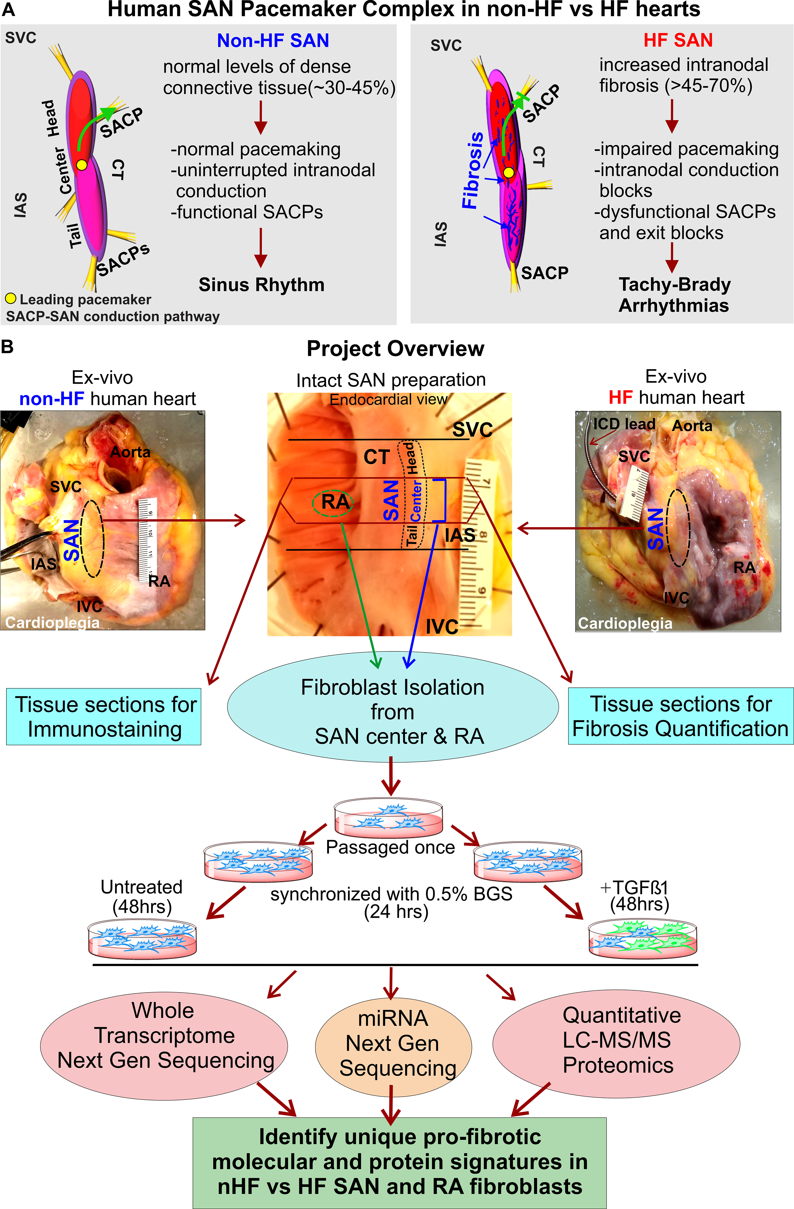
Up to 50% of the adult human sinoatrial node (SAN) is composed of dense connective tissue. Cardiac diseases including heart failure (HF) may increase fibrosis within the SAN pacemaker complex, leading to impaired automaticity and conduction of electric activity to the atria. Unlike the role of cardiac fibroblasts in pathologic fibrotic remodeling and tissue repair, nothing is known about fibroblasts that maintain the inherently fibrotic SAN environment.
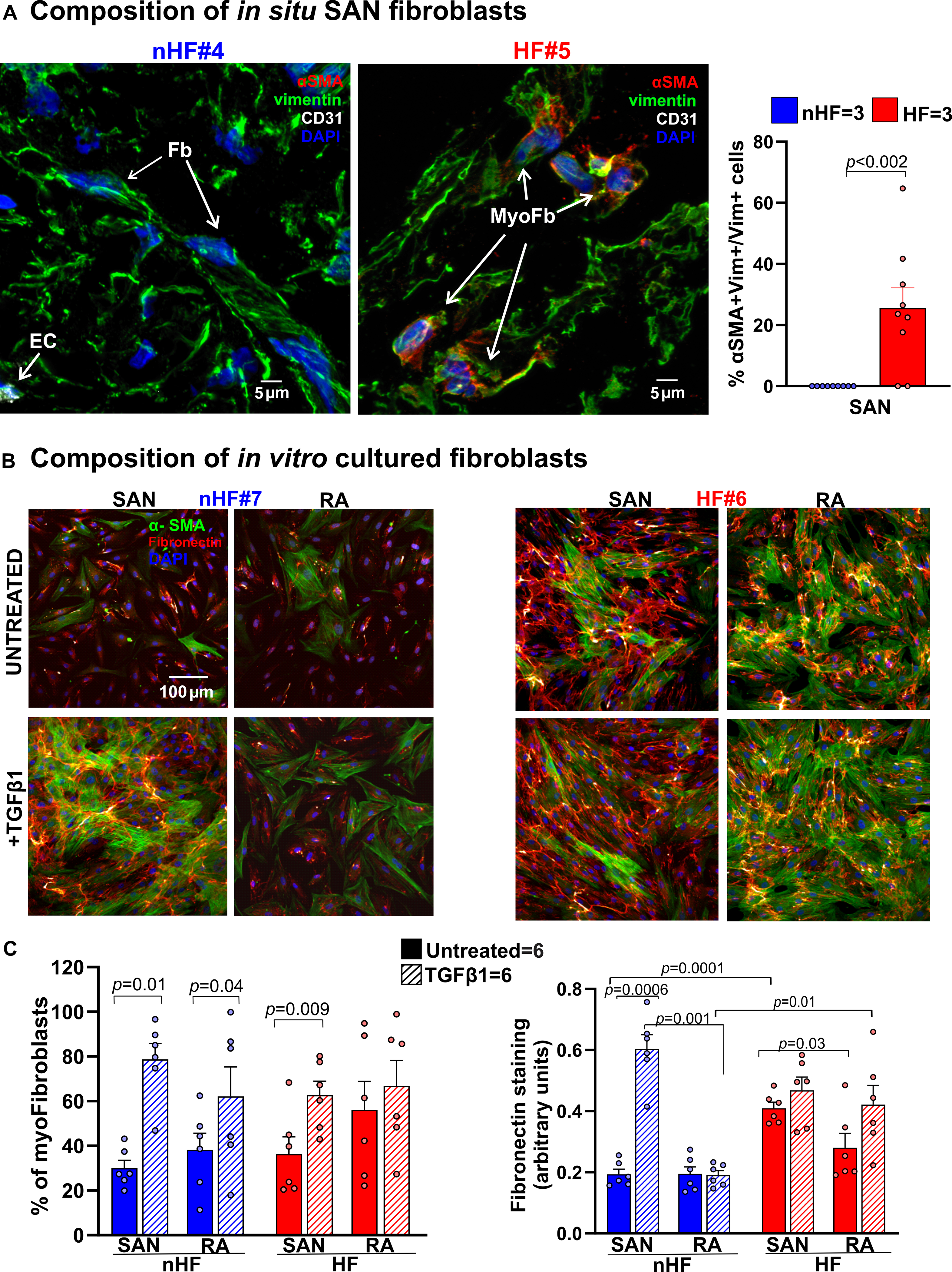
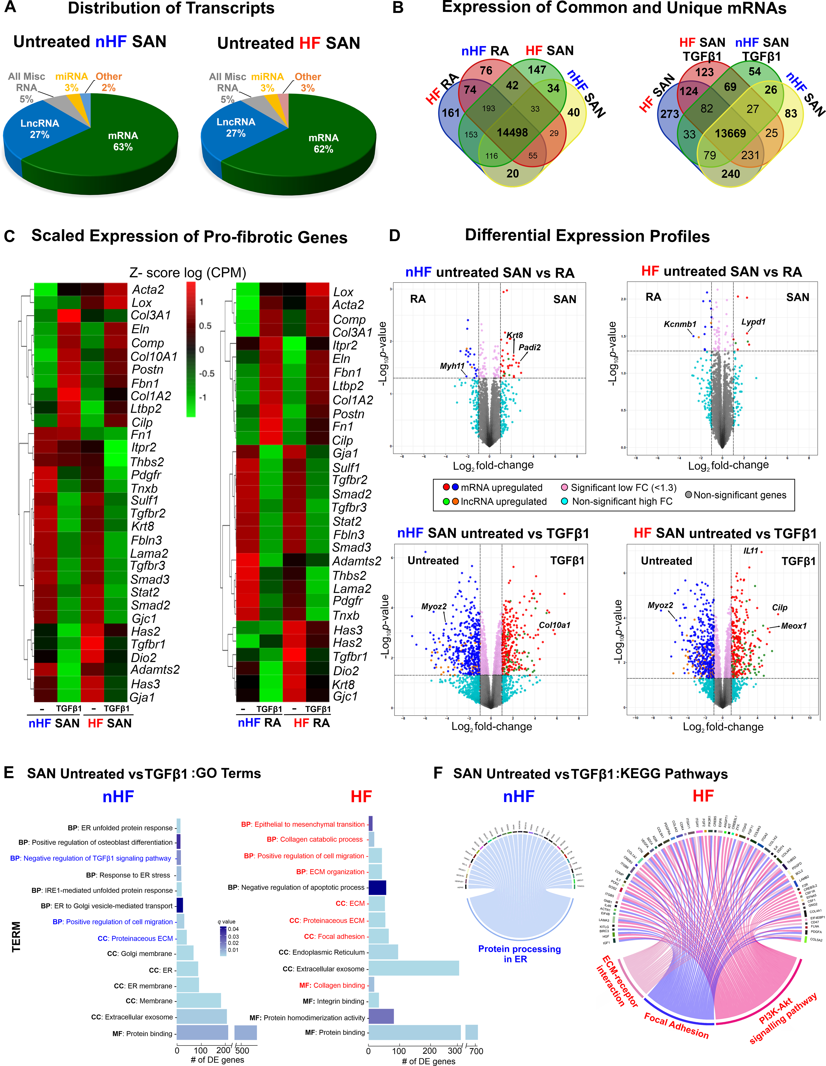
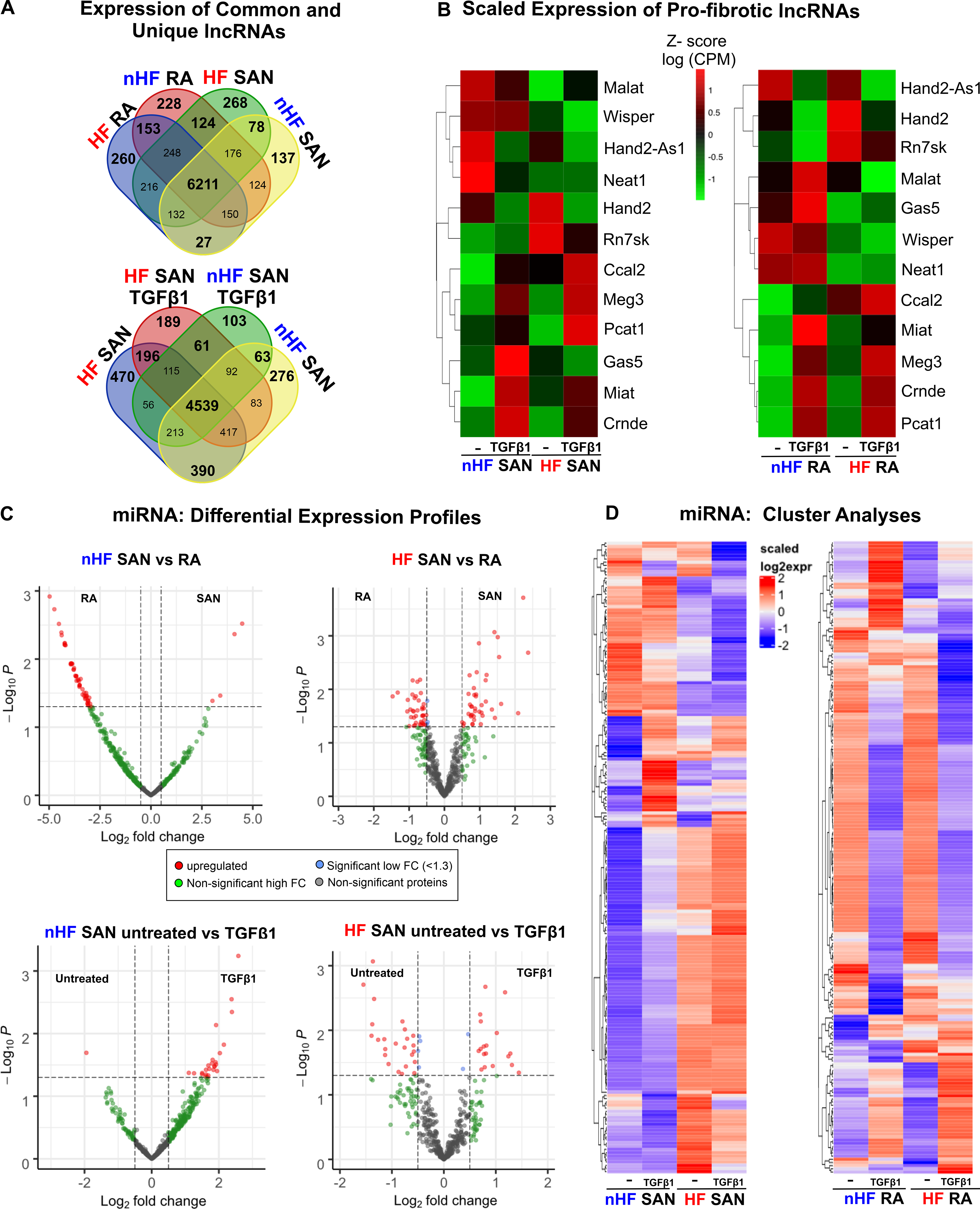
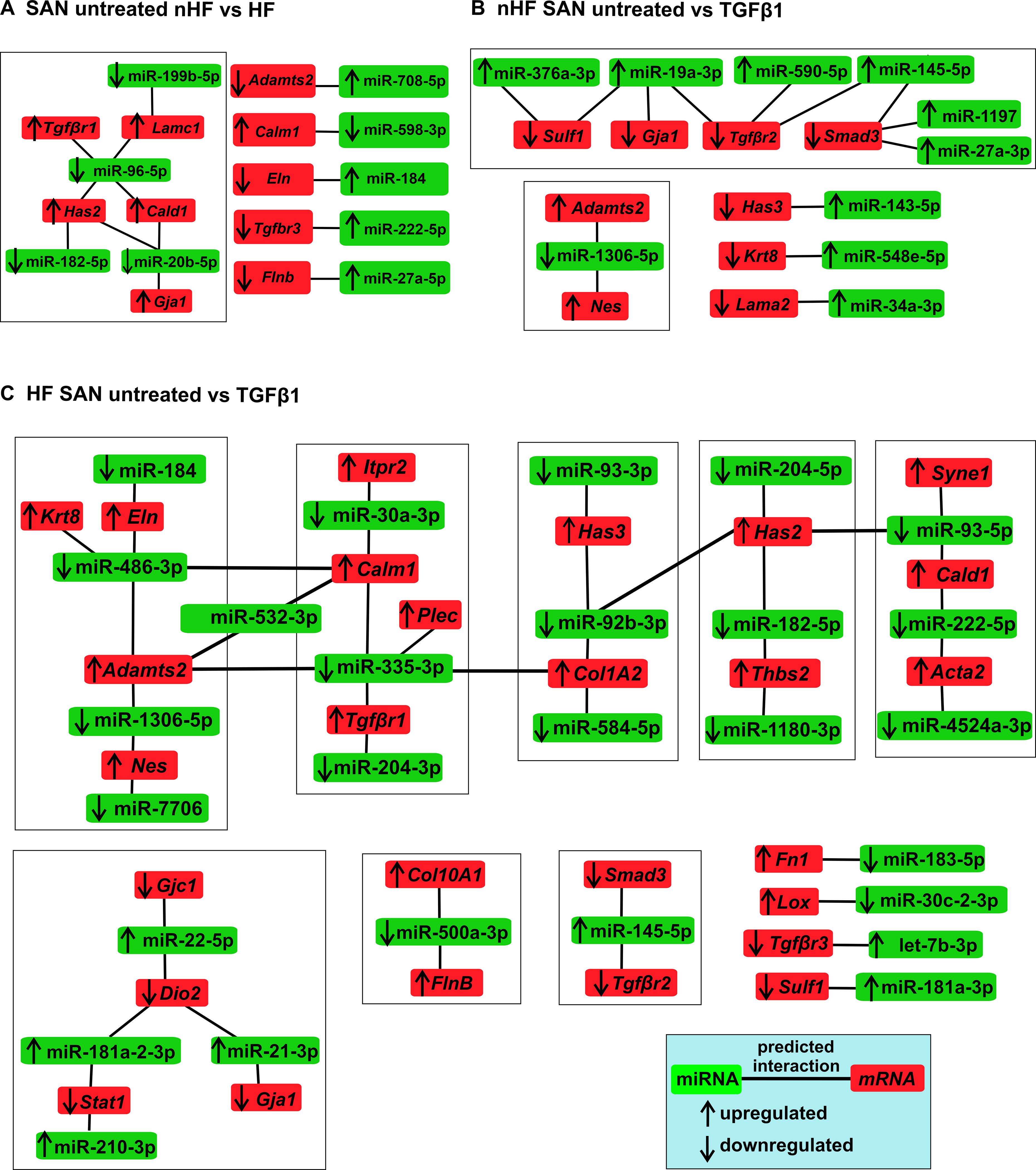
Intact SAN pacemaker complex was dissected from cardioplegically arrested explanted nonfailing hearts (non-HF; n=22; 48.7±3.1 years of age) and human failing hearts (n=16; 54.9±2.6 years of age). Connective tissue content was quantified from Masson trichrome-stained head-center and center-tail SAN sections. Expression of extracellular matrix proteins, including collagens 1 and 3A1, CILP1 (cartilage intermediate layer protein 1), and POSTN (periostin), and fibroblast and myofibroblast numbers were quantified by in situ and in vitro immunolabeling. Fibroblasts from the central intramural SAN pacemaker compartment (≈10×5×2 mm) and right atria were isolated, cultured, passaged once, and treated ± transforming growth factor β1 and subjected to comprehensive high-throughput next-generation sequencing of whole transcriptome, microRNA, and proteomic analyses.
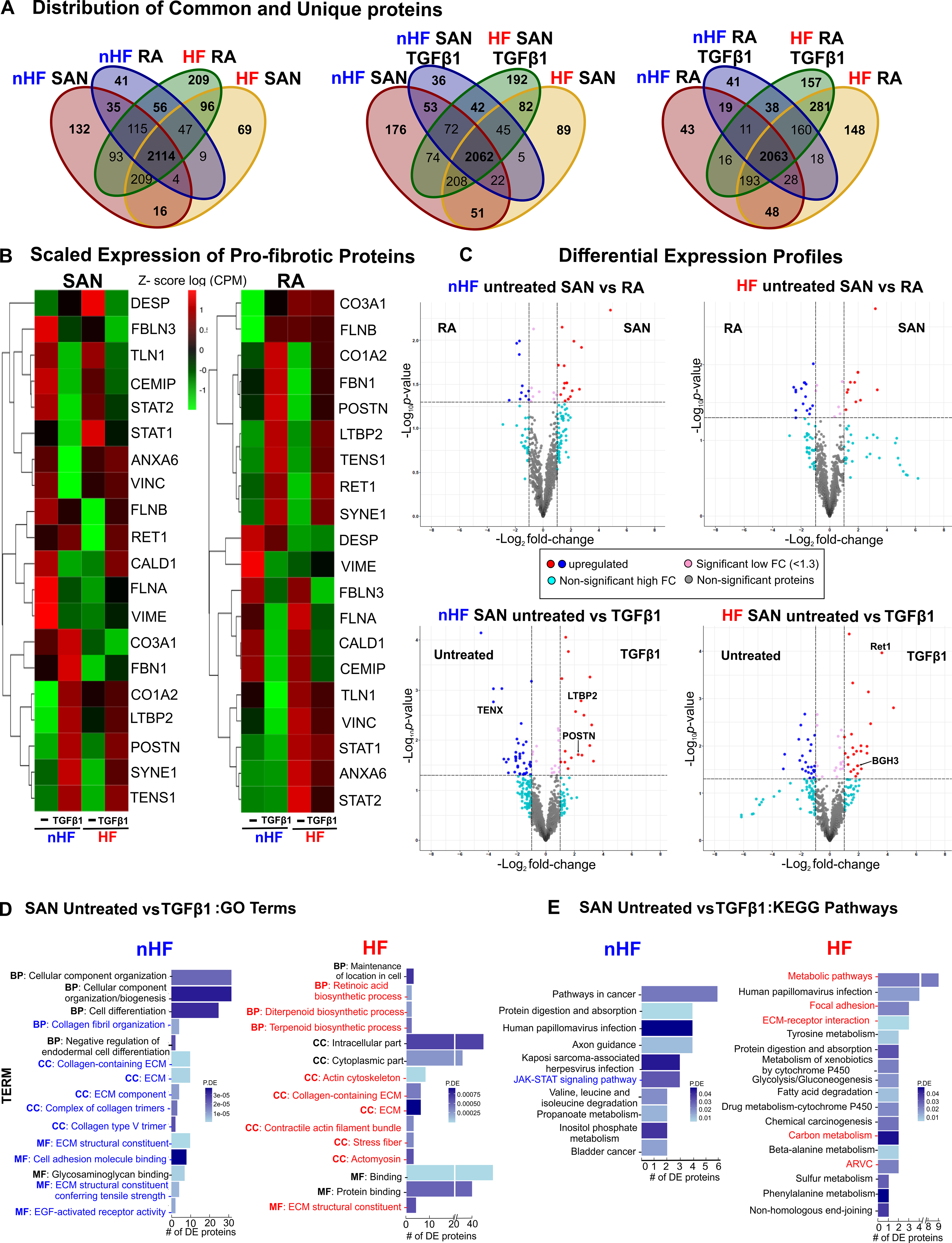
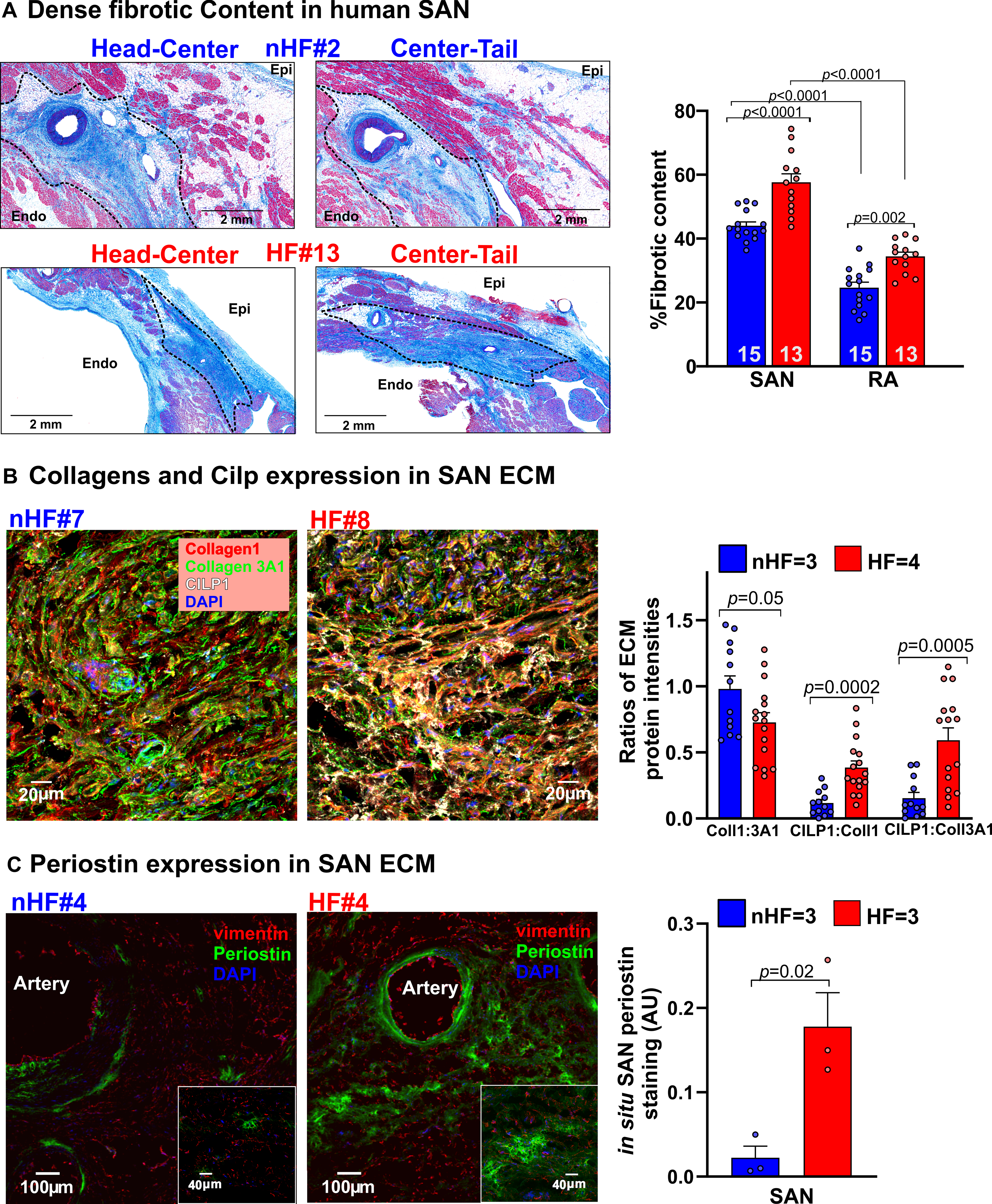
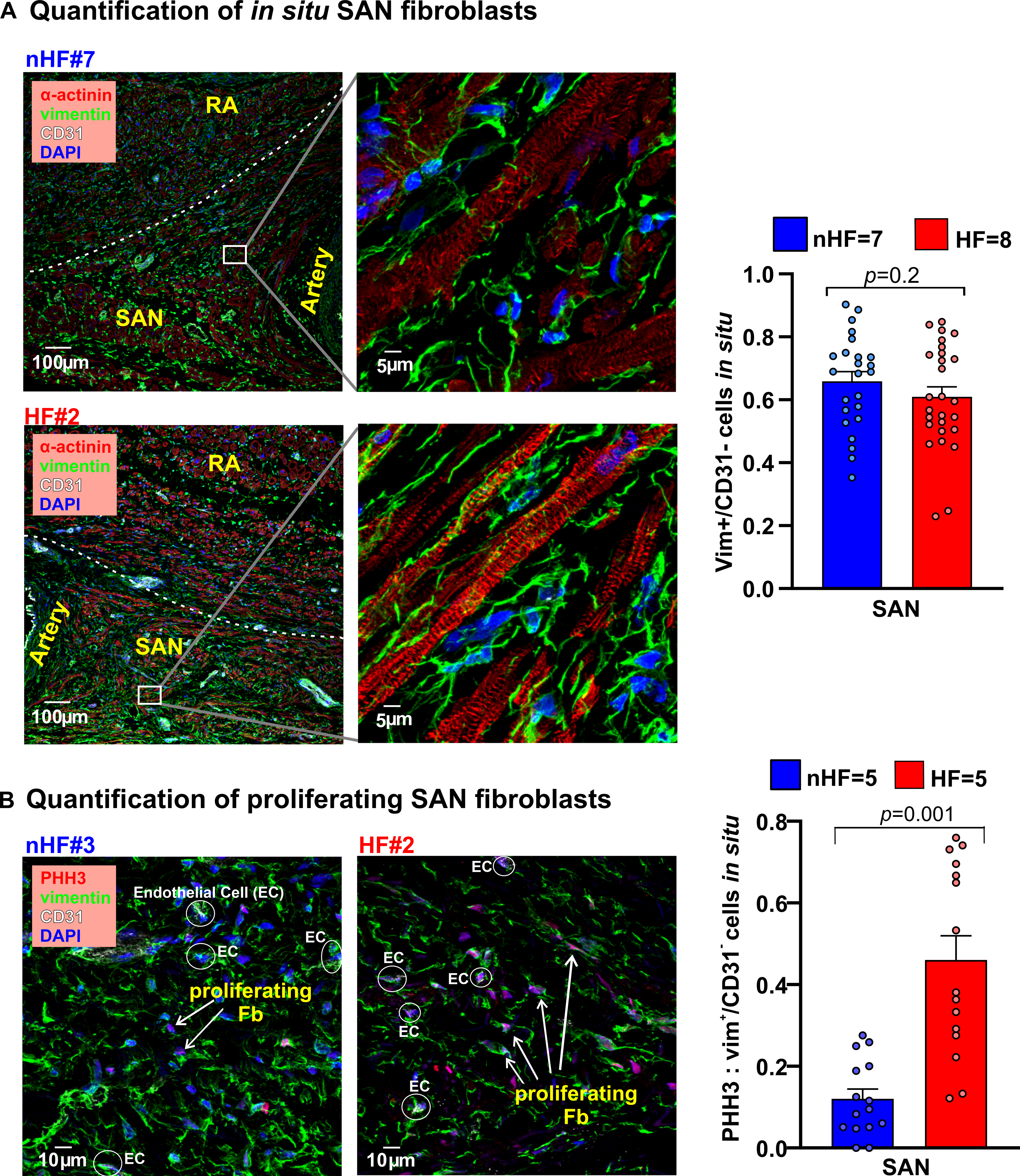
Intranodal fibrotic content was significantly higher in SAN pacemaker complex from HF versus non-HF hearts (57.7±2.6% versus 44.0±1.2%; <0.0001). Proliferating phosphorylated histone 3/vimentin/CD31 (cluster of differentiation 31) fibroblasts were higher in HF SAN. Vimentin/α-smooth muscle actin/CD31 myofibroblasts along with increased interstitial POSTN expression were found only in HF SAN. RNA sequencing and proteomic analyses identified unique differences in mRNA, long noncoding RNA, microRNA, and proteomic profiles between non-HF and HF SAN and right atria fibroblasts and transforming growth factor β1-induced myofibroblasts. Specifically, proteins and signaling pathways associated with extracellular matrix flexibility, stiffness, focal adhesion, and metabolism were altered in HF SAN fibroblasts compared with non-HF SAN.
This study revealed increased SAN-specific fibrosis with presence of myofibroblasts, CILP1, and POSTN-positive interstitial fibrosis only in HF versus non-HF human hearts. Comprehensive proteotranscriptomic profiles of SAN fibroblasts identified upregulation of genes and proteins promoting stiffer SAN extracellular matrix in HF hearts. Fibroblast-specific profiles generated by our proteotranscriptomic analyses of the human SAN provide a comprehensive framework for future studies to investigate the role of SAN-specific fibrosis in cardiac rhythm regulation and arrhythmias.
成人窦房结(SAN)的 50%可能由致密结缔组织组成。包括心力衰竭(HF)在内的心脏疾病可能会增加 SAN 起搏复合体中的纤维化,导致电活动自动性和传导到心房受损。与心脏成纤维细胞在病理性纤维化重塑和组织修复中的作用不同,维持固有纤维化 SAN 环境的成纤维细胞的作用尚不清楚。
从心脏停搏的心脏中分离出完整的 SAN 起搏复合体体外非衰竭心脏(非 HF;n=22;年龄 48.7±3.1 岁)和人类衰竭心脏(n=16;年龄 54.9±2.6 岁)。从 Masson 三色染色的头中心和中心尾 SAN 切片中定量计算结缔组织含量。通过原位和体外免疫标记定量测定细胞外基质蛋白,包括胶原蛋白 1 和 3A1、CILP1(软骨中间层蛋白 1)和 POSTN(骨粘连蛋白)的表达,以及成纤维细胞和肌成纤维细胞的数量。从中央壁内 SAN 起搏间隔(≈10×5×2mm)和右心房分离、培养、传代一次,并在转化生长因子β1存在或不存在的情况下进行全面的高通量下一代测序,以进行全转录组、microRNA 和蛋白质组分析。
HF 组 SAN 起搏复合体中的节内纤维化含量明显高于非 HF 组(57.7±2.6%对 44.0±1.2%;<0.0001)。HF SAN 中增殖的磷酸化组蛋白 3/波形蛋白/CD31(分化群 31)成纤维细胞较多。仅在 HF SAN 中发现了伴有间质 POSTN 表达增加的波形蛋白/α-平滑肌肌动蛋白/CD31 肌成纤维细胞。RNA 测序和蛋白质组学分析显示,非 HF 和 HF SAN 以及右心房成纤维细胞和转化生长因子β1 诱导的肌成纤维细胞之间的 mRNA、长非编码 RNA、microRNA 和蛋白质组谱存在独特差异。具体而言,与非 HF SAN 相比,HF SAN 成纤维细胞中与细胞外基质灵活性、刚性、焦点附着和代谢相关的蛋白质和信号通路发生改变。
本研究显示,与非 HF 人心脏相比,HF 心脏的 SAN 中仅存在 SAN 特异性纤维化、肌成纤维细胞、CILP1 和 POSTN 阳性间质纤维化。SAN 成纤维细胞的综合蛋白质组学特征确定了 HF 心脏中促进 SAN 细胞外基质变硬的基因和蛋白质的上调。我们对人类 SAN 的蛋白质组学分析生成的成纤维细胞特异性图谱为未来研究 SAN 特异性纤维化在心脏节律调节和心律失常中的作用提供了一个全面的框架。