• 文献检索
  • 文档翻译
  • 深度研究
  • 学术资讯
  • Suppr Zotero 插件Zotero 插件
  • 邀请有礼
  • 套餐&价格
  • 历史记录
应用&插件
Suppr Zotero 插件Zotero 插件浏览器插件Mac 客户端Windows 客户端微信小程序
定价
高级版会员购买积分包购买API积分包
服务
文献检索文档翻译深度研究API 文档MCP 服务
关于我们
关于 Suppr公司介绍联系我们用户协议隐私条款
关注我们

Suppr 超能文献

核心技术专利:CN118964589B侵权必究
粤ICP备2023148730 号-1Suppr @ 2026

文献检索

告别复杂PubMed语法,用中文像聊天一样搜索,搜遍4000万医学文献。AI智能推荐,让科研检索更轻松。

立即免费搜索

文件翻译

保留排版,准确专业,支持PDF/Word/PPT等文件格式,支持 12+语言互译。

免费翻译文档

深度研究

AI帮你快速写综述,25分钟生成高质量综述,智能提取关键信息,辅助科研写作。

立即免费体验

单细胞多组学分析 COVID-19 中的免疫反应。

Single-cell multi-omics analysis of the immune response in COVID-19.

机构信息

Biosciences Institute, Newcastle University, Newcastle upon Tyne, UK.

Wellcome - MRC Cambridge Stem Cell Institute, University of Cambridge, Cambridge, UK.

出版信息

Nat Med. 2021 May;27(5):904-916. doi: 10.1038/s41591-021-01329-2. Epub 2021 Apr 20.

DOI:10.1038/s41591-021-01329-2
PMID:33879890
原文链接:https://pmc.ncbi.nlm.nih.gov/articles/PMC8121667/
Abstract

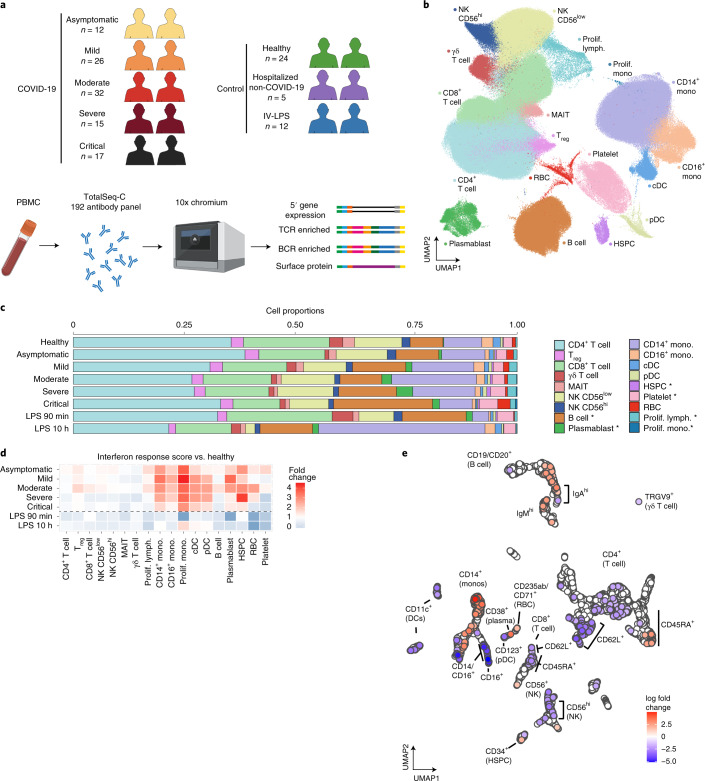
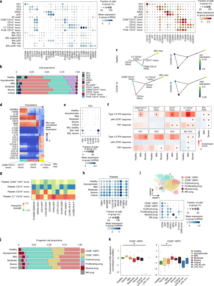
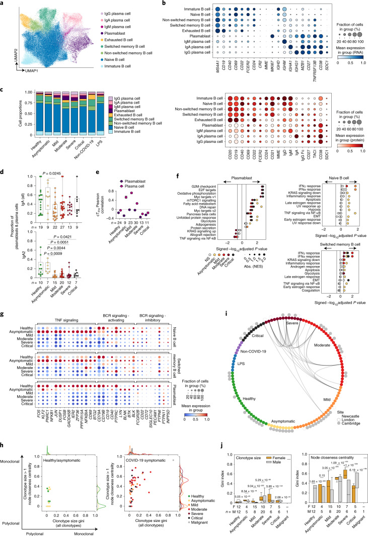
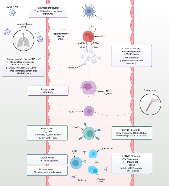
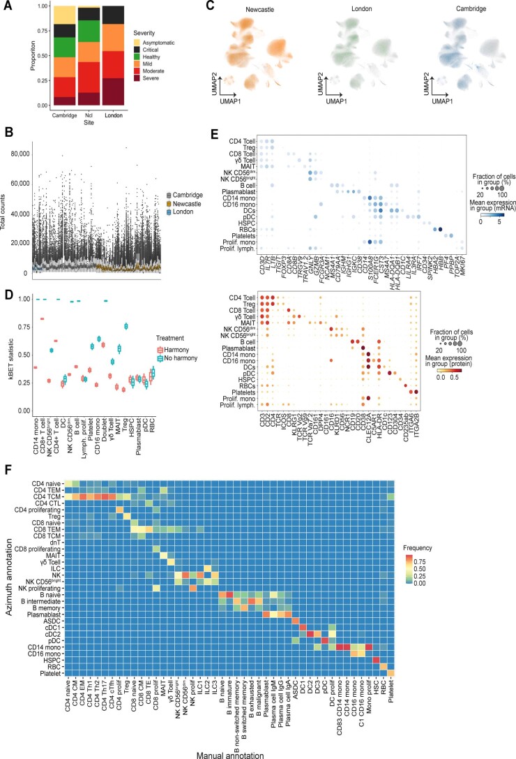
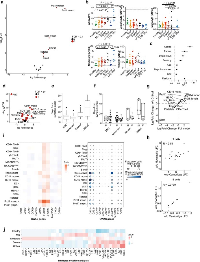
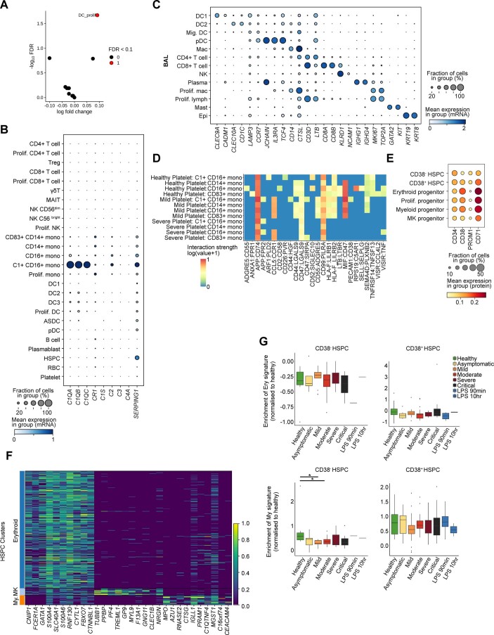
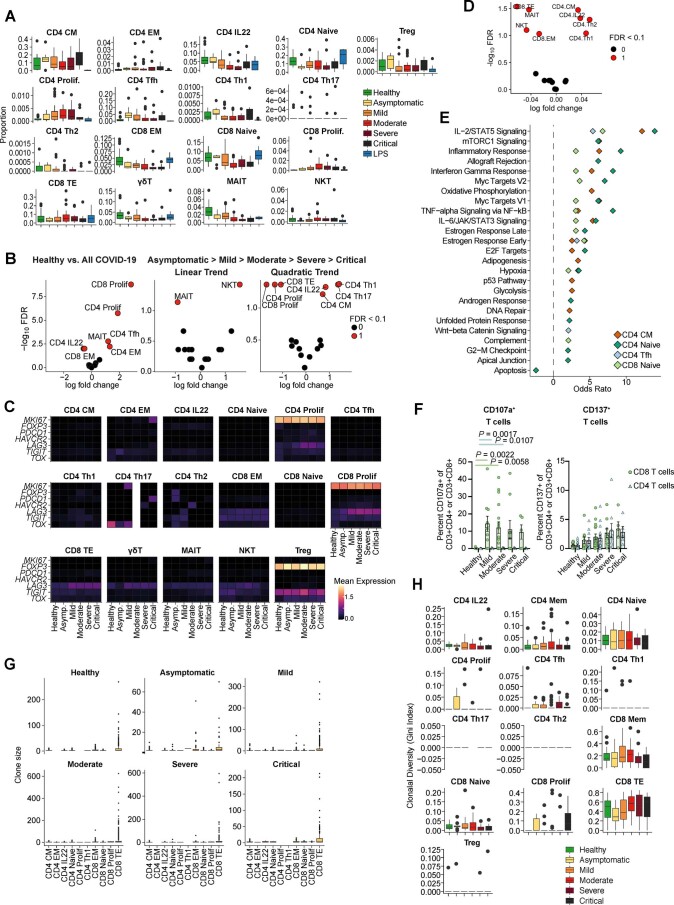
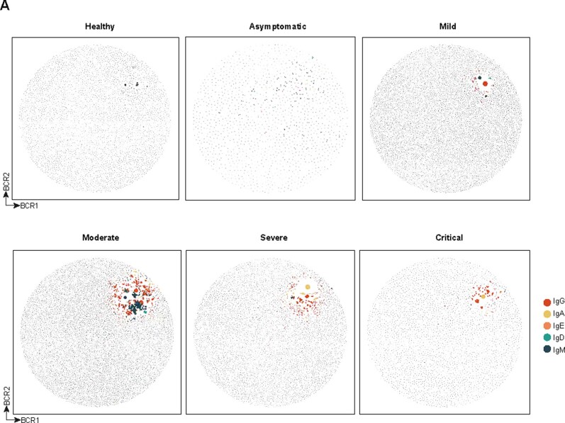
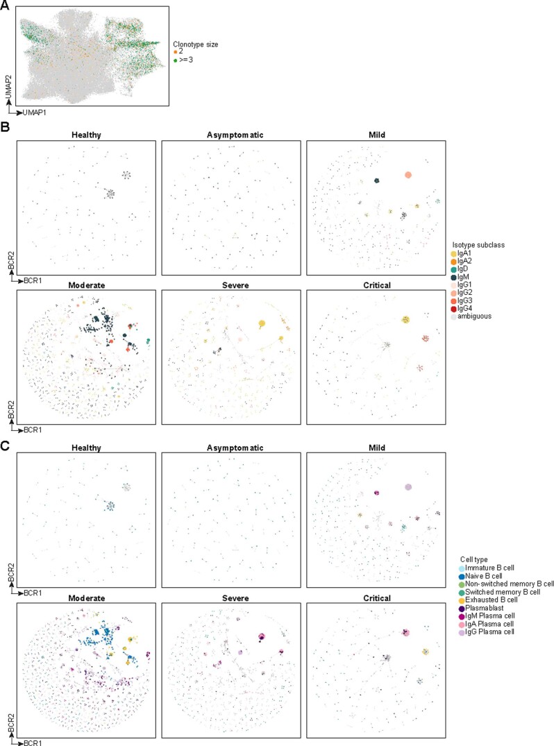
Analysis of human blood immune cells provides insights into the coordinated response to viral infections such as severe acute respiratory syndrome coronavirus 2, which causes coronavirus disease 2019 (COVID-19). We performed single-cell transcriptome, surface proteome and T and B lymphocyte antigen receptor analyses of over 780,000 peripheral blood mononuclear cells from a cross-sectional cohort of 130 patients with varying severities of COVID-19. We identified expansion of nonclassical monocytes expressing complement transcripts (CD16C1QA/B/C) that sequester platelets and were predicted to replenish the alveolar macrophage pool in COVID-19. Early, uncommitted CD34 hematopoietic stem/progenitor cells were primed toward megakaryopoiesis, accompanied by expanded megakaryocyte-committed progenitors and increased platelet activation. Clonally expanded CD8 T cells and an increased ratio of CD8 effector T cells to effector memory T cells characterized severe disease, while circulating follicular helper T cells accompanied mild disease. We observed a relative loss of IgA2 in symptomatic disease despite an overall expansion of plasmablasts and plasma cells. Our study highlights the coordinated immune response that contributes to COVID-19 pathogenesis and reveals discrete cellular components that can be targeted for therapy.

摘要

对人类血液免疫细胞的分析为深入了解病毒感染(如导致 2019 冠状病毒病(COVID-19)的严重急性呼吸综合征冠状病毒 2)的协调反应提供了线索。我们对来自 130 名 COVID-19 患者的横断面队列的超过 780,000 个外周血单核细胞进行了单细胞转录组、表面蛋白质组和 T 淋巴细胞和 B 淋巴细胞抗原受体分析。我们鉴定出了表达补体转录本(CD16C1QA/B/C)的非经典单核细胞的扩增,这些细胞会隔离血小板,并预计会补充 COVID-19 中的肺泡巨噬细胞池。早期未定型的 CD34 造血干细胞/祖细胞被诱导向巨核细胞生成,伴随着巨核细胞定向祖细胞的扩增和血小板激活的增加。克隆性扩增的 CD8 T 细胞和 CD8 效应 T 细胞与效应记忆 T 细胞的比值增加是严重疾病的特征,而循环滤泡辅助 T 细胞伴随着轻症疾病。尽管浆母细胞和浆细胞总体扩增,但我们观察到在有症状疾病中 IgA2 的相对缺失。我们的研究强调了导致 COVID-19 发病机制的协调免疫反应,并揭示了可作为治疗靶点的离散细胞成分。

https://cdn.ncbi.nlm.nih.gov/pmc/blobs/0591/8121667/85ce767520f8/41591_2021_1329_Fig14_ESM.jpg
https://cdn.ncbi.nlm.nih.gov/pmc/blobs/0591/8121667/3278425f3d7e/41591_2021_1329_Fig1_HTML.jpg
https://cdn.ncbi.nlm.nih.gov/pmc/blobs/0591/8121667/b34251bb1991/41591_2021_1329_Fig2_HTML.jpg
https://cdn.ncbi.nlm.nih.gov/pmc/blobs/0591/8121667/421bc5ef6e96/41591_2021_1329_Fig3_HTML.jpg
https://cdn.ncbi.nlm.nih.gov/pmc/blobs/0591/8121667/4e51db78616f/41591_2021_1329_Fig4_HTML.jpg
https://cdn.ncbi.nlm.nih.gov/pmc/blobs/0591/8121667/1e00f3ad064c/41591_2021_1329_Fig5_HTML.jpg
https://cdn.ncbi.nlm.nih.gov/pmc/blobs/0591/8121667/a5875e3db57d/41591_2021_1329_Fig6_ESM.jpg
https://cdn.ncbi.nlm.nih.gov/pmc/blobs/0591/8121667/9ede92cf0dfc/41591_2021_1329_Fig7_ESM.jpg
https://cdn.ncbi.nlm.nih.gov/pmc/blobs/0591/8121667/f83d0c7ce355/41591_2021_1329_Fig8_ESM.jpg
https://cdn.ncbi.nlm.nih.gov/pmc/blobs/0591/8121667/d9918102723d/41591_2021_1329_Fig9_ESM.jpg
https://cdn.ncbi.nlm.nih.gov/pmc/blobs/0591/8121667/acd5e8ced580/41591_2021_1329_Fig10_ESM.jpg
https://cdn.ncbi.nlm.nih.gov/pmc/blobs/0591/8121667/a5de59ecf3b5/41591_2021_1329_Fig11_ESM.jpg
https://cdn.ncbi.nlm.nih.gov/pmc/blobs/0591/8121667/febfc2b222cf/41591_2021_1329_Fig12_ESM.jpg
https://cdn.ncbi.nlm.nih.gov/pmc/blobs/0591/8121667/27db97549867/41591_2021_1329_Fig13_ESM.jpg
https://cdn.ncbi.nlm.nih.gov/pmc/blobs/0591/8121667/85ce767520f8/41591_2021_1329_Fig14_ESM.jpg

相似文献

1
Single-cell multi-omics analysis of the immune response in COVID-19.单细胞多组学分析 COVID-19 中的免疫反应。
Nat Med. 2021 May;27(5):904-916. doi: 10.1038/s41591-021-01329-2. Epub 2021 Apr 20.
2
Dynamic blood single-cell immune responses in patients with COVID-19.COVID-19 患者的动态血液单细胞免疫反应。
Signal Transduct Target Ther. 2021 Mar 6;6(1):110. doi: 10.1038/s41392-021-00526-2.
3
Single-cell multi-omics reveals dyssynchrony of the innate and adaptive immune system in progressive COVID-19.单细胞多组学揭示了进展期新冠肺炎中固有免疫系统和适应性免疫系统的不同步性。
Nat Commun. 2022 Jan 21;13(1):440. doi: 10.1038/s41467-021-27716-4.
4
Single-cell immune profiling reveals distinct immune response in asymptomatic COVID-19 patients.单细胞免疫谱分析揭示无症状 COVID-19 患者的独特免疫反应。
Signal Transduct Target Ther. 2021 Sep 16;6(1):342. doi: 10.1038/s41392-021-00753-7.
5
A Single-Cell Atlas of Lymphocyte Adaptive Immune Repertoires and Transcriptomes Reveals Age-Related Differences in Convalescent COVID-19 Patients.单细胞图谱揭示了恢复期 COVID-19 患者的淋巴细胞适应性免疫反应和转录组的年龄相关差异。
Front Immunol. 2021 Jul 12;12:701085. doi: 10.3389/fimmu.2021.701085. eCollection 2021.
6
Integrating single-cell sequencing data with GWAS summary statistics reveals CD16+monocytes and memory CD8+T cells involved in severe COVID-19.单细胞测序数据与 GWAS 汇总统计数据的整合揭示了 CD16+单核细胞和记忆 CD8+T 细胞参与严重 COVID-19。
Genome Med. 2022 Feb 17;14(1):16. doi: 10.1186/s13073-022-01021-1.
7
Complement activation induces excessive T cell cytotoxicity in severe COVID-19.补体激活可导致重症 COVID-19 中 T 细胞过度细胞毒性。
Cell. 2022 Feb 3;185(3):493-512.e25. doi: 10.1016/j.cell.2021.12.040. Epub 2021 Dec 28.
8
Single-cell epigenomic landscape of peripheral immune cells reveals establishment of trained immunity in individuals convalescing from COVID-19.外周免疫细胞的单细胞表观基因组景观揭示了 COVID-19 康复个体中训练免疫的建立。
Nat Cell Biol. 2021 Jun;23(6):620-630. doi: 10.1038/s41556-021-00690-1. Epub 2021 Jun 9.
9
Single-Cell Analysis Reveals the Immune Characteristics of Myeloid Cells and Memory T Cells in Recovered COVID-19 Patients With Different Severities.单细胞分析揭示了不同严重程度的 COVID-19 恢复期患者中髓系细胞和记忆 T 细胞的免疫特征。
Front Immunol. 2022 Jan 3;12:781432. doi: 10.3389/fimmu.2021.781432. eCollection 2021.
10
Integrated longitudinal immunophenotypic, transcriptional and repertoire analyses delineate immune responses in COVID-19 patients.整合纵向免疫表型、转录组和库分析描绘了 COVID-19 患者的免疫反应。
Sci Immunol. 2021 Aug 10;6(62). doi: 10.1126/sciimmunol.abg5021.

引用本文的文献

1
Scvi-hub: an actionable repository for model-driven single-cell analysis.Scvi-hub:一个用于模型驱动单细胞分析的可操作资源库。
Nat Methods. 2025 Sep 8. doi: 10.1038/s41592-025-02799-9.
2
Reproducible single-cell annotation of programs underlying T cell subsets, activation states and functions.对T细胞亚群、激活状态和功能背后的程序进行可重复的单细胞注释。
Nat Methods. 2025 Sep 3. doi: 10.1038/s41592-025-02793-1.
3
scELMo: Embeddings from Language Models are Good Learners for Single-cell Data Analysis.scELMo:来自语言模型的嵌入是单细胞数据分析的优秀学习者。

本文引用的文献

1
COVID-19 immune features revealed by a large-scale single-cell transcriptome atlas.大规模单细胞转录组图谱揭示的新冠病毒免疫特征
Cell. 2021 Nov 11;184(23):5838. doi: 10.1016/j.cell.2021.10.023.
2
Time-resolved systems immunology reveals a late juncture linked to fatal COVID-19.时间分辨系统免疫学揭示了与致命 COVID-19 相关的晚期节点。
Cell. 2021 Apr 1;184(7):1836-1857.e22. doi: 10.1016/j.cell.2021.02.018. Epub 2021 Feb 10.
3
Highly functional virus-specific cellular immune response in asymptomatic SARS-CoV-2 infection.无症状 SARS-CoV-2 感染中具有高功能的病毒特异性细胞免疫应答。
bioRxiv. 2025 Aug 23:2023.12.07.569910. doi: 10.1101/2023.12.07.569910.
4
An interpretable and adaptive autoencoder for efficient tissue deconvolution.一种用于高效组织解卷积的可解释且自适应的自动编码器。
Nucleic Acids Res. 2025 Aug 27;53(16). doi: 10.1093/nar/gkaf821.
5
Wnt signaling - using the bloodstream to send a message.Wnt信号传导——利用血液循环来传递信息。
Cell Mol Life Sci. 2025 Aug 29;82(1):322. doi: 10.1007/s00018-025-05863-x.
6
Single-cell multi-omics-based immune temporal network resolution in sepsis: unravelling molecular mechanisms and precise therapeutic targets.脓毒症中基于单细胞多组学的免疫时间网络解析:揭示分子机制和精确治疗靶点
Front Immunol. 2025 Aug 11;16:1616794. doi: 10.3389/fimmu.2025.1616794. eCollection 2025.
7
scIVNL-seq resolves in vivo single-cell RNA dynamics of immune cells during Salmonella infection.scIVNL-seq解析了沙门氏菌感染期间免疫细胞的体内单细胞RNA动态变化。
Nat Commun. 2025 Aug 26;16(1):7937. doi: 10.1038/s41467-025-63155-1.
8
The role of complement in long COVID pathogenesis.补体在新冠后综合征发病机制中的作用。
JCI Insight. 2025 Aug 22;10(16). doi: 10.1172/jci.insight.194314.
9
LLM-based cell type annotation harmonization across single-cell studies using GCTHarmony.使用GCTHarmony在单细胞研究中基于大语言模型的细胞类型注释协调
Res Sq. 2025 Aug 12:rs.3.rs-7151095. doi: 10.21203/rs.3.rs-7151095/v1.
10
Distinct platelet transcriptomic heterogeneity in respiratory illnesses and modulation by corticosteroids and immune antibody treatments.呼吸道疾病中独特的血小板转录组异质性以及皮质类固醇和免疫抗体治疗的调节作用。
J Transl Med. 2025 Aug 19;23(1):935. doi: 10.1186/s12967-025-06845-0.
J Exp Med. 2021 May 3;218(5). doi: 10.1084/jem.20202617.
4
Distinct developmental pathways from blood monocytes generate human lung macrophage diversity.血液单核细胞分化为人类肺部巨噬细胞的不同发育途径产生了多样性。
Immunity. 2021 Feb 9;54(2):259-275.e7. doi: 10.1016/j.immuni.2020.12.003. Epub 2020 Dec 30.
5
Genetic mechanisms of critical illness in COVID-19.新型冠状病毒肺炎危重症的遗传机制。
Nature. 2021 Mar;591(7848):92-98. doi: 10.1038/s41586-020-03065-y. Epub 2020 Dec 11.
6
Temporal and spatial heterogeneity of host response to SARS-CoV-2 pulmonary infection.宿主对 SARS-CoV-2 肺部感染的反应的时空异质性。
Nat Commun. 2020 Dec 9;11(1):6319. doi: 10.1038/s41467-020-20139-7.
7
Longitudinal Multi-omics Analyses Identify Responses of Megakaryocytes, Erythroid Cells, and Plasmablasts as Hallmarks of Severe COVID-19.纵向多组学分析鉴定出巨核细胞、红细胞和浆细胞反应为严重 COVID-19 的特征。
Immunity. 2020 Dec 15;53(6):1296-1314.e9. doi: 10.1016/j.immuni.2020.11.017. Epub 2020 Nov 26.
8
Extrafollicular B cell responses correlate with neutralizing antibodies and morbidity in COVID-19.滤泡外 B 细胞反应与 COVID-19 中的中和抗体和发病相关。
Nat Immunol. 2020 Dec;21(12):1506-1516. doi: 10.1038/s41590-020-00814-z. Epub 2020 Oct 7.
9
Antigen-Specific Adaptive Immunity to SARS-CoV-2 in Acute COVID-19 and Associations with Age and Disease Severity.急性 COVID-19 患者中针对 SARS-CoV-2 的抗原特异性适应性免疫反应,及其与年龄和疾病严重程度的关联。
Cell. 2020 Nov 12;183(4):996-1012.e19. doi: 10.1016/j.cell.2020.09.038. Epub 2020 Sep 16.
10
COVID-19 vaccine BNT162b1 elicits human antibody and T1 T cell responses.COVID-19 疫苗 BNT162b1 可引发人体抗体和 T1 T 细胞应答。
Nature. 2020 Oct;586(7830):594-599. doi: 10.1038/s41586-020-2814-7. Epub 2020 Sep 30.