NAFLD Research Center, Department of Medicine, University of California San Diego, La Jolla, California, USA.
Department of Gastroenterology and Hepatology, Musashino Red Cross Hospital, Musashino, Japan.
Gut. 2022 May;71(5):983-990. doi: 10.1136/gutjnl-2021-324264. Epub 2021 Apr 21.
Emerging data suggest that a 30% relative decline in liver fat, as assessed by MRI-proton density fat fraction (MRI-PDFF), may be associated with Non-Alcoholic Fatty Liver Disease Activity Score improvement, but the association between decline in MRI-PDFF and fibrosis regression is not known. Therefore, we aimed to examine the association between ≥30% relative decline in MRI-PDFF and fibrosis regression in non-alcoholic fatty liver disease (NAFLD).
This prospective study included 100 well-characterised patients with biopsy-proven NAFLD with paired contemporaneous MRI-PDFF assessment at two time points. MRI-PDFF response was defined as ≥30% relative decline in MRI-PDFF. The was ≥1 stage histological fibrosis regression.
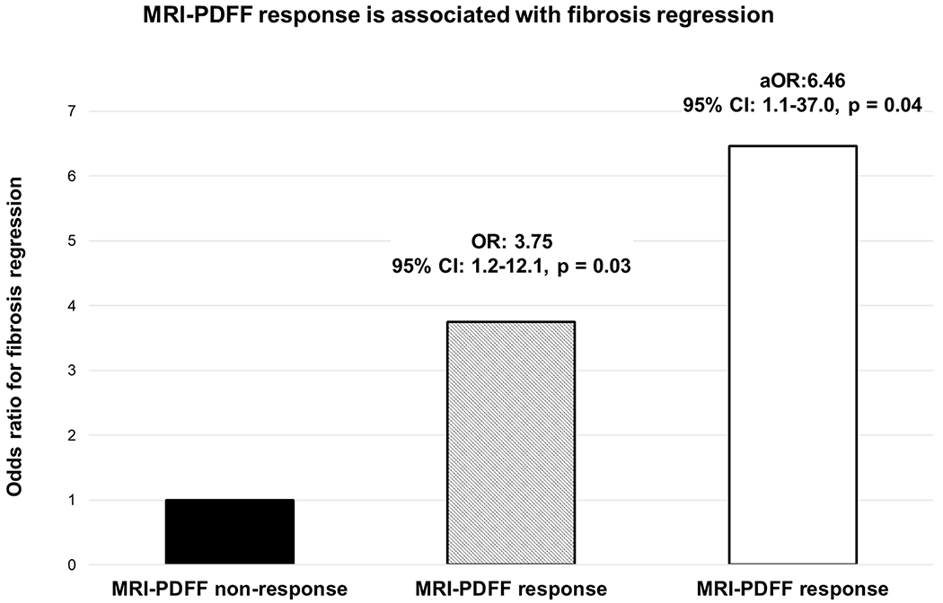
The median (IQR) age was 54 (43-62) years and body mass index was 31.9 (29-36) kg/m. In multivariable-adjusted logistic regression analysis (adjusted for age, gender, diabetes status, race/ethnicity, interval between biopsies, gamma-glutamyl transferase, liver stiffness by magnetic resonance elastography and change in platelet counts), MRI-PDFF response was an independent predictor of fibrosis regression with an adjusted OR of 6.46 (95% CI 1.1 to 37.0, p=0.04). The proportion of patients with MRI-PDFF response with fibrosis regression, no change in fibrosis and fibrosis progression was 40.0%, 24.6% and 13.0%, respectively, and the proportion of patients with MRI-PDFF response increased with fibrosis regression (p=0.03).
≥30% reduction in MRI-PDFF in early phase trials can provide a useful estimate of odds of ≥1 stage improvement in fibrosis. These data may be helpful in sample size estimation in non-alcoholic steatohepatitis trials.
新出现的数据表明,磁共振质子密度脂肪分数(MRI-PDFF)评估的肝脂肪减少 30%,可能与非酒精性脂肪性肝病活动评分改善相关,但 MRI-PDFF 下降与纤维化消退之间的关系尚不清楚。因此,我们旨在研究 MRI-PDFF 下降≥30%与非酒精性脂肪性肝病(NAFLD)纤维化消退之间的关系。
这项前瞻性研究纳入了 100 例经活检证实的 NAFLD 患者,这些患者在两个时间点进行了配对的同期 MRI-PDFF 评估。MRI-PDFF 反应定义为 MRI-PDFF 下降≥30%。主要终点是≥1 期组织学纤维化消退。
中位(IQR)年龄为 54(43-62)岁,体重指数为 31.9(29-36)kg/m。在多变量调整的逻辑回归分析中(调整年龄、性别、糖尿病状态、种族/民族、活检间隔、γ-谷氨酰转移酶、磁共振弹性成像的肝硬度和血小板计数变化),MRI-PDFF 反应是纤维化消退的独立预测因子,调整后的比值比为 6.46(95%CI 1.1-37.0,p=0.04)。MRI-PDFF 反应患者中纤维化消退、纤维化无变化和纤维化进展的比例分别为 40.0%、24.6%和 13.0%,MRI-PDFF 反应的比例随着纤维化消退而增加(p=0.03)。
早期试验中 MRI-PDFF 减少≥30%可以提供纤维化改善≥1 期的可能性的有用估计。这些数据可能有助于非酒精性脂肪性肝炎试验的样本量估计。