• 文献检索
  • 文档翻译
  • 深度研究
  • 学术资讯
  • Suppr Zotero 插件Zotero 插件
  • 邀请有礼
  • 套餐&价格
  • 历史记录
应用&插件
Suppr Zotero 插件Zotero 插件浏览器插件Mac 客户端Windows 客户端微信小程序
定价
高级版会员购买积分包购买API积分包
服务
文献检索文档翻译深度研究API 文档MCP 服务
关于我们
关于 Suppr公司介绍联系我们用户协议隐私条款
关注我们

Suppr 超能文献

核心技术专利:CN118964589B侵权必究
粤ICP备2023148730 号-1Suppr @ 2026

文献检索

告别复杂PubMed语法,用中文像聊天一样搜索,搜遍4000万医学文献。AI智能推荐,让科研检索更轻松。

立即免费搜索

文件翻译

保留排版,准确专业,支持PDF/Word/PPT等文件格式,支持 12+语言互译。

免费翻译文档

深度研究

AI帮你快速写综述,25分钟生成高质量综述,智能提取关键信息,辅助科研写作。

立即免费体验

五分钟内完成内淋巴积水的磁共振成像

MR Imaging of Endolymphatic Hydrops in Five Minutes.

机构信息

Department of Radiology, Nagoya University Graduate School of Medicine.

Canon Medical Systems.

出版信息

Magn Reson Med Sci. 2022 Jul 1;21(3):401-405. doi: 10.2463/mrms.ici.2021-0022. Epub 2021 Apr 24.

DOI:10.2463/mrms.ici.2021-0022
PMID:33896892
原文链接:https://pmc.ncbi.nlm.nih.gov/articles/PMC9316136/
Abstract

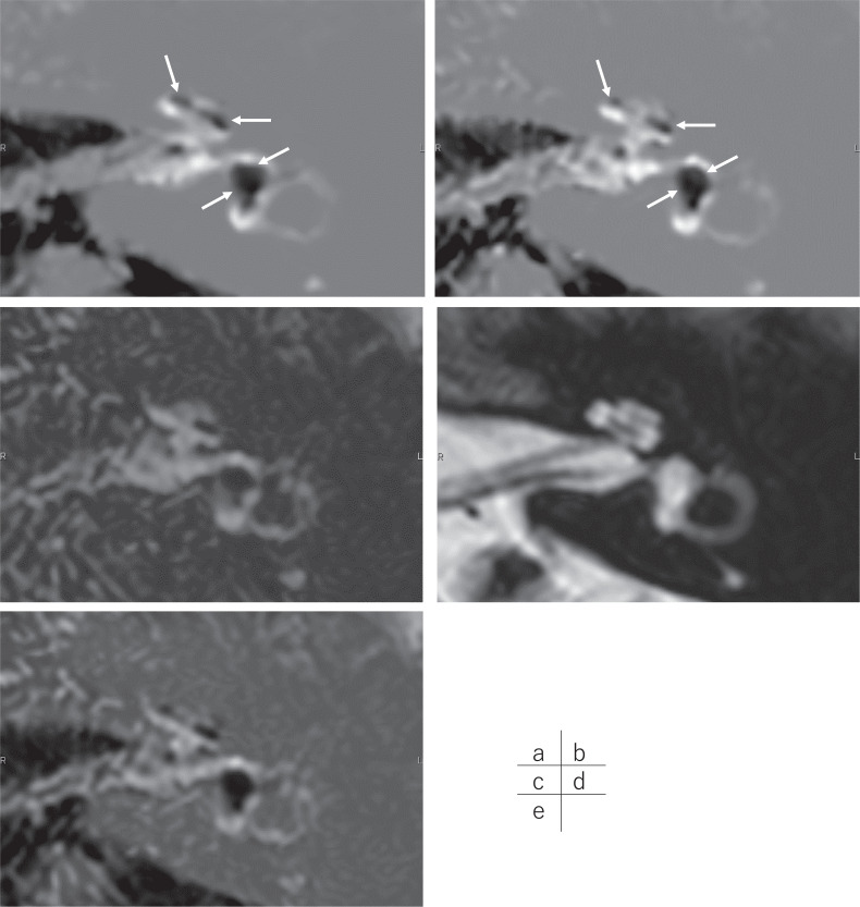
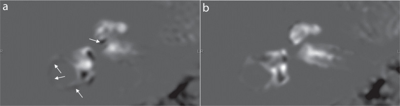
In this study, we present images acquired by a fast-imaging method for the evaluation of endolymphatic hydrops after intravenous administration of a single dose of gadolinium-based contrast agent. We utilized the hybrid of reversed image of MR cisternography and a positive perilymph signal by heavily T2- weighted 3D-fluid attenuated inversion recovery-multiplied by T2 (HYDROPS2-Mi2) method combined with deep learning reconstruction denoising. The scan time for the fast protocol was approximately 5 mins, which is far shorter than previously reported scan times. The fast acquisition provides similar image quality and less motion artifacts compared to the longer method.

摘要

在这项研究中,我们展示了一种快速成像方法获取的图像,用于评估单次静脉注射钆基造影剂后内淋巴积水的情况。我们利用 MR 脑池造影术反转图像和重度 T2 加权 3D 液体衰减反转恢复-多重 T2(HYDROPS2-Mi2)方法与深度学习重建去噪相结合的正外淋巴信号的混合体。快速协议的扫描时间约为 5 分钟,远短于先前报道的扫描时间。与较长的方法相比,快速采集提供了相似的图像质量和更少的运动伪影。

https://cdn.ncbi.nlm.nih.gov/pmc/blobs/24cc/9316136/b3e39d8ae1d0/mrms-21-401-g2.jpg
https://cdn.ncbi.nlm.nih.gov/pmc/blobs/24cc/9316136/66f5b7826d7c/mrms-21-401-g1.jpg
https://cdn.ncbi.nlm.nih.gov/pmc/blobs/24cc/9316136/b3e39d8ae1d0/mrms-21-401-g2.jpg
https://cdn.ncbi.nlm.nih.gov/pmc/blobs/24cc/9316136/66f5b7826d7c/mrms-21-401-g1.jpg
https://cdn.ncbi.nlm.nih.gov/pmc/blobs/24cc/9316136/b3e39d8ae1d0/mrms-21-401-g2.jpg

相似文献

1
MR Imaging of Endolymphatic Hydrops in Five Minutes.五分钟内完成内淋巴积水的磁共振成像
Magn Reson Med Sci. 2022 Jul 1;21(3):401-405. doi: 10.2463/mrms.ici.2021-0022. Epub 2021 Apr 24.
2
Quantification of Endolymphatic Space Volume after Intravenous Administration of a Single Dose of Gadolinium-based Contrast Agent: 3D-real Inversion Recovery versus HYDROPS-Mi2.单次静脉注射钆类造影剂后内淋巴间隙容积的定量分析:3D 真实反转恢复与 HYDROPS-Mi2 的比较。
Magn Reson Med Sci. 2020 May 1;19(2):119-124. doi: 10.2463/mrms.mp.2019-0013. Epub 2019 May 7.
3
MR Imaging of Endolymphatic Hydrops: Utility of iHYDROPS-Mi2 Combined with Deep Learning Reconstruction Denoising.内淋巴积水的磁共振成像:iHYDROPS-Mi2 联合深度学习重建降噪的应用。
Magn Reson Med Sci. 2021 Sep 1;20(3):272-279. doi: 10.2463/mrms.mp.2020-0082. Epub 2020 Aug 21.
4
Magnetic resonance imaging of endolymphatic hydrops: a comparison of methods with and without gadolinium-based contrast agent administration.内淋巴积水的磁共振成像:有无钆基造影剂给药方法的比较。
Nagoya J Med Sci. 2023 May;85(2):299-309. doi: 10.18999/nagjms.85.2.299.
5
Improved HYDROPS: Imaging of Endolymphatic Hydrops after Intravenous Administration of Gadolinium.改良的HYDROPS:静脉注射钆后内淋巴积水的成像
Magn Reson Med Sci. 2017 Oct 10;16(4):357-361. doi: 10.2463/mrms.tn.2016-0126. Epub 2017 May 22.
6
Relationship between Contrast Enhancement of the Perivascular Space in the Basal Ganglia and Endolymphatic Volume Ratio.基底节区血管周围间隙增强与内淋巴管容积比的关系。
Magn Reson Med Sci. 2018 Jan 10;17(1):67-72. doi: 10.2463/mrms.mp.2017-0001. Epub 2017 Jun 8.
7
Semi-quantification of endolymphatic size on MR imaging after intravenous injection of single-dose gadodiamide: comparison between two types of processing strategies.静脉注射单剂量钆喷酸葡胺后磁共振成像中内淋巴液大小的半定量分析:两种处理策略的比较。
Magn Reson Med Sci. 2013 Dec 25;12(4):261-9. doi: 10.2463/mrms.2013-0019. Epub 2013 Oct 29.
8
Imaging of Ménière's disease by subtraction of MR cisternography from positive perilymph image.MR 池造影术减去正迷路造影术对梅尼埃病的影像学研究。
Magn Reson Med Sci. 2012;11(4):303-9. doi: 10.2463/mrms.11.303.
9
[Optimization of delivering minimum Gd-DTPA at the posterior upper point on tympanic medial wall and hT2W-3D-FLAIR sequence for detecting endolymphatic hydrops].[鼓膜内侧壁后上方点最小剂量钆喷酸葡胺给药及hT2W-3D-FLAIR序列用于检测内淋巴积水的优化]
Zhonghua Er Bi Yan Hou Tou Jing Wai Ke Za Zhi. 2018 Dec 7;53(12):931-938. doi: 10.3760/cma.j.issn.1673-0860.2018.12.011.
10
Pitfalls of Using T2-contrast Enhancement Techniques in 3D-FLAIR to Detect Endolymphatic Hydrops.使用 3D-FLAIR 的 T2 对比增强技术检测内淋巴积水的陷阱。
Magn Reson Med Sci. 2023 Jul 1;22(3):335-344. doi: 10.2463/mrms.mp.2022-0017. Epub 2022 May 10.

引用本文的文献

1
Thin-slice 2D MR Imaging of the Shoulder Joint Using Denoising Deep Learning Reconstruction Provides Higher Image Quality Than 3D MR Imaging.使用去噪深度学习重建的肩关节二维薄层磁共振成像比三维磁共振成像提供更高的图像质量。
Magn Reson Med Sci. 2024 May 22. doi: 10.2463/mrms.mp.2023-0115.
2
Fairness of artificial intelligence in healthcare: review and recommendations.人工智能在医疗保健中的公平性:综述与建议。
Jpn J Radiol. 2024 Jan;42(1):3-15. doi: 10.1007/s11604-023-01474-3. Epub 2023 Aug 4.
3
Iterative Denoising Accelerated 3D FLAIR Sequence for Hydrops MR Imaging at 3T.

本文引用的文献

1
Diagnostic and therapeutic strategies for Meniere's disease of the Japan Society for Equilibrium Research.日本平衡研究学会梅尼埃病的诊断与治疗策略
Auris Nasus Larynx. 2021 Feb;48(1):15-22. doi: 10.1016/j.anl.2020.10.009. Epub 2020 Oct 29.
2
MR Imaging of Endolymphatic Hydrops: Utility of iHYDROPS-Mi2 Combined with Deep Learning Reconstruction Denoising.内淋巴积水的磁共振成像:iHYDROPS-Mi2 联合深度学习重建降噪的应用。
Magn Reson Med Sci. 2021 Sep 1;20(3):272-279. doi: 10.2463/mrms.mp.2020-0082. Epub 2020 Aug 21.
3
The Correlation Between Endolymphatic Hydrops and Clinical Features of Meniere Disease.
3T 下用于水脑症磁共振成像的迭代去噪加速 3D FLAIR 序列。
AJNR Am J Neuroradiol. 2023 Sep;44(9):1064-1069. doi: 10.3174/ajnr.A7953. Epub 2023 Aug 3.
4
Current State of Artificial Intelligence in Clinical Applications for Head and Neck MR Imaging.人工智能在头颈部磁共振成像临床应用中的现状。
Magn Reson Med Sci. 2023 Oct 1;22(4):401-414. doi: 10.2463/mrms.rev.2023-0047. Epub 2023 Aug 1.
5
Clinical applications of artificial intelligence in liver imaging.人工智能在肝脏成像中的临床应用。
Radiol Med. 2023 Jun;128(6):655-667. doi: 10.1007/s11547-023-01638-1. Epub 2023 May 10.
6
The Glymphatic System in Humans: Investigations With Magnetic Resonance Imaging.人体中的类淋巴系统:磁共振成像研究
Invest Radiol. 2024 Jan 1;59(1):1-12. doi: 10.1097/RLI.0000000000000969. Epub 2023 Mar 13.
7
Assessing the usefulness of inner ear contrast-enhanced magnetic resonance imaging and pure-tone audiometry in estimating the prognostic value of hearing improvement in patients with definite Meniere's disease.评估内耳对比增强磁共振成像和纯音测听在预测明确梅尼埃病患者听力改善预后价值中的作用。
Eur Arch Otorhinolaryngol. 2023 Apr;280(4):2051-2055. doi: 10.1007/s00405-022-07770-4. Epub 2022 Dec 10.
8
Diagnostic advantage of thin slice 2D MRI and multiplanar reconstruction of the knee joint using deep learning based denoising approach.基于深度学习去噪方法的膝关节薄层二维 MRI 和多平面重建的诊断优势。
Sci Rep. 2022 Jun 20;12(1):10362. doi: 10.1038/s41598-022-14190-1.
内淋巴积水与梅尼埃病临床特征的相关性。
Laryngoscope. 2021 Jan;131(1):E144-E150. doi: 10.1002/lary.28576. Epub 2020 Feb 21.
4
The vestibular aqueduct sign: Magnetic resonance imaging can detect abnormalities in both ears of patients with unilateral Meniere's disease.前庭水管征:磁共振成像可检测单侧梅尼埃病患者双耳的异常。
J Neuroradiol. 2020 Mar;47(2):174-179. doi: 10.1016/j.neurad.2018.10.003. Epub 2018 Nov 10.
5
In vivo Assessment of an Endolymphatic Hydrops Gradient Along the Cochlea in Patients With Menière's Disease by Magnetic Resonance Imaging-A Pilot Study.通过磁共振成像对梅尼埃病患者耳蜗内内淋巴积水梯度的体内评估-一项初步研究。
Otol Neurotol. 2018 Dec;39(10):e1091-e1099. doi: 10.1097/MAO.0000000000002016.
6
Improved 3D-real Inversion Recovery: A Robust Imaging Technique for Endolymphatic Hydrops after Intravenous Administration of Gadolinium.改进的三维真实反转恢复:一种静脉注射钆后用于内淋巴积水的稳健成像技术。
Magn Reson Med Sci. 2019 Jan 10;18(1):105-108. doi: 10.2463/mrms.bc.2017-0158. Epub 2018 Mar 7.
7
Volume Quantification of Endolymph after Intravenous Administration of a Single Dose of Gadolinium Contrast Agent: Comparison of 18- versus 8-minute Imaging Protocols.静脉注射单剂量钆对比剂后内淋巴的容积定量:18分钟与8分钟成像方案的比较
Magn Reson Med Sci. 2015;14(4):257-62. doi: 10.2463/mrms.2014-0118. Epub 2015 Mar 31.
8
Time course for measuring endolymphatic size in healthy volunteers following intravenous administration of gadoteridol.静脉注射钆特醇后,健康志愿者内淋巴大小测量的时间进程。
Magn Reson Med Sci. 2014;13(2):73-80. doi: 10.2463/mrms.2013-0080. Epub 2014 Apr 28.
9
Visualization of endolymphatic hydrops with MR imaging in patients with Ménière's disease and related pathologies: current status of its methods and clinical significance.梅尼埃病及相关病变患者内淋巴积水的磁共振成像可视化:其方法的现状及临床意义
Jpn J Radiol. 2014 Apr;32(4):191-204. doi: 10.1007/s11604-014-0290-4. Epub 2014 Feb 6.
10
Imaging of Ménière's disease by subtraction of MR cisternography from positive perilymph image.MR 池造影术减去正迷路造影术对梅尼埃病的影像学研究。
Magn Reson Med Sci. 2012;11(4):303-9. doi: 10.2463/mrms.11.303.