• 文献检索
  • 文档翻译
  • 深度研究
  • 学术资讯
  • Suppr Zotero 插件Zotero 插件
  • 邀请有礼
  • 套餐&价格
  • 历史记录
应用&插件
Suppr Zotero 插件Zotero 插件浏览器插件Mac 客户端Windows 客户端微信小程序
定价
高级版会员购买积分包购买API积分包
服务
文献检索文档翻译深度研究API 文档MCP 服务
关于我们
关于 Suppr公司介绍联系我们用户协议隐私条款
关注我们

Suppr 超能文献

核心技术专利:CN118964589B侵权必究
粤ICP备2023148730 号-1Suppr @ 2026

文献检索

告别复杂PubMed语法,用中文像聊天一样搜索,搜遍4000万医学文献。AI智能推荐,让科研检索更轻松。

立即免费搜索

文件翻译

保留排版,准确专业,支持PDF/Word/PPT等文件格式,支持 12+语言互译。

免费翻译文档

深度研究

AI帮你快速写综述,25分钟生成高质量综述,智能提取关键信息,辅助科研写作。

立即免费体验

改良的HYDROPS:静脉注射钆后内淋巴积水的成像

Improved HYDROPS: Imaging of Endolymphatic Hydrops after Intravenous Administration of Gadolinium.

作者信息

Naganawa Shinji, Kawai Hisashi, Taoka Toshiaki, Sone Michihiko

机构信息

Department of Radiology, Nagoya University Graduate School of Medicine.

Department of Otorhinolaryngology, Nagoya University Graduate School of Medicine.

出版信息

Magn Reson Med Sci. 2017 Oct 10;16(4):357-361. doi: 10.2463/mrms.tn.2016-0126. Epub 2017 May 22.

DOI:10.2463/mrms.tn.2016-0126
PMID:28529249
原文链接:https://pmc.ncbi.nlm.nih.gov/articles/PMC5743528/
Abstract

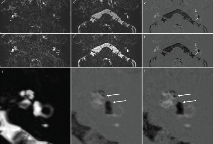
To improve the imaging protocol for the evaluation of endolymphatic hydrops after intravenous administration of a gadolinium-based contrast agent, we modified our previously reported hybrid of reversed image of positive endolymph signal and native image of positive perilymph signal (HYDROPS) method. Although the scan time of the new protocol was half that of the previous one, there were no significant differences between two protocols in the mean contrast noise ratio between the endolymph and perilymph and the area ratio of the endolymph size values in nine patients.

摘要

为了改进静脉注射钆基造影剂后评估内淋巴积水的成像方案,我们对之前报道的内淋巴阳性信号反转图像与外淋巴阳性信号原始图像相结合的(HYDROPS)方法进行了改进。尽管新方案的扫描时间是之前方案的一半,但在9名患者中,两种方案在内淋巴和外淋巴之间的平均对比噪声比以及内淋巴大小值的面积比方面没有显著差异。

https://cdn.ncbi.nlm.nih.gov/pmc/blobs/c18a/5743528/77b99cf4f129/mrms-16-357-g1.jpg

相似文献

1
Improved HYDROPS: Imaging of Endolymphatic Hydrops after Intravenous Administration of Gadolinium.改良的HYDROPS:静脉注射钆后内淋巴积水的成像
Magn Reson Med Sci. 2017 Oct 10;16(4):357-361. doi: 10.2463/mrms.tn.2016-0126. Epub 2017 May 22.
2
MR Imaging of Endolymphatic Hydrops in Five Minutes.五分钟内完成内淋巴积水的磁共振成像
Magn Reson Med Sci. 2022 Jul 1;21(3):401-405. doi: 10.2463/mrms.ici.2021-0022. Epub 2021 Apr 24.
3
Separate visualization of endolymphatic space, perilymphatic space and bone by a single pulse sequence; 3D-inversion recovery imaging utilizing real reconstruction after intratympanic Gd-DTPA administration at 3 Tesla.通过单一脉冲序列分别显示内淋巴间隙、外淋巴间隙和骨质;在3特斯拉场强下经鼓室内注射钆喷酸葡胺后利用真实重建的三维反转恢复成像。
Eur Radiol. 2008 May;18(5):920-4. doi: 10.1007/s00330-008-0854-8. Epub 2008 Mar 7.
4
Semi-quantification of endolymphatic size on MR imaging after intravenous injection of single-dose gadodiamide: comparison between two types of processing strategies.静脉注射单剂量钆喷酸葡胺后磁共振成像中内淋巴液大小的半定量分析:两种处理策略的比较。
Magn Reson Med Sci. 2013 Dec 25;12(4):261-9. doi: 10.2463/mrms.2013-0019. Epub 2013 Oct 29.
5
[Optimization of delivering minimum Gd-DTPA at the posterior upper point on tympanic medial wall and hT2W-3D-FLAIR sequence for detecting endolymphatic hydrops].[鼓膜内侧壁后上方点最小剂量钆喷酸葡胺给药及hT2W-3D-FLAIR序列用于检测内淋巴积水的优化]
Zhonghua Er Bi Yan Hou Tou Jing Wai Ke Za Zhi. 2018 Dec 7;53(12):931-938. doi: 10.3760/cma.j.issn.1673-0860.2018.12.011.
6
Volume Quantification of Endolymph after Intravenous Administration of a Single Dose of Gadolinium Contrast Agent: Comparison of 18- versus 8-minute Imaging Protocols.静脉注射单剂量钆对比剂后内淋巴的容积定量:18分钟与8分钟成像方案的比较
Magn Reson Med Sci. 2015;14(4):257-62. doi: 10.2463/mrms.2014-0118. Epub 2015 Mar 31.
7
Imaging of endolymphatic and perilymphatic fluid after intravenous administration of single-dose gadodiamide.静脉单次注射钆喷酸葡胺后内淋巴液和外淋巴液的影像学表现。
Magn Reson Med Sci. 2012;11(2):145-50. doi: 10.2463/mrms.11.145.
8
Semi-quantitative evaluation of endolymphatic hydrops by bilateral intratympanic gadolinium-based contrast agent (GBCA) administration with MRI for Meniere's disease.通过双侧鼓室内注射钆基造影剂(GBCA)并结合MRI对梅尼埃病内淋巴积水进行半定量评估。
Acta Otolaryngol. 2010;130(1):10-6. doi: 10.3109/00016480902858881.
9
[Magnetic resonance imaging study of endolymphatic hydrops based on 3D-FLAIR sequence after single-dose intravenous gadolinium injection].基于单剂量静脉注射钆后3D-FLAIR序列的内淋巴积水磁共振成像研究
Zhonghua Yi Xue Za Zhi. 2019 Jan 29;99(5):333-337. doi: 10.3760/cma.j.issn.0376-2491.2019.05.003.
10
[Visualization of endolymphatic hydrops in 3D-FLAIR MRI after intratympanic Gd-DTPA administration in Meniere's disease patients].[梅尼埃病患者鼓室内注射钆喷酸葡胺后3D-FLAIR MRI对内淋巴积水的可视化]
Zhonghua Er Bi Yan Hou Tou Jing Wai Ke Za Zhi. 2013 Aug;48(8):628-33.

引用本文的文献

1
A modern conceptual framework for study and treatment of Meniere's disease.梅尼埃病研究与治疗的现代概念框架。
Front Neurol. 2025 May 16;16:1607435. doi: 10.3389/fneur.2025.1607435. eCollection 2025.
2
New non-contrast MRI of endolymphatic hydrops in Ménière's disease considering inversion time.考虑反转时间的梅尼埃病内淋巴积水的新型非对比磁共振成像
Laryngoscope Investig Otolaryngol. 2024 Aug 10;9(4):e1314. doi: 10.1002/lio2.1314. eCollection 2024 Aug.
3
An unenhanced 3D-FLAIR sequence using long repetition time and constant flip angle to image endolymphatic hydrops.

本文引用的文献

1
Gd-based Contrast Enhancement of the Perivascular Spaces in the Basal Ganglia.钆增强基底节区血管周围间隙的影像学表现
Magn Reson Med Sci. 2017 Jan 10;16(1):61-65. doi: 10.2463/mrms.mp.2016-0039. Epub 2016 Jul 12.
2
Meniere's disease.梅尼埃病。
Nat Rev Dis Primers. 2016 May 12;2:16028. doi: 10.1038/nrdp.2016.28.
3
Simple Estimation of the Endolymphatic Volume Ratio after Intravenous Administration of a Single-dose of Gadolinium Contrast.静脉注射单剂量钆对比剂后内淋巴体积比的简易估算
一种使用长重复时间和恒定翻转角的非增强3D-FLAIR序列来成像内淋巴积水。
Eur Radiol. 2025 Jan;35(1):29-37. doi: 10.1007/s00330-024-10906-1. Epub 2024 Jul 10.
4
Direct Visualization of Tracer Permeation into the Endolymph in Human Patients Using MR Imaging.使用磁共振成像直接可视化示踪剂渗透到人类患者内淋巴中的情况。
Magn Reson Med Sci. 2025 Apr 1;24(2):253-261. doi: 10.2463/mrms.mp.2024-0011. Epub 2024 Apr 3.
5
Current State of Artificial Intelligence in Clinical Applications for Head and Neck MR Imaging.人工智能在头颈部磁共振成像临床应用中的现状。
Magn Reson Med Sci. 2023 Oct 1;22(4):401-414. doi: 10.2463/mrms.rev.2023-0047. Epub 2023 Aug 1.
6
Functional and Molecular Markers for Hearing Loss and Vertigo Attacks in Meniere's Disease.梅尼埃病听力损失和眩晕发作的功能和分子标志物。
Int J Mol Sci. 2023 Jan 28;24(3):2504. doi: 10.3390/ijms24032504.
7
Assessing the usefulness of inner ear contrast-enhanced magnetic resonance imaging and pure-tone audiometry in estimating the prognostic value of hearing improvement in patients with definite Meniere's disease.评估内耳对比增强磁共振成像和纯音测听在预测明确梅尼埃病患者听力改善预后价值中的作用。
Eur Arch Otorhinolaryngol. 2023 Apr;280(4):2051-2055. doi: 10.1007/s00405-022-07770-4. Epub 2022 Dec 10.
8
Correlation of clinical parameters with endolymphatic hydrops on MRI in Meniere's disease.梅尼埃病中临床参数与MRI上内淋巴积水的相关性
Front Neurol. 2022 Jul 25;13:937703. doi: 10.3389/fneur.2022.937703. eCollection 2022.
9
Editorial: Imaging of the Vestibular System.社论:前庭系统的影像学
Front Neurol. 2022 Jun 23;13:937955. doi: 10.3389/fneur.2022.937955. eCollection 2022.
10
IE-Vnet: Deep Learning-Based Segmentation of the Inner Ear's Total Fluid Space.IE-Vnet:基于深度学习的内耳全液腔分割
Front Neurol. 2022 May 11;13:663200. doi: 10.3389/fneur.2022.663200. eCollection 2022.
Magn Reson Med Sci. 2016 Oct 11;15(4):379-385. doi: 10.2463/mrms.mp.2015-0175. Epub 2016 Mar 21.
4
Heavily T₂-Weighted 3D-FLAIR Improves the Detection of Cochlear Lymph Fluid Signal Abnormalities in Patients with Sudden Sensorineural Hearing Loss.重T₂加权三维液体衰减反转恢复序列(3D-FLAIR)可提高突发性感音神经性听力损失患者耳蜗淋巴液信号异常的检出率。
Magn Reson Med Sci. 2016;15(2):203-11. doi: 10.2463/mrms.mp.2015-0065. Epub 2015 Nov 20.
5
The Technical and Clinical Features of 3D-FLAIR in Neuroimaging.神经影像学中三维液体衰减反转恢复序列(3D-FLAIR)的技术与临床特征
Magn Reson Med Sci. 2015;14(2):93-106. doi: 10.2463/mrms.2014-0132. Epub 2015 Mar 31.
6
Volume Quantification of Endolymph after Intravenous Administration of a Single Dose of Gadolinium Contrast Agent: Comparison of 18- versus 8-minute Imaging Protocols.静脉注射单剂量钆对比剂后内淋巴的容积定量:18分钟与8分钟成像方案的比较
Magn Reson Med Sci. 2015;14(4):257-62. doi: 10.2463/mrms.2014-0118. Epub 2015 Mar 31.
7
Imaging of endolymphatic hydrops in 10 minutes: a new strategy to reduce scan time to one third.10分钟内完成内淋巴积水成像:将扫描时间缩短至三分之一的新策略
Magn Reson Med Sci. 2015;14(1):77-83. doi: 10.2463/mrms.2014-0065. Epub 2014 Dec 15.
8
Time course for measuring endolymphatic size in healthy volunteers following intravenous administration of gadoteridol.静脉注射钆特醇后,健康志愿者内淋巴大小测量的时间进程。
Magn Reson Med Sci. 2014;13(2):73-80. doi: 10.2463/mrms.2013-0080. Epub 2014 Apr 28.
9
Serial scans in healthy volunteers following intravenous administration of gadoteridol: time course of contrast enhancement in various cranial fluid spaces.静脉注射钆特醇后对健康志愿者进行的系列扫描:不同颅腔液腔中对比增强的时间进程。
Magn Reson Med Sci. 2014;13(1):7-13. doi: 10.2463/mrms.2013-0056. Epub 2014 Jan 31.
10
Semi-quantification of endolymphatic size on MR imaging after intravenous injection of single-dose gadodiamide: comparison between two types of processing strategies.静脉注射单剂量钆喷酸葡胺后磁共振成像中内淋巴液大小的半定量分析:两种处理策略的比较。
Magn Reson Med Sci. 2013 Dec 25;12(4):261-9. doi: 10.2463/mrms.2013-0019. Epub 2013 Oct 29.