Tsutsui Kenta, Florio Maria Cristina, Yang Annie, Wirth Ashley N, Yang Dongmei, Kim Mary S, Ziman Bruce D, Bychkov Rostislav, Monfredi Oliver J, Maltsev Victor A, Lakatta Edward G
Laboratory of Cardiovascular Science, Biomedical Research Center, Intramural Research Program, National Institute on Aging, NIH, Baltimore, MD, United States.
Department of Cardiovascular Medicine, Faculty of Medicine, Saitama Medical University International Medical Center, Saitama, Japan.
Front Physiol. 2021 Apr 9;12:596832. doi: 10.3389/fphys.2021.596832. eCollection 2021.
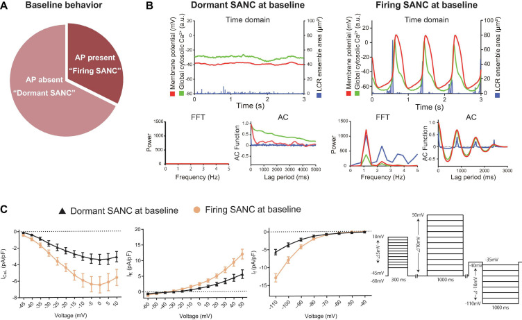
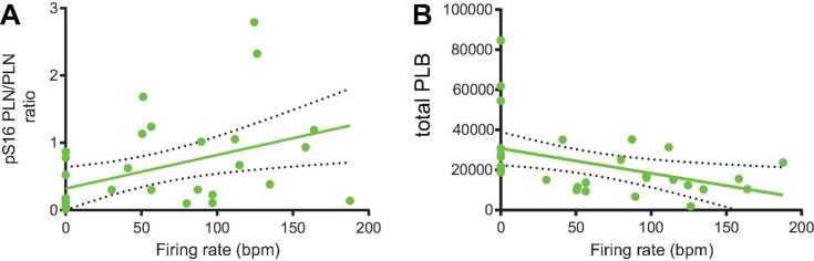
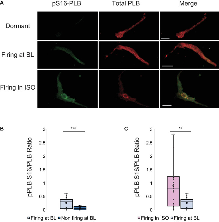
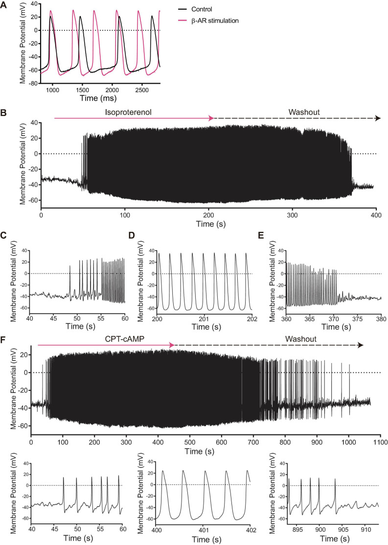
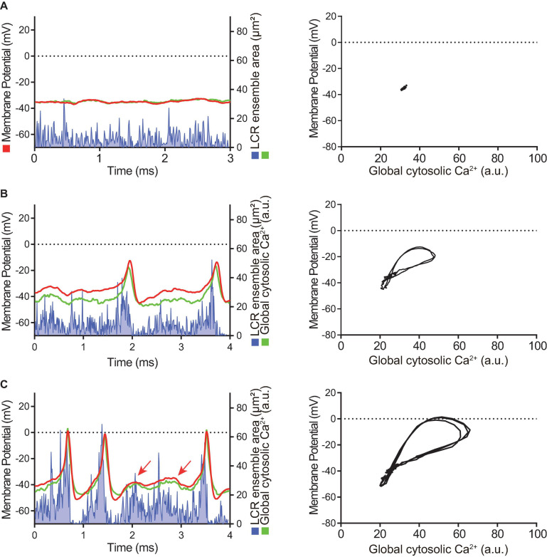
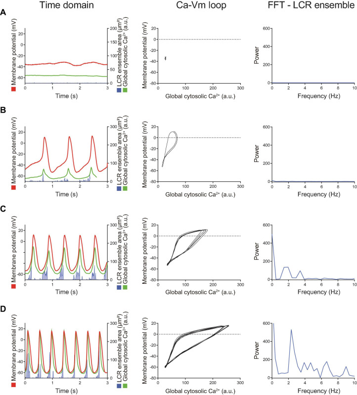
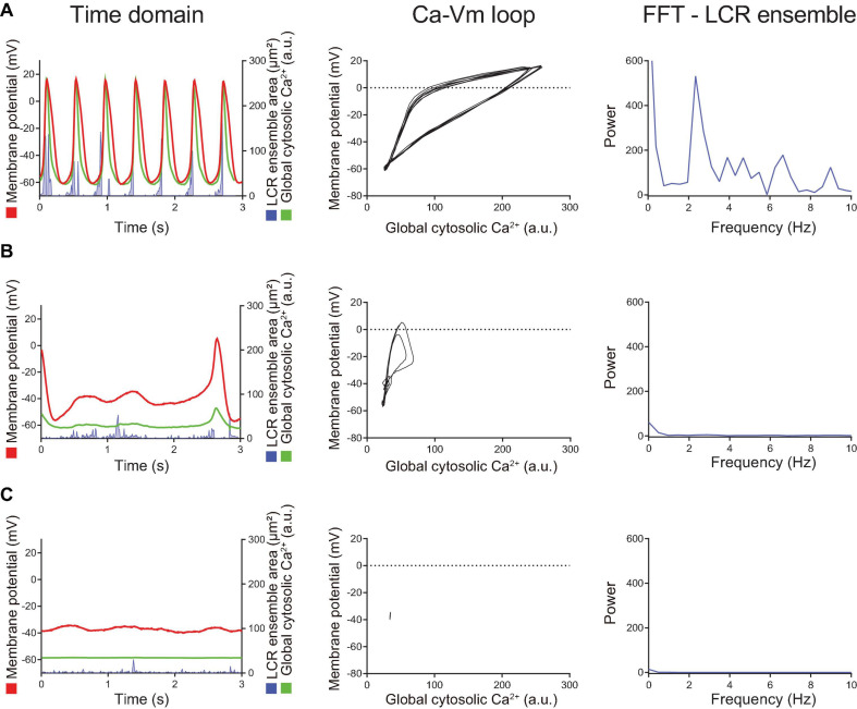
Action potential (AP) firing rate and rhythm of sinoatrial nodal cells (SANC) are controlled by synergy between intracellular rhythmic local Ca releases (LCRs) ("Ca clock") and sarcolemmal electrogenic mechanisms ("membrane clock"). However, some SANC do not fire APs (dormant SANC). Prior studies have shown that β-adrenoceptor stimulation can restore AP firing in these cells. Here we tested whether this relates to improvement of synchronization of clock coupling. We characterized membrane potential, ion currents, Ca dynamics, and phospholamban (PLB) phosphorylation, regulating Ca pump in enzymatically isolated single guinea pig SANC prior to, during, and following β-adrenoceptor stimulation (isoproterenol) or application of cell-permeant cAMP (CPT-cAMP). Phosphorylation of PLB (Serine 16) was quantified in the same cells following Ca measurement. In dormant SANC LCRs were small and disorganized at baseline, membrane potential was depolarized (-38 ± 1 mV, = 46), and I, I, and I densities were smaller vs SANC firing APs. β-adrenoceptor stimulation or application of CPT-cAMP led to spontaneous AP generation in 44 and 46% of dormant SANC, respectively. The initial response was an increase in size, rhythmicity and synchronization of LCRs, paralleled with membrane hyperpolarization and small amplitude APs (rate ∼1 Hz). During the transition to steady-state AP firing, LCR size further increased, while LCR period shortened. LCRs became more synchronized resulting in the growth of an ensemble LCR signal peaked in late diastole, culminating in AP ignition; the rate of diastolic depolarization, AP amplitude, and AP firing rate increased. I, I, and I amplitudes in dormant SANC increased in response to β-adrenoceptor stimulation. During washout, all changes reversed in order. Total PLB was higher, but the ratio of phosphorylated PLB (Serine 16) to total PLB was lower in dormant SANC. β-adrenoceptor stimulation increased this ratio in AP-firing cells. Thus, transition of dormant SANC to AP firing is linked to the increased functional coupling of membrane and Ca clock proteins. The transition occurs via (i) an increase in cAMP-mediated phosphorylation of PLB accelerating Ca pumping, (ii) increased spatiotemporal LCR synchronization, yielding a larger diastolic LCR ensemble signal resulting in an earlier increase in diastolic I; and (iii) increased current densities of I, I, and I.
窦房结细胞(SANC)的动作电位(AP)发放频率和节律受细胞内节律性局部钙释放(LCRs)(“钙钟”)与肌膜电生机制(“膜钟”)之间协同作用的控制。然而,一些SANC并不发放AP(静止SANC)。先前的研究表明,β-肾上腺素能受体刺激可恢复这些细胞的AP发放。在此,我们测试了这是否与时钟耦合同步性的改善有关。我们对酶分离的单个豚鼠SANC在β-肾上腺素能受体刺激(异丙肾上腺素)之前、期间和之后或应用细胞渗透性cAMP(CPT-cAMP)期间的膜电位、离子电流、钙动力学和受磷蛋白(PLB)磷酸化进行了表征,后者可调节钙泵。在测量钙之后,对相同细胞中PLB(丝氨酸16)的磷酸化进行了定量。在静止SANC中,LCRs在基线时较小且无组织,膜电位去极化(-38±1 mV,n = 46),与发放AP的SANC相比,I、I和I密度较小。β-肾上腺素能受体刺激或应用CPT-cAMP分别导致44%和46%的静止SANC产生自发性AP。最初的反应是LCRs的大小、节律性和同步性增加,同时伴有膜超极化和小幅度AP(频率约为1 Hz)。在向稳态AP发放转变的过程中,LCR大小进一步增加,而LCR周期缩短。LCRs变得更加同步,导致在舒张末期达到峰值的整体LCR信号增强,最终引发AP;舒张期去极化速率、AP幅度和AP发放频率增加。静止SANC中的I、I和I幅度响应β-肾上腺素能受体刺激而增加。在洗脱过程中,所有变化按顺序逆转。静止SANC中的总PLB较高,但磷酸化PLB(丝氨酸16)与总PLB的比率较低。β-肾上腺素能受体刺激增加了发放AP细胞中的这一比率。因此,静止SANC向AP发放的转变与膜和钙钟蛋白功能耦合的增加有关。这种转变通过以下方式发生:(i)cAMP介导的PLB磷酸化增加,加速钙泵活动;(ii)时空LCR同步性增加,产生更大的舒张期LCR整体信号,导致舒张期I更早增加;以及(iii)I、I和I的电流密度增加。