Amsterdam Cardiovascular Sciences, Department of Vascular Surgery, Amsterdam University Medical Centers, Location VUmc, Amsterdam, The Netherlands.
Amsterdam Cardiovascular Sciences, Department of Physiology, Amsterdam University Medical Centers, Location VUmc, Amsterdam, The Netherlands.
J Endovasc Ther. 2021 Aug;28(4):604-613. doi: 10.1177/15266028211009272. Epub 2021 Apr 26.
Abdominal aortic aneurysms (AAAs) are associated with overall high mortality in case of rupture. Since the pathophysiology is unclear, no adequate pharmacological therapy exists. Smooth muscle cells (SMCs) dysfunction and extracellular matrix (ECM) degradation have been proposed as underlying causes. We investigated SMC spatial organization and SMC-ECM interactions in our novel 3-dimensional (3D) vascular model. We validated our model for future use by comparing it to existing 2-dimensional (2D) cell culture. Our model can be used for translational studies of SMC and their role in AAA pathophysiology.
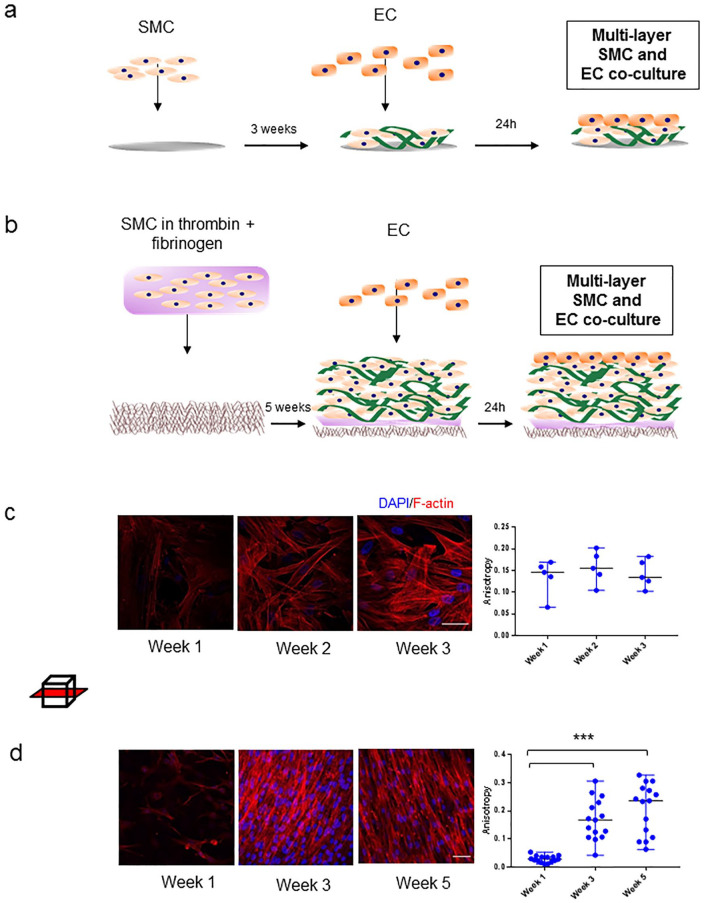
SMC isolated from the medial layer of were the aortic wall of controls and AAA patients seeded on electrospun poly-lactide--glycolide scaffolds and cultured for 5 weeks, after which endothelial cells (EC) are added. Cell morphology, orientation, mechanical properties and ECM production were quantified for validation and comparison between controls and patients.
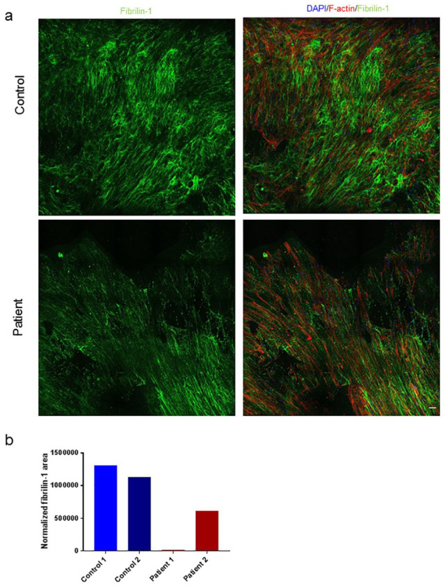
We show that cultured SMC proliferate into multiple layers after 5 weeks in culture and produce ECM proteins, mimicking their behavior in the medial aortic layer. EC attach to multilayered SMC, mimicking layer interactions. The novel SMC model exhibits viscoelastic properties comparable to biological vessels; cytoskeletal organization increases during the 5 weeks in culture; increased cytoskeletal alignment and decreased ECM production indicate different organization of AAA patients' cells compared with control.
We present a valuable preclinical model of AAA constructed with patient specific cells with applications in both translational research and therapeutic developments. We observed SMC spatial reorganization in a time course of 5 weeks in our robust, patient-specific model of SMC-EC organization and ECM production.
腹主动脉瘤(AAA)破裂时总死亡率很高。由于病理生理学不清楚,因此没有足够的药物治疗方法。平滑肌细胞(SMC)功能障碍和细胞外基质(ECM)降解被认为是潜在的原因。我们在新的 3 维(3D)血管模型中研究了 SMC 的空间组织和 SMC-ECM 相互作用。通过将其与现有的 2 维(2D)细胞培养进行比较,我们验证了该模型以供将来使用。我们的模型可用于 SMC 的转化研究及其在 AAA 病理生理学中的作用。
从控制组和 AAA 患者的主动脉壁中分离出中膜层的 SMC,接种到电纺聚乳酸-乙交酯支架上,并培养 5 周,然后添加内皮细胞(EC)。为了进行验证和比较,对细胞形态,定向,机械性能和 ECM 产生进行了量化。
我们表明,在培养 5 周后,培养的 SMC 增殖成多层,并产生 ECM 蛋白,模拟其在主动脉中层的行为。EC 附着在多层 SMC 上,模拟层间相互作用。新型 SMC 模型表现出与生物血管相当的粘弹性特性;细胞骨架组织在培养的 5 周内增加;细胞骨架排列的增加和 ECM 产生的减少表明与对照组相比,AAA 患者的细胞具有不同的组织。
我们提出了一种使用具有患者特异性细胞的有价值的 AAA 临床前模型,可应用于转化研究和治疗开发。我们在我们强大的,基于患者的 SMC-EC 组织和 ECM 产生的 5 周时间过程中观察到 SMC 空间重新组织。