• 文献检索
  • 文档翻译
  • 深度研究
  • 学术资讯
  • Suppr Zotero 插件Zotero 插件
  • 邀请有礼
  • 套餐&价格
  • 历史记录
应用&插件
Suppr Zotero 插件Zotero 插件浏览器插件Mac 客户端Windows 客户端微信小程序
定价
高级版会员购买积分包购买API积分包
服务
文献检索文档翻译深度研究API 文档MCP 服务
关于我们
关于 Suppr公司介绍联系我们用户协议隐私条款
关注我们

Suppr 超能文献

核心技术专利:CN118964589B侵权必究
粤ICP备2023148730 号-1Suppr @ 2026

文献检索

告别复杂PubMed语法,用中文像聊天一样搜索,搜遍4000万医学文献。AI智能推荐,让科研检索更轻松。

立即免费搜索

文件翻译

保留排版,准确专业,支持PDF/Word/PPT等文件格式,支持 12+语言互译。

免费翻译文档

深度研究

AI帮你快速写综述,25分钟生成高质量综述,智能提取关键信息,辅助科研写作。

立即免费体验

中心凹厚度及视网膜外层完整性对葡萄膜炎性黄斑水肿视觉预后的影响

The impact of central foveal thickness and integrity of the outer retinal layers in the visual outcome of uveitic macular edema.

作者信息

Alvarez-Guzman Carlos, Bustamante-Arias Andres, Colorado-Zavala Maria F, Rodriguez-Garcia Alejandro

机构信息

Tecnologico de Monterrey, School of Medicine and Health Sciences, Institute of Ophthalmology and Visual Sciences, Ocular Immunology & Uveitis Service, Monterrey, Mexico.

Hospital Zambrano Hellion, TecSalud, Av. Batallon de San Patricio No. 112. Col. Real de San Agustin, San Pedro Garza Garcia, N.L., C.P. 66278, Mexico.

出版信息

Int J Retina Vitreous. 2021 Apr 27;7(1):36. doi: 10.1186/s40942-021-00306-8.

DOI:10.1186/s40942-021-00306-8
PMID:33902745
原文链接:https://pmc.ncbi.nlm.nih.gov/articles/PMC8077744/
Abstract

BACKGROUND

To analyze the relationship between the central foveal thickness (CFT) and the integrity of the ellipsoid portion of inner segments (EPIS) and interdigitating zone (IZ) retinal layers in the visual outcome of uveitic macular edema (UME).

METHODS

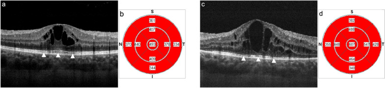
Prospective, observational, and cross-sectional study of eyes with UME. Spectral-domain optical coherence tomography (SD-OCT) macular morphological pattern, CFT, and integrity of the outer retinal layers were analyzed. We arranged the data by EPIS or IZ integrity and contrasted it with student t-test (quantitative variables) and Fisher exact test or χ² distribution (categorical variables) to evaluate visual impairment and retinal measures. Receiver operator curve (ROC) estimation and logistic regression (probit) assessed if the sample´s variance could be associated with IZ or EPIS integrity.

RESULTS

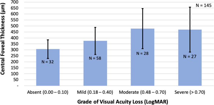
We included 145 SD-OCT macular scans from 45 patients at different stages of UME. Cystoid macular edema (CME) increased the risk of severe (P ≤ 0.0162) and moderate visual loss (P ≤ 0.0032). The highest CFT values occurred in patients with moderate (478.11 ± 167.62 μm) and severe (449.4 ± 224.86 μm) visual loss. Of all morphological patterns of macular edema, only CME showed a statistically significant relationship with severe visual impairment (44.92%, p = 0.0035, OR 4.29 [1.62-11.4]). Likewise, an increased probability of severe visual loss correlated negatively with both, IZ (37.93%, P ≤ 0.001, OR 10.02) and EPIS (38.98%, P ≤ 0.001, OR 13.1) disruption. A CFT > 337 μm showed a higher probability of IZ (AUROC = 0.7341, SEN 77.59%, ESP 65.52) and EPIS (AUROC = 0.7489, SEN 76.37%, ESP 65.12%) loss of integrity. Moreover, when BCVA reached 0.44 LogMAR (≤ 20/50 Snellen eq.), it was more likely to have IZ (AUROC = 0.8706, ESP 88.51%, SEN 77.59%) and EPIS (AUROC = 0.8898, ESP 88.3%, SEN 76.27) disruption.

CONCLUSIONS

Significantly increased CFT has a higher probability for EPIS and IZ disruption, which significantly increases the risk for irreversible visual loss in eyes with UME. Evaluating these layers' integrity by optical coherence tomography helps predict the visual outcome and make the right therapeutic decisions. Trial registration The study was registered on April 13, 2020, at the Instituto Tecnologico y de Estudios Superiores de Monterrey Research Committee (License No. COFEPRIS 20 CI 19 039 002), project registration No. P000338-CAVICaREMU-CI-CR002, and the Ethics Committee (License No. CONBIOETICA 19 CEI 011-2016-10-17), project registration No. P000338-CAVICaREMU-CEIC-CR002.

摘要

背景

分析葡萄膜炎性黄斑水肿(UME)患者的中心凹厚度(CFT)与视网膜内层椭圆体带(EPIS)和指状交叉区(IZ)视网膜层完整性之间的关系,及其对视觉预后的影响。

方法

对UME患者进行前瞻性、观察性横断面研究。分析黄斑部形态学模式、CFT及外层视网膜层的完整性。根据EPIS或IZ的完整性对数据进行整理,并采用学生t检验(定量变量)、Fisher精确检验或χ²分布(分类变量)进行对比,以评估视力损害和视网膜测量结果。采用受试者工作特征曲线(ROC)估计和逻辑回归(概率单位)分析样本方差是否与IZ或EPIS完整性相关。

结果

纳入45例不同阶段UME患者的145幅黄斑部SD-OCT扫描图像。黄斑囊样水肿(CME)增加了严重视力丧失(P≤0.0162)和中度视力丧失(P≤0.0032)的风险。中度视力丧失(478.11±167.62μm)和严重视力丧失(449.4±224.86μm)患者的CFT值最高。在所有黄斑水肿形态中,只有CME与严重视力损害存在统计学显著相关性(44.92%,p = 0.0035,OR 4.29 [1.62 - 11.4])。同样,严重视力丧失的可能性增加与IZ(37.93%,P≤0.001,OR 10.02)和EPIS(38.98%,P≤0.001,OR 13.1)破坏均呈负相关。CFT>337μm时,IZ(曲线下面积=0.7341,敏感度77.59%,特异度65.52%)和EPIS(曲线下面积=0.7489,敏感度76.37%,特异度65.12%)完整性丧失的可能性更高。此外,当最佳矫正视力(BCVA)达到0.44 LogMAR(≤20/50 Snellen视力表等效值)时,IZ(曲线下面积=0.8706,特异度88.51%,敏感度77.59%)和EPIS(曲线下面积=0.8898,特异度88.3%,敏感度76.27%)破坏的可能性更大。

结论

CFT显著增加时,EPIS和IZ破坏的可能性更高,这显著增加了UME患者不可逆视力丧失的风险。通过光学相干断层扫描评估这些层的完整性有助于预测视觉预后并做出正确的治疗决策。试验注册 本研究于2020年4月13日在蒙特雷理工学院研究委员会注册(许可证编号:COFEPRIS 20 CI 19 039 002),项目注册号:P000338 - CAVICaREMU - CI - CR002,以及伦理委员会(许可证编号:CONBIOETICA 19 CEI 011 - 2016 - 10 - 17),项目注册号:P000338 - CAVICaREMU - CEIC - CR002。

https://cdn.ncbi.nlm.nih.gov/pmc/blobs/9292/8077744/45e20e84f335/40942_2021_306_Fig4_HTML.jpg
https://cdn.ncbi.nlm.nih.gov/pmc/blobs/9292/8077744/6d39876e5110/40942_2021_306_Fig1_HTML.jpg
https://cdn.ncbi.nlm.nih.gov/pmc/blobs/9292/8077744/2d6746833fc1/40942_2021_306_Fig2_HTML.jpg
https://cdn.ncbi.nlm.nih.gov/pmc/blobs/9292/8077744/946938313ba2/40942_2021_306_Fig3_HTML.jpg
https://cdn.ncbi.nlm.nih.gov/pmc/blobs/9292/8077744/45e20e84f335/40942_2021_306_Fig4_HTML.jpg

相似文献

1
The impact of central foveal thickness and integrity of the outer retinal layers in the visual outcome of uveitic macular edema.中心凹厚度及视网膜外层完整性对葡萄膜炎性黄斑水肿视觉预后的影响
Int J Retina Vitreous. 2021 Apr 27;7(1):36. doi: 10.1186/s40942-021-00306-8.
2
Correlation between Visual Acuity, Inner Segment/Outer Segment Junction, and Cone Outer Segment Tips Line Integrity in Uveitic Macular Edema.葡萄膜炎性黄斑水肿中视力、内节/外节连接部及视锥细胞外节顶端线完整性之间的相关性
Biomed Res Int. 2015;2015:853728. doi: 10.1155/2015/853728. Epub 2015 Jun 8.
3
Fundus autofluorescence and spectral domain optical coherence tomography in uveitic macular edema.葡萄膜炎性黄斑水肿中的眼底自发荧光和光谱域光学相干断层扫描
Graefes Arch Clin Exp Ophthalmol. 2009 Dec;247(12):1685-9. doi: 10.1007/s00417-009-1149-8. Epub 2009 Aug 11.
4
Visual acuity and microperimetric mapping of lesion area in eyes with inflammatory cystoid macular oedema.炎症性囊样黄斑水肿眼的视力和病变区微视野图。
Acta Ophthalmol. 2014 Jun;92(4):332-8. doi: 10.1111/aos.12201. Epub 2013 Jun 27.
5
Spectral-domain optical coherence tomography in uveitic macular edema: morphological features and prognostic factors.频域光相干断层扫描在葡萄膜炎性黄斑水肿中的应用:形态学特征和预后因素。
Ophthalmologica. 2012;228(1):13-8. doi: 10.1159/000337234. Epub 2012 Apr 14.
6
Association of Disorganization of Retinal Inner Layers With Visual Acuity In Eyes With Uveitic Cystoid Macular Edema.葡萄膜炎性黄斑囊样水肿患者视网膜内层结构紊乱与视力的相关性
Am J Ophthalmol. 2017 May;177:116-125. doi: 10.1016/j.ajo.2017.02.017. Epub 2017 Feb 22.
7
Spectral-Domain OCT Predictors of Visual Outcomes after Ranibizumab Treatment for Macular Edema Resulting from Retinal Vein Occlusion.雷珠单抗治疗视网膜静脉阻塞所致黄斑水肿后视力预后的频域光学相干断层扫描预测指标
Ophthalmol Retina. 2020 Jan;4(1):67-76. doi: 10.1016/j.oret.2019.08.009. Epub 2019 Aug 28.
8
[Correlation of retinal sensitivity, visual acuity and central macular thickness in different types of diabetic macular edema].[不同类型糖尿病性黄斑水肿中视网膜敏感度、视力与黄斑中心厚度的相关性]
Zhonghua Yan Ke Za Zhi. 2013 Dec;49(12):1081-8.
9
Defining Cystoid Macular Degeneration in Diabetic Macular Edema: An OCT-Based Single-center Study.糖尿病性黄斑水肿中囊样黄斑变性的定义:一项基于光学相干断层扫描的单中心研究。
Turk J Ophthalmol. 2019 Dec 31;49(6):315-322. doi: 10.4274/tjo.galenos.2019.22687.
10
Patterns of macular edema in patients with uveitis: qualitative and quantitative assessment using optical coherence tomography.葡萄膜炎患者黄斑水肿的模式:使用光学相干断层扫描进行定性和定量评估
Ophthalmology. 2004 May;111(5):946-53. doi: 10.1016/j.ophtha.2003.08.037.

引用本文的文献

1
Macular Thickness and Volume by Spectral-Domain Optical Coherence Tomography and their Related Factors in the Elderly Population.老年人群中通过频域光学相干断层扫描测量的黄斑厚度和体积及其相关因素
J Curr Ophthalmol. 2024 Aug 10;35(4):362-368. doi: 10.4103/joco.joco_153_23. eCollection 2023 Oct-Dec.
2
Measurement of Foveal Retinal Thickness in Myopic Patients Using Different Display Modes on Optical Coherence Tomography: A Retrospective, Cross-Sectional Study.使用光学相干断层扫描不同显示模式测量近视患者的黄斑视网膜厚度:一项回顾性横断面研究。
Ophthalmol Ther. 2023 Feb;12(1):167-178. doi: 10.1007/s40123-022-00584-x. Epub 2022 Oct 26.

本文引用的文献

1
Spectral-domain optical coherence tomography findings of the macula in 500 consecutive patients with uveitis.500例葡萄膜炎连续患者黄斑区的频域光学相干断层扫描结果
Eye (Lond). 2016 Nov;30(11):1415-1423. doi: 10.1038/eye.2016.133. Epub 2016 Jul 8.
2
Uveitic macular edema.葡萄膜炎性黄斑水肿
Eye (Lond). 2016 Oct;30(10):1277-1292. doi: 10.1038/eye.2016.115. Epub 2016 Jun 3.
3
Structure-function Relationships in Uveitic Cystoid Macular Edema: Using En Face Optical Coherence Tomography to Predict Vision.葡萄膜炎性黄斑囊样水肿的结构-功能关系:使用正面光学相干断层扫描预测视力。
Ocul Immunol Inflamm. 2016 Jun;24(3):274-81. doi: 10.3109/09273948.2015.1056535. Epub 2015 Oct 15.
4
Correlation between Visual Acuity, Inner Segment/Outer Segment Junction, and Cone Outer Segment Tips Line Integrity in Uveitic Macular Edema.葡萄膜炎性黄斑水肿中视力、内节/外节连接部及视锥细胞外节顶端线完整性之间的相关性
Biomed Res Int. 2015;2015:853728. doi: 10.1155/2015/853728. Epub 2015 Jun 8.
5
Correlation between foveal cone outer segment tips line and visual recovery after epiretinal membrane surgery.黄斑区锥体细胞外节端线与视网膜前膜手术后视力恢复的相关性。
Invest Ophthalmol Vis Sci. 2013 Nov 5;54(12):7302-8. doi: 10.1167/iovs.13-12702.
6
Causes of visual loss associated with uveitis in a singapore tertiary eye center.新加坡一家三级眼科中心葡萄膜炎相关的视力丧失的原因。
Ocul Immunol Inflamm. 2013 Aug;21(4):264-9. doi: 10.3109/09273948.2013.774424. Epub 2013 Apr 25.
7
Association between external limiting membrane status and visual acuity in diabetic macular oedema.糖尿病性黄斑水肿中外膜限制膜状态与视力的关系。
Br J Ophthalmol. 2013 Feb;97(2):228-32. doi: 10.1136/bjophthalmol-2011-301418. Epub 2012 Nov 21.
8
Characterization of serous retinal detachments in uveitis patients with optical coherence tomography.利用光学相干断层扫描技术对葡萄膜炎患者浆液性视网膜脱离的特征分析
J Ophthalmic Inflamm Infect. 2012 Dec;2(4):191-7. doi: 10.1007/s12348-012-0084-8. Epub 2012 Jun 2.
9
Morphometric spectral-domain optical coherence tomography features of epiretinal membrane correlate with visual acuity in patients with uveitis.后发性葡萄膜炎患者的视网膜前膜形态计量谱域光相干断层扫描特征与视力相关。
Am J Ophthalmol. 2012 Jul;154(1):78-86.e1. doi: 10.1016/j.ajo.2012.01.032. Epub 2012 Apr 26.
10
The impact of macular edema on visual function in intermediate, posterior, and panuveitis.中间型、后部和全葡萄膜炎对视力功能的影响。
Ocul Immunol Inflamm. 2012 Jun;20(3):171-81. doi: 10.3109/09273948.2012.658467. Epub 2012 Apr 24.