Department of Pathology, Memorial Sloan Kettering Cancer Center, New York, NY, USA.
Mod Pathol. 2021 Aug;34(8):1487-1494. doi: 10.1038/s41379-021-00807-9. Epub 2021 Apr 26.
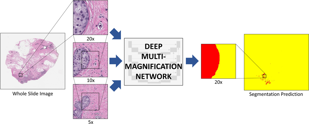
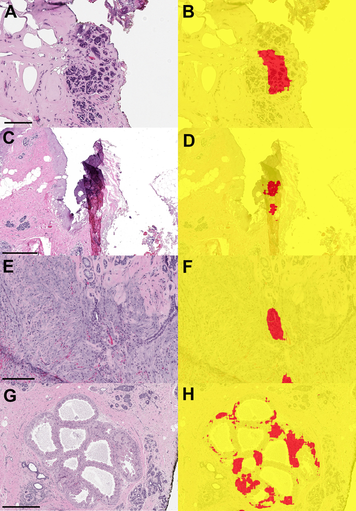
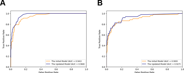
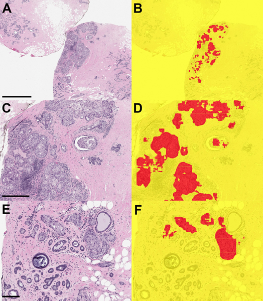
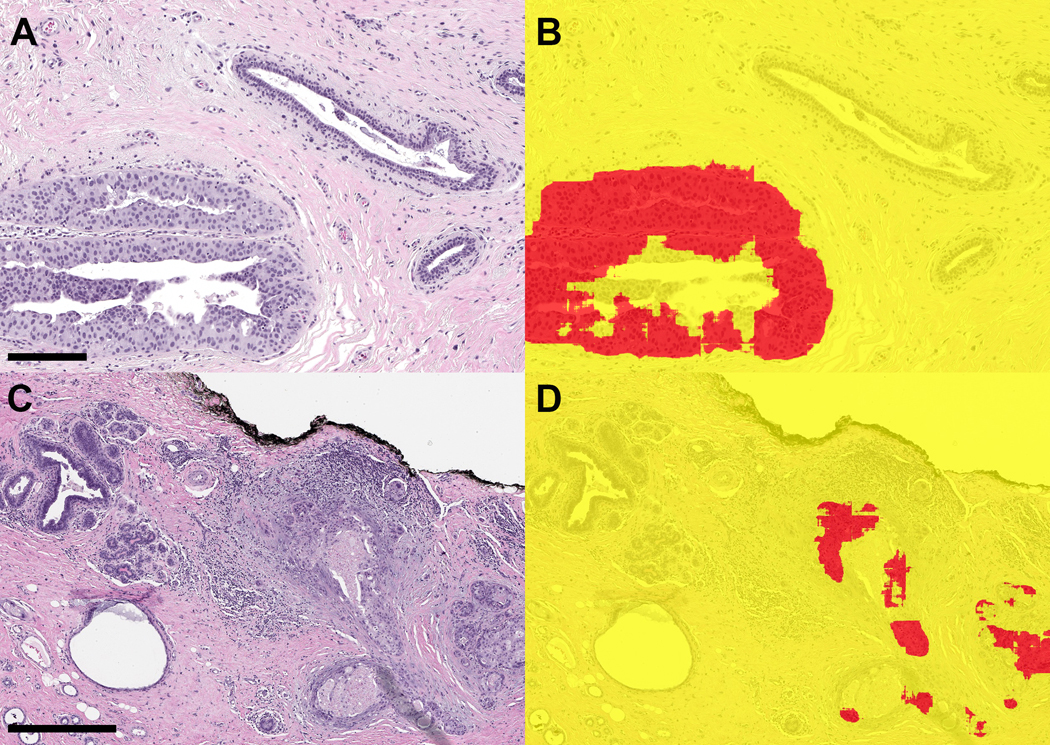
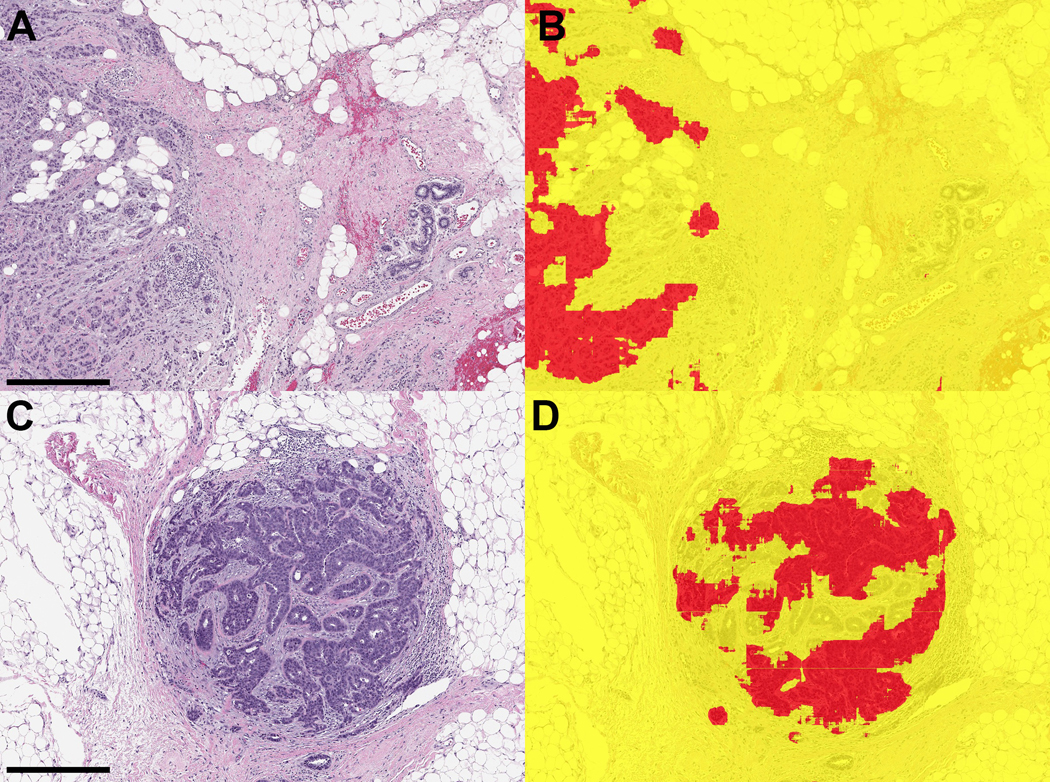
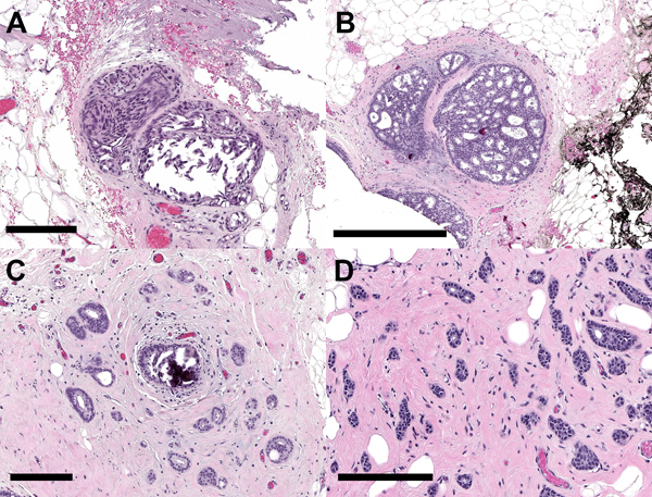
The surgical margin status of breast lumpectomy specimens for invasive carcinoma and ductal carcinoma in situ (DCIS) guides clinical decisions, as positive margins are associated with higher rates of local recurrence. The "cavity shave" method of margin assessment has the benefits of allowing the surgeon to orient shaved margins intraoperatively and the pathologist to assess one inked margin per specimen. We studied whether a deep convolutional neural network, a deep multi-magnification network (DMMN), could accurately segment carcinoma from benign tissue in whole slide images (WSIs) of shave margin slides, and therefore serve as a potential screening tool to improve the efficiency of microscopic evaluation of these specimens. Applying the pretrained DMMN model, or the initial model, to a validation set of 408 WSIs (348 benign, 60 with carcinoma) achieved an area under the curve (AUC) of 0.941. After additional manual annotations and fine-tuning of the model, the updated model achieved an AUC of 0.968 with sensitivity set at 100% and corresponding specificity of 78%. We applied the initial model and updated model to a testing set of 427 WSIs (374 benign, 53 with carcinoma) which showed AUC values of 0.900 and 0.927, respectively. Using the pixel classification threshold selected from the validation set, the model achieved a sensitivity of 92% and specificity of 78%. The four false-negative classifications resulted from two small foci of DCIS (1 mm, 0.5 mm) and two foci of well-differentiated invasive carcinoma (3 mm, 1.5 mm). This proof-of-principle study demonstrates that a DMMN machine learning model can segment invasive carcinoma and DCIS in surgical margin specimens with high accuracy and has the potential to be used as a screening tool for pathologic assessment of these specimens.
乳腺肿瘤切除术标本中浸润性癌和导管原位癌(DCIS)的手术切缘状态指导临床决策,因为阳性切缘与更高的局部复发率相关。评估切缘的“腔面刮除”方法具有允许外科医生在手术中对刮除的切缘进行定位以及病理学家对每个标本评估一个墨染切缘的优势。我们研究了深度卷积神经网络(一种深度多倍放大网络,DMMN)是否可以准确地对切除边缘玻片的全玻片图像(WSI)中的癌组织与良性组织进行分割,从而作为一种潜在的筛选工具,以提高这些标本的显微镜评估效率。将预训练的 DMMN 模型或初始模型应用于 408 张 WSI(348 张良性,60 张癌性)的验证集,获得曲线下面积(AUC)为 0.941。在对模型进行额外的手动注释和微调后,更新后的模型的 AUC 为 0.968,灵敏度设置为 100%,相应的特异性为 78%。我们将初始模型和更新模型应用于 427 张 WSI(374 张良性,53 张癌性)的测试集,分别得到 AUC 值 0.900 和 0.927。使用从验证集选择的像素分类阈值,模型的灵敏度为 92%,特异性为 78%。四次假阴性分类结果归因于两个小的 DCIS 病灶(1mm,0.5mm)和两个分化良好的浸润性癌病灶(3mm,1.5mm)。这项初步研究表明,DMMN 机器学习模型可以高精度地对手术切缘标本中的浸润性癌和 DCIS 进行分割,并且有可能作为这些标本病理评估的筛选工具。