Department of Pathology, Memorial Sloan Kettering Cancer Center, New York, NY 10065 USA.
Department of Pathology, Memorial Sloan Kettering Cancer Center, New York, NY 10065 USA.
Comput Med Imaging Graph. 2021 Mar;88:101866. doi: 10.1016/j.compmedimag.2021.101866. Epub 2021 Jan 12.
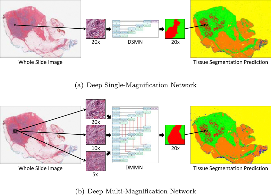
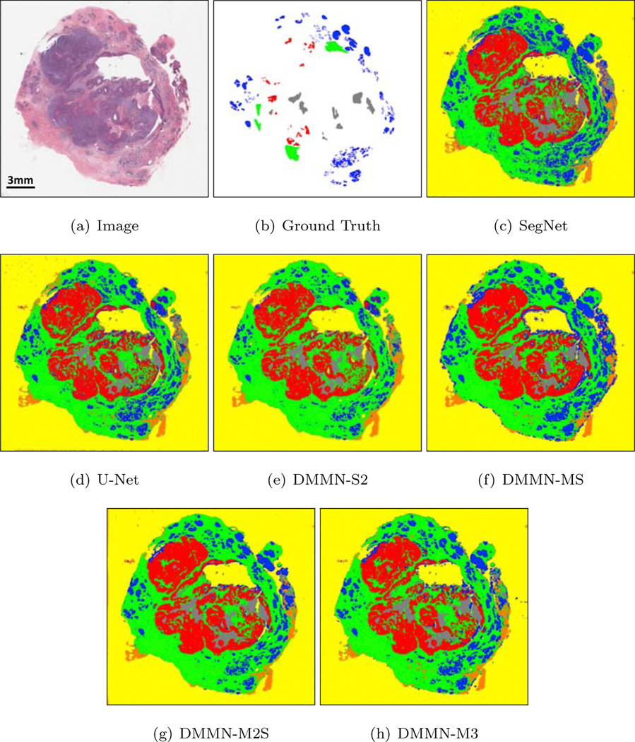
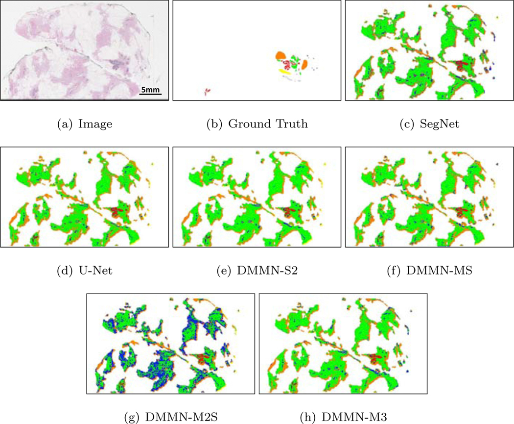
Pathologic analysis of surgical excision specimens for breast carcinoma is important to evaluate the completeness of surgical excision and has implications for future treatment. This analysis is performed manually by pathologists reviewing histologic slides prepared from formalin-fixed tissue. In this paper, we present Deep Multi-Magnification Network trained by partial annotation for automated multi-class tissue segmentation by a set of patches from multiple magnifications in digitized whole slide images. Our proposed architecture with multi-encoder, multi-decoder, and multi-concatenation outperforms other single and multi-magnification-based architectures by achieving the highest mean intersection-over-union, and can be used to facilitate pathologists' assessments of breast cancer.
乳腺癌外科切除标本的病理分析对于评估手术切除的完整性非常重要,并且对未来的治疗有影响。这种分析是由病理学家手动进行的,他们查看从福尔马林固定组织制备的组织学幻灯片。在本文中,我们提出了一种由部分注释训练的深度多放大网络,用于通过数字化全切片图像中来自多个放大倍数的一组斑块自动进行多类组织分割。我们提出的具有多编码器、多解码器和多连接的体系结构优于其他基于单和多放大倍数的体系结构,实现了最高的平均交并比,可用于辅助病理学家评估乳腺癌。