Couture Heather D, Williams Lindsay A, Geradts Joseph, Nyante Sarah J, Butler Ebonee N, Marron J S, Perou Charles M, Troester Melissa A, Niethammer Marc
1Department of Computer Science, University of North Carolina at Chapel Hill, Chapel Hill, NC 27599 USA.
2Department of Epidemiology, University of North Carolina at Chapel Hill, Chapel Hill, NC 27599 USA.
NPJ Breast Cancer. 2018 Sep 3;4:30. doi: 10.1038/s41523-018-0079-1. eCollection 2018.
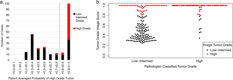
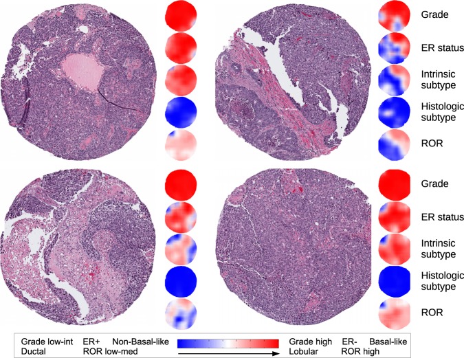
RNA-based, multi-gene molecular assays are available and widely used for patients with ER-positive/HER2-negative breast cancers. However, RNA-based genomic tests can be costly and are not available in many countries. Methods for inferring molecular subtype from histologic images may identify patients most likely to benefit from further genomic testing. To identify patients who could benefit from molecular testing based on H&E stained histologic images, we developed an image analysis approach using deep learning. A training set of 571 breast tumors was used to create image-based classifiers for tumor grade, ER status, PAM50 intrinsic subtype, histologic subtype, and risk of recurrence score (ROR-PT). The resulting classifiers were applied to an independent test set ( = 288), and accuracy, sensitivity, and specificity of each was assessed on the test set. Histologic image analysis with deep learning distinguished low-intermediate vs. high tumor grade (82% accuracy), ER status (84% accuracy), Basal-like vs. non-Basal-like (77% accuracy), Ductal vs. Lobular (94% accuracy), and high vs. low-medium ROR-PT score (75% accuracy). Sampling considerations in the training set minimized bias in the test set. Incorrect classification of ER status was significantly more common for Luminal B tumors. These data provide proof of principle that molecular marker status, including a critical clinical biomarker (i.e., ER status), can be predicted with accuracy >75% based on H&E features. Image-based methods could be promising for identifying patients with a greater need for further genomic testing, or in place of classically scored variables typically accomplished using human-based scoring.
基于RNA的多基因分子检测方法已可用于雌激素受体阳性/人表皮生长因子受体2阴性乳腺癌患者,并得到广泛应用。然而,基于RNA的基因组检测成本高昂,且在许多国家无法获得。从组织学图像推断分子亚型的方法可能会识别出最有可能从进一步基因组检测中获益的患者。为了识别那些可基于苏木精和伊红(H&E)染色的组织学图像从分子检测中获益的患者,我们开发了一种使用深度学习的图像分析方法。使用一个包含571个乳腺肿瘤的训练集来创建基于图像的肿瘤分级、雌激素受体状态、PAM50内在亚型、组织学亚型和复发风险评分(ROR-PT)的分类器。将所得分类器应用于一个独立的测试集(n = 288),并在测试集上评估每个分类器的准确性、敏感性和特异性。深度学习的组织学图像分析能够区分低-中级与高级肿瘤分级(准确率82%)、雌激素受体状态(准确率84%)、基底样与非基底样(准确率77%)、导管型与小叶型(准确率94%)以及高与低-中ROR-PT评分(准确率75%)。训练集中的抽样考虑将测试集中的偏差降至最低。Luminal B肿瘤雌激素受体状态的错误分类明显更为常见。这些数据提供了原理证明,即基于H&E特征可以准确预测分子标志物状态,包括关键的临床生物标志物(即雌激素受体状态),准确率>75%。基于图像的方法对于识别更需要进一步基因组检测的患者,或替代通常使用基于人工评分完成的经典评分变量可能很有前景。