• 文献检索
  • 文档翻译
  • 深度研究
  • 学术资讯
  • Suppr Zotero 插件Zotero 插件
  • 邀请有礼
  • 套餐&价格
  • 历史记录
应用&插件
Suppr Zotero 插件Zotero 插件浏览器插件Mac 客户端Windows 客户端微信小程序
定价
高级版会员购买积分包购买API积分包
服务
文献检索文档翻译深度研究API 文档MCP 服务
关于我们
关于 Suppr公司介绍联系我们用户协议隐私条款
关注我们

Suppr 超能文献

核心技术专利:CN118964589B侵权必究
粤ICP备2023148730 号-1Suppr @ 2026

文献检索

告别复杂PubMed语法,用中文像聊天一样搜索,搜遍4000万医学文献。AI智能推荐,让科研检索更轻松。

立即免费搜索

文件翻译

保留排版,准确专业,支持PDF/Word/PPT等文件格式,支持 12+语言互译。

免费翻译文档

深度研究

AI帮你快速写综述,25分钟生成高质量综述,智能提取关键信息,辅助科研写作。

立即免费体验

用于锥形束CT肺肿瘤分割的深度跨模态(MR-CT)诱导蒸馏学习

Deep cross-modality (MR-CT) educed distillation learning for cone beam CT lung tumor segmentation.

作者信息

Jiang Jue, Riyahi Alam Sadegh, Chen Ishita, Zhang Perry, Rimner Andreas, Deasy Joseph O, Veeraraghavan Harini

机构信息

Department of Medical Physics, Memorial Sloan Kettering Cancer Center, 1275 York Avenue, New York, NY, 1006, USA.

Department of Radiation Oncology, Memorial Sloan Kettering Cancer Center, 1275 York Avenue, New York, NY, 1006, USA.

出版信息

Med Phys. 2021 Jul;48(7):3702-3713. doi: 10.1002/mp.14902. Epub 2021 May 25.

DOI:10.1002/mp.14902
PMID:33905558
原文链接:https://pmc.ncbi.nlm.nih.gov/articles/PMC9662709/
Abstract

PURPOSE

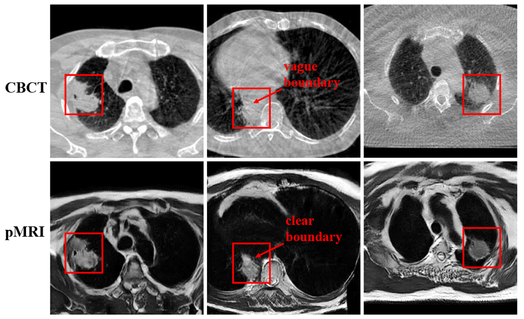
Despite the widespread availability of in-treatment room cone beam computed tomography (CBCT) imaging, due to the lack of reliable segmentation methods, CBCT is only used for gross set up corrections in lung radiotherapies. Accurate and reliable auto-segmentation tools could potentiate volumetric response assessment and geometry-guided adaptive radiation therapies. Therefore, we developed a new deep learning CBCT lung tumor segmentation method.

METHODS

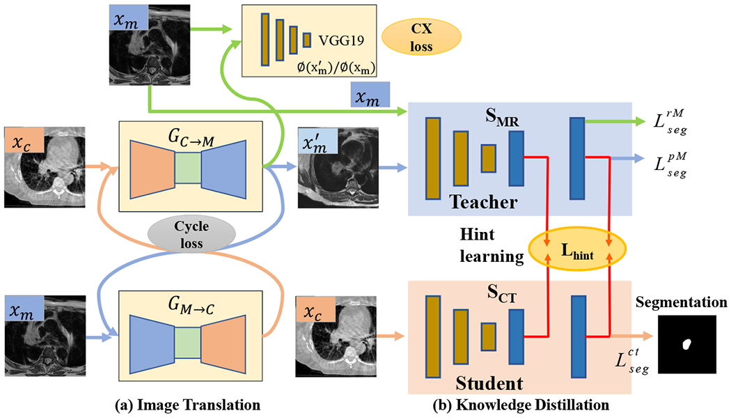
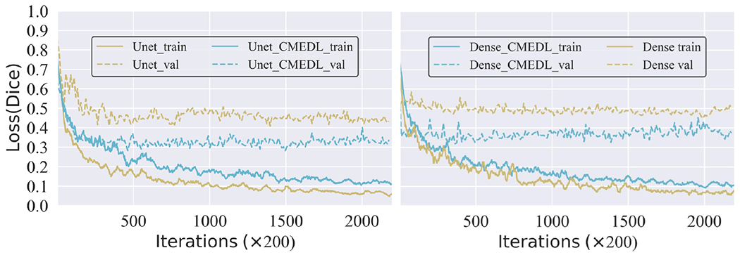
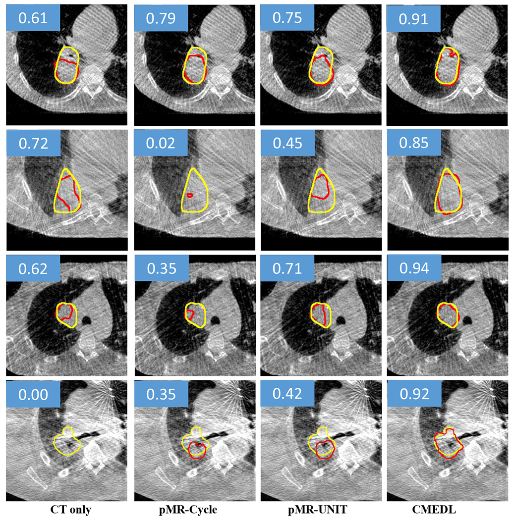
The key idea of our approach called cross-modality educed distillation (CMEDL) is to use magnetic resonance imaging (MRI) to guide a CBCT segmentation network training to extract more informative features during training. We accomplish this by training an end-to-end network comprised of unpaired domain adaptation (UDA) and cross-domain segmentation distillation networks (SDNs) using unpaired CBCT and MRI datasets. UDA approach uses CBCT and MRI that are not aligned and may arise from different sets of patients. The UDA network synthesizes pseudo MRI from CBCT images. The SDN consists of teacher MRI and student CBCT segmentation networks. Feature distillation regularizes the student network to extract CBCT features that match the statistical distribution of MRI features extracted by the teacher network and obtain better differentiation of tumor from background. The UDA network was implemented with a cycleGAN improved with contextual losses separately on Unet and dense fully convolutional segmentation networks (DenseFCN). Performance comparisons were done against CBCT only using 2D and 3D networks. We also compared against an alternative framework that used UDA with MR segmentation network, whereby segmentation was done on the synthesized pseudo MRI representation. All networks were trained with 216 weekly CBCTs and 82 T2-weighted turbo spin echo MRI acquired from different patient cohorts. Validation was done on 20 weekly CBCTs from patients not used in training. Independent testing was done on 38 weekly CBCTs from patients not used in training or validation. Segmentation accuracy was measured using surface Dice similarity coefficient (SDSC) and Hausdroff distance at 95th percentile (HD95) metrics.

RESULTS

The CMEDL approach significantly improved (p < 0.001) the accuracy of both Unet (SDSC of 0.83 ± 0.08; HD95 of 7.69 ± 7.86 mm) and DenseFCN (SDSC of 0.75 ± 0.13; HD95 of 11.42 ± 9.87 mm) over CBCT only 2DUnet (SDSC of 0.69 ± 0.11; HD95 of 21.70 ± 16.34 mm), 3D Unet (SDSC of 0.72 ± 0.20; HD95 15.01 ± 12.98 mm), and DenseFCN (SDSC of 0.66 ± 0.15; HD95 of 22.15 ± 17.19 mm) networks. The alternate framework using UDA with the MRI network was also more accurate than the CBCT only methods but less accurate the CMEDL approach.

CONCLUSIONS

Our results demonstrate feasibility of the introduced CMEDL approach to produce reasonably accurate lung cancer segmentation from CBCT images. Further validation on larger datasets is necessary for clinical translation.

摘要

目的

尽管治疗室内锥形束计算机断层扫描(CBCT)成像已广泛应用,但由于缺乏可靠的分割方法,CBCT仅用于肺癌放疗中的大体摆位校正。准确可靠的自动分割工具可增强容积反应评估和几何形状引导的自适应放射治疗。因此,我们开发了一种新的深度学习CBCT肺肿瘤分割方法。

方法

我们的方法称为跨模态诱导蒸馏(CMEDL),其关键思想是在训练期间使用磁共振成像(MRI)来指导CBCT分割网络训练,以提取更多信息特征。我们通过使用未配对的CBCT和MRI数据集训练一个由未配对域自适应(UDA)和跨域分割蒸馏网络(SDN)组成的端到端网络来实现这一点。UDA方法使用未对齐且可能来自不同患者组的CBCT和MRI。UDA网络从CBCT图像合成伪MRI。SDN由教师MRI和学生CBCT分割网络组成。特征蒸馏使学生网络正规化,以提取与教师网络提取的MRI特征统计分布相匹配的CBCT特征,并更好地将肿瘤与背景区分开来。UDA网络在Unet和密集全卷积分割网络(DenseFCN)上分别使用上下文损失改进的cycleGAN实现。仅使用2D和3D网络对CBCT进行性能比较。我们还与使用UDA和MR分割网络的替代框架进行了比较,该框架在合成的伪MRI表示上进行分割。所有网络均使用从不同患者队列中获取的216份每周CBCT和82份T2加权快速自旋回波MRI进行训练。对未用于训练的患者的20份每周CBCT进行验证。对未用于训练或验证的患者的38份每周CBCT进行独立测试。使用表面骰子相似系数(SDSC)和第95百分位数的豪斯多夫距离(HD95)指标测量分割准确性。

结果

与仅使用CBCT的2D Unet(SDSC为0.69±0.11;HD95为21.70±16.34mm)、3D Unet(SDSC为0.72±0.20;HD95为15.01±12.98mm)和DenseFCN(SDSC为0.66±0.15;HD95为22.15±17.19mm)网络相比,CMEDL方法显著提高了(p<0.001)Unet(SDSC为0.83±0.08;HD95为7.69±7.86mm)和DenseFCN(SDSC为0.75±0.13;HD95为11.42±9.87mm)的准确性。使用UDA和MRI网络的替代框架也比仅使用CBCT的方法更准确,但比CMEDL方法准确性低。

结论

我们的结果证明了引入的CMEDL方法从CBCT图像中进行合理准确的肺癌分割的可行性。需要在更大的数据集上进行进一步验证以实现临床转化。

https://cdn.ncbi.nlm.nih.gov/pmc/blobs/e37b/9662709/7027ea2e3666/nihms-1844961-f0006.jpg
https://cdn.ncbi.nlm.nih.gov/pmc/blobs/e37b/9662709/1fdc096b4410/nihms-1844961-f0001.jpg
https://cdn.ncbi.nlm.nih.gov/pmc/blobs/e37b/9662709/87d4148b24d2/nihms-1844961-f0002.jpg
https://cdn.ncbi.nlm.nih.gov/pmc/blobs/e37b/9662709/f00be21c5233/nihms-1844961-f0003.jpg
https://cdn.ncbi.nlm.nih.gov/pmc/blobs/e37b/9662709/ea99d281e43b/nihms-1844961-f0004.jpg
https://cdn.ncbi.nlm.nih.gov/pmc/blobs/e37b/9662709/01cbcb0222cd/nihms-1844961-f0005.jpg
https://cdn.ncbi.nlm.nih.gov/pmc/blobs/e37b/9662709/7027ea2e3666/nihms-1844961-f0006.jpg

相似文献

1
Deep cross-modality (MR-CT) educed distillation learning for cone beam CT lung tumor segmentation.用于锥形束CT肺肿瘤分割的深度跨模态(MR-CT)诱导蒸馏学习
Med Phys. 2021 Jul;48(7):3702-3713. doi: 10.1002/mp.14902. Epub 2021 May 25.
2
Unpaired Cross-Modality Educed Distillation (CMEDL) for Medical Image Segmentation.用于医学图像分割的非配对跨模态导出蒸馏(CMEDL)
IEEE Trans Med Imaging. 2022 May;41(5):1057-1068. doi: 10.1109/TMI.2021.3132291. Epub 2022 May 2.
3
Integrating cross-modality hallucinated MRI with CT to aid mediastinal lung tumor segmentation.将跨模态幻觉磁共振成像与计算机断层扫描相结合以辅助纵隔肺肿瘤分割。
Med Image Comput Comput Assist Interv. 2019 Oct;11769:221-229. doi: 10.1007/978-3-030-32226-7_25. Epub 2019 Oct 10.
4
Cross-modality (CT-MRI) prior augmented deep learning for robust lung tumor segmentation from small MR datasets.跨模态(CT-MRI)先验增强深度学习在从小的 MRI 数据集稳健的肺肿瘤分割。
Med Phys. 2019 Oct;46(10):4392-4404. doi: 10.1002/mp.13695. Epub 2019 Aug 20.
5
Self-derived organ attention for unpaired CT-MRI deep domain adaptation based MRI segmentation.基于未配对 CT-MRI 深度域自适应的自我衍生器官注意力的 MRI 分割。
Phys Med Biol. 2020 Oct 7;65(20):205001. doi: 10.1088/1361-6560/ab9fca.
6
Progressively refined deep joint registration segmentation (ProRSeg) of gastrointestinal organs at risk: Application to MRI and cone-beam CT.渐进式精细化深度联合注册分割(ProRSeg)胃肠道危险器官:MRI 和锥形束 CT 的应用。
Med Phys. 2023 Aug;50(8):4758-4774. doi: 10.1002/mp.16527. Epub 2023 Jun 2.
7
Pelvic multi-organ segmentation on cone-beam CT for prostate adaptive radiotherapy.用于前列腺自适应放疗的锥形束CT盆腔多器官分割
Med Phys. 2020 Aug;47(8):3415-3422. doi: 10.1002/mp.14196. Epub 2020 May 11.
8
Lung tumor segmentation in 4D CT images using motion convolutional neural networks.使用运动卷积神经网络进行 4D CT 图像中的肺部肿瘤分割。
Med Phys. 2021 Nov;48(11):7141-7153. doi: 10.1002/mp.15204. Epub 2021 Sep 13.
9
Comparing different CT, PET and MRI multi-modality image combinations for deep learning-based head and neck tumor segmentation.比较基于深度学习的头颈部肿瘤分割的不同 CT、PET 和 MRI 多模态图像组合。
Acta Oncol. 2021 Nov;60(11):1399-1406. doi: 10.1080/0284186X.2021.1949034. Epub 2021 Jul 15.
10
Synthetic CT-aided multiorgan segmentation for CBCT-guided adaptive pancreatic radiotherapy.基于合成 CT 的多器官自动勾画在锥形束 CT 引导下自适应胰腺放疗中的应用。
Med Phys. 2021 Nov;48(11):7063-7073. doi: 10.1002/mp.15264. Epub 2021 Oct 13.

引用本文的文献

1
Artificial intelligence for treatment delivery: image-guided radiotherapy.用于治疗的人工智能:图像引导放射治疗
Strahlenther Onkol. 2025 Mar;201(3):283-297. doi: 10.1007/s00066-024-02277-9. Epub 2024 Aug 13.
2
Artificial intelligence applied to image-guided radiation therapy (IGRT): a systematic review by the Young Group of the Italian Association of Radiotherapy and Clinical Oncology (yAIRO).人工智能在图像引导放射治疗(IGRT)中的应用:意大利放射治疗和临床肿瘤学协会青年组(yAIRO)的系统评价。
Radiol Med. 2024 Jan;129(1):133-151. doi: 10.1007/s11547-023-01708-4. Epub 2023 Sep 23.
3
Research on Segmentation Technology in Lung Cancer Radiotherapy Based on Deep Learning.

本文引用的文献

1
Evaluation of measures for assessing time-saving of automatic organ-at-risk segmentation in radiotherapy.放射治疗中自动危及器官分割时间节省评估措施的评价
Phys Imaging Radiat Oncol. 2019 Dec 17;13:1-6. doi: 10.1016/j.phro.2019.12.001. eCollection 2020 Jan.
2
Quantification of accumulated dose and associated anatomical changes of esophagus using weekly Magnetic Resonance Imaging acquired during radiotherapy of locally advanced lung cancer.在局部晚期肺癌放疗期间,利用每周一次的磁共振成像对食管累积剂量及相关解剖学变化进行量化。
Phys Imaging Radiat Oncol. 2020 Jan;13:36-43. doi: 10.1016/j.phro.2020.03.002. Epub 2020 Mar 26.
3
基于深度学习的肺癌放射治疗分割技术研究。
Curr Med Imaging. 2023;19(11):1231-1244. doi: 10.2174/1573405619666230123104243.
4
Gross Tumor Volume Segmentation for Stage III NSCLC Radiotherapy Using 3D ResSE-Unet.使用3D ResSE-Unet进行III期非小细胞肺癌放疗的肿瘤总体积分割
Technol Cancer Res Treat. 2022 Jan-Dec;21:15330338221090847. doi: 10.1177/15330338221090847.
5
One shot PACS: Patient specific Anatomic Context and Shape prior aware recurrent registration-segmentation of longitudinal thoracic cone beam CTs.单次成像的PACS:基于患者特定解剖背景和形状先验的纵向胸部锥形束CT的递归配准分割
IEEE Trans Med Imaging. 2022 Feb 25;PP. doi: 10.1109/TMI.2022.3154934.
Pelvic multi-organ segmentation on cone-beam CT for prostate adaptive radiotherapy.
用于前列腺自适应放疗的锥形束CT盆腔多器官分割
Med Phys. 2020 Aug;47(8):3415-3422. doi: 10.1002/mp.14196. Epub 2020 May 11.
4
Cancer statistics, 2020.癌症统计数据,2020 年。
CA Cancer J Clin. 2020 Jan;70(1):7-30. doi: 10.3322/caac.21590. Epub 2020 Jan 8.
5
Long-Term Results of NRG Oncology RTOG 0617: Standard- Versus High-Dose Chemoradiotherapy With or Without Cetuximab for Unresectable Stage III Non-Small-Cell Lung Cancer.NRG肿瘤学RTOG 0617的长期结果:不可切除的III期非小细胞肺癌采用标准剂量与高剂量放化疗联合或不联合西妥昔单抗的疗效对比
J Clin Oncol. 2020 Mar 1;38(7):706-714. doi: 10.1200/JCO.19.01162. Epub 2019 Dec 16.
6
Anatomical Adaptation-Early Clinical Evidence of Benefit and Future Needs in Lung Cancer.解剖学适应性——肺癌早期临床获益证据及未来需求
Semin Radiat Oncol. 2019 Jul;29(3):274-283. doi: 10.1016/j.semradonc.2019.02.009.
7
Adaptive Radiotherapy for Anatomical Changes.自适应放疗应对解剖学变化。
Semin Radiat Oncol. 2019 Jul;29(3):245-257. doi: 10.1016/j.semradonc.2019.02.007.
8
Tumor-aware, Adversarial Domain Adaptation from CT to MRI for Lung Cancer Segmentation.用于肺癌分割的从CT到MRI的肿瘤感知对抗域适应
Med Image Comput Comput Assist Interv. 2018 Sep;11071:777-785. doi: 10.1007/978-3-030-00934-2_86.
9
Multiple Resolution Residually Connected Feature Streams for Automatic Lung Tumor Segmentation From CT Images.多分辨率残差连接特征流用于从 CT 图像中自动分割肺肿瘤。
IEEE Trans Med Imaging. 2019 Jan;38(1):134-144. doi: 10.1109/TMI.2018.2857800. Epub 2018 Jul 23.
10
A longitudinal four-dimensional computed tomography and cone beam computed tomography dataset for image-guided radiation therapy research in lung cancer.用于肺癌图像引导放射治疗研究的纵向四维计算机断层扫描和锥形束计算机断层扫描数据集。
Med Phys. 2017 Feb;44(2):762-771. doi: 10.1002/mp.12059. Epub 2017 Feb 2.