Department of Medical Physics, Memorial Sloan Kettering Cancer Center, New York, New York, USA.
Department of Radiation Oncology, Memorial Sloan Kettering Cancer Center, New York, New York, USA.
Med Phys. 2023 Aug;50(8):4758-4774. doi: 10.1002/mp.16527. Epub 2023 Jun 2.
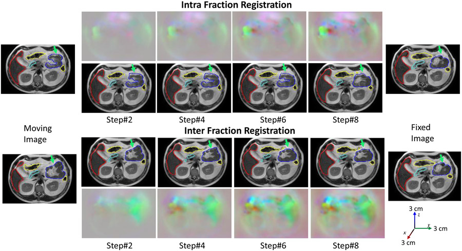
Adaptive radiation treatment (ART) for locally advanced pancreatic cancer (LAPC) requires consistently accurate segmentation of the extremely mobile gastrointestinal (GI) organs at risk (OAR) including the stomach, duodenum, large and small bowel. Also, due to lack of sufficiently accurate and fast deformable image registration (DIR), accumulated dose to the GI OARs is currently only approximated, further limiting the ability to more precisely adapt treatments.
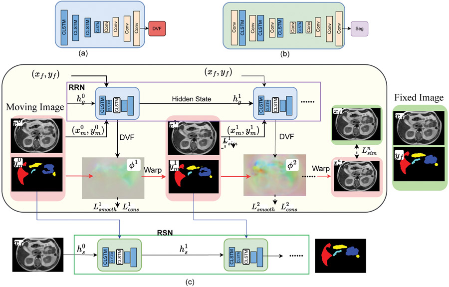
Develop a 3-D Progressively refined joint Registration-Segmentation (ProRSeg) deep network to deformably align and segment treatment fraction magnetic resonance images (MRI)s, then evaluate segmentation accuracy, registration consistency, and feasibility for OAR dose accumulation.
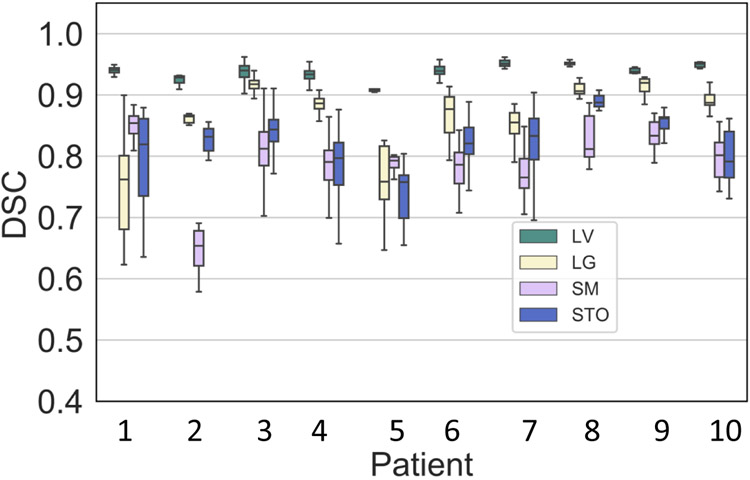
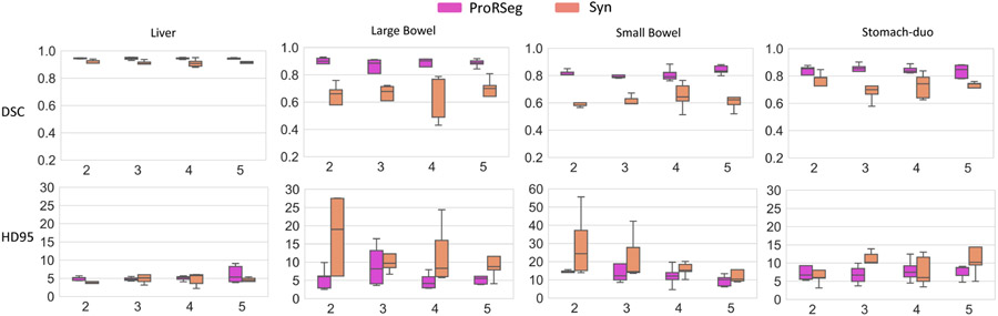
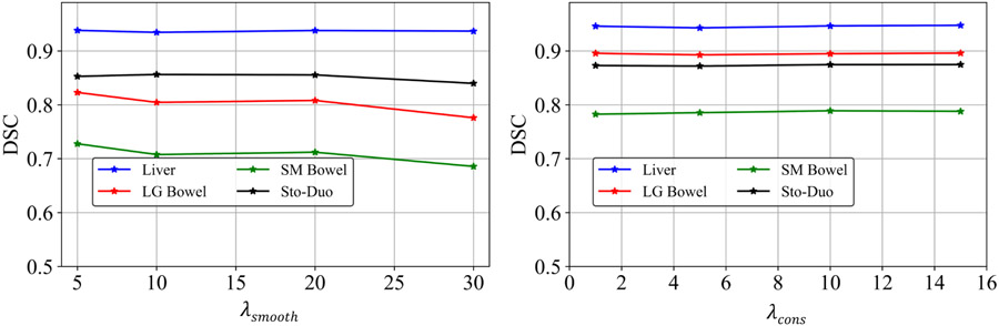
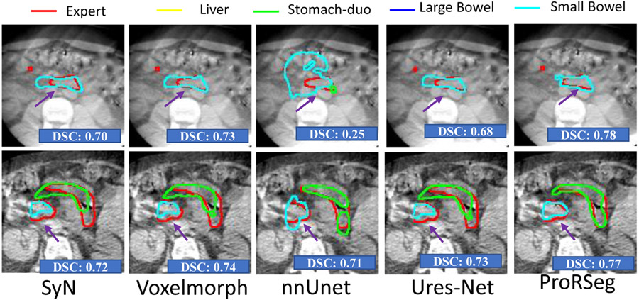
ProRSeg was trained using five-fold cross-validation with 110 T2-weighted MRI acquired at five treatment fractions from 10 different patients, taking care that same patient scans were not placed in training and testing folds. Segmentation accuracy was measured using Dice similarity coefficient (DSC) and Hausdorff distance at 95th percentile (HD95). Registration consistency was measured using coefficient of variation (CV) in displacement of OARs. Statistical comparison to other deep learning and iterative registration methods were done using the Kruskal-Wallis test, followed by pair-wise comparisons with Bonferroni correction applied for multiple testing. Ablation tests and accuracy comparisons against multiple methods were done. Finally, applicability of ProRSeg to segment cone-beam CT (CBCT) scans was evaluated on a publicly available dataset of 80 scans using five-fold cross-validation.
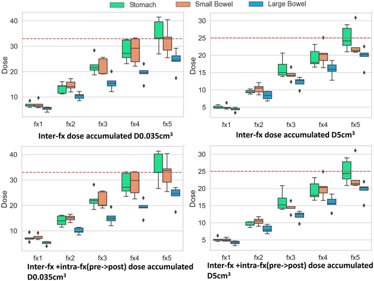
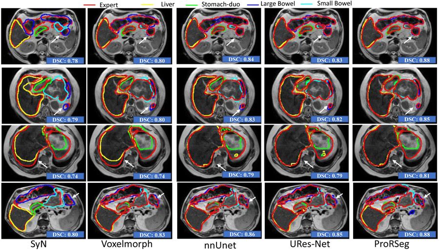
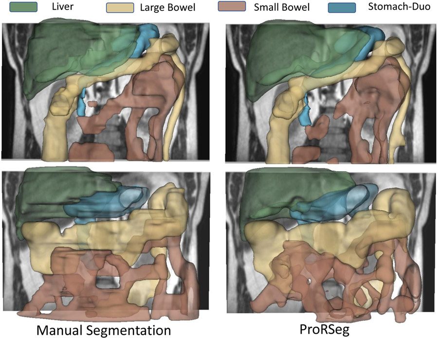
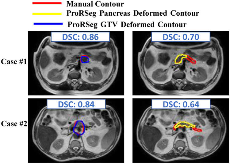
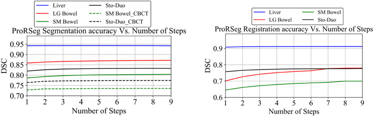
ProRSeg processed 3D volumes (128 × 192 × 128) in 3 s on a NVIDIA Tesla V100 GPU. It's segmentations were significantly more accurate ( ) than compared methods, achieving a DSC of 0.94 ±0.02 for liver, 0.88±0.04 for large bowel, 0.78±0.03 for small bowel and 0.82±0.04 for stomach-duodenum from MRI. ProRSeg achieved a DSC of 0.72±0.01 for small bowel and 0.76±0.03 for stomach-duodenum from public CBCT dataset. ProRSeg registrations resulted in the lowest CV in displacement (stomach-duodenum : 0.75%, : 0.73%, and : 0.81%; small bowel : 0.80%, : 0.80%, and : 0.68%; large bowel : 0.71%, : 0.81%, and : 0.75%). ProRSeg based dose accumulation accounting for intra-fraction (pre-treatment to post-treatment MRI scan) and inter-fraction motion showed that the organ dose constraints were violated in four patients for stomach-duodenum and for three patients for small bowel. Study limitations include lack of independent testing and ground truth phantom datasets to measure dose accumulation accuracy.
ProRSeg produced more accurate and consistent GI OARs segmentation and DIR of MRI and CBCTs compared to multiple methods. Preliminary results indicates feasibility for OAR dose accumulation using ProRSeg.
局部晚期胰腺癌(LAPC)的适应性放疗(ART)需要对包括胃、十二指肠、大小肠在内的极移动胃肠道(GI)危及器官(OAR)进行始终如一地准确分割。此外,由于缺乏足够准确和快速的可变形图像配准(DIR),目前只能对 GI OAR 的累积剂量进行近似计算,这进一步限制了更精确地调整治疗的能力。
开发一种 3D 渐进式精细联合注册-分割(ProRSeg)深度网络,以对治疗分次磁共振成像(MRI)进行可变形配准和分割,然后评估分割准确性、注册一致性以及对 OAR 剂量积累的可行性。
ProRSeg 使用五折交叉验证进行训练,使用来自 10 名不同患者的 5 个治疗分次的 110 个 T2 加权 MRI,特别注意不要将同一患者的扫描放在训练和测试折之间。使用 Dice 相似系数(DSC)和第 95 百分位数的 Hausdorff 距离(HD95)测量分割准确性。使用 OAR 位移的变异系数(CV)测量注册一致性。使用 Kruskal-Wallis 检验对其他深度学习和迭代注册方法进行了统计学比较,然后对 Bonferroni 校正进行了两两比较,以进行多次测试。进行了消融测试和与多种方法的准确性比较。最后,使用五折交叉验证在一个公开的 80 个扫描的数据集上评估了 ProRSeg 对锥形束 CT(CBCT)扫描的分割适用性。
ProRSeg 在 NVIDIA Tesla V100 GPU 上以 3 秒的速度处理 3D 体积(128×192×128)。与其他方法相比,它的分割结果更准确( ),在 MRI 上获得了肝脏的 DSC 为 0.94±0.02,大肠为 0.88±0.04,小肠为 0.78±0.03,胃十二指肠为 0.82±0.04。从公共 CBCT 数据集获得了小肠的 DSC 为 0.72±0.01 和胃十二指肠的 DSC 为 0.76±0.03。ProRSeg 配准的结果是位移 CV 最低(胃十二指肠 :0.75%, :0.73%, :0.81%;小肠 :0.80%, :0.80%, :0.68%;大肠 :0.71%, :0.81%, :0.75%)。基于 ProRSeg 的剂量积累考虑了分次内(治疗前至治疗后 MRI 扫描)和分次间运动,结果表明有四名患者的胃十二指肠和三名患者的小肠违反了器官剂量限制。研究的局限性包括缺乏独立的测试和地面真实体模数据集来测量剂量积累的准确性。
ProRSeg 与多种方法相比,在 MRI 和 CBCT 的 GI OAR 分割和 DIR 方面产生了更准确和一致的结果。初步结果表明,使用 ProRSeg 进行 OAR 剂量积累是可行的。