• 文献检索
  • 文档翻译
  • 深度研究
  • 学术资讯
  • Suppr Zotero 插件Zotero 插件
  • 邀请有礼
  • 套餐&价格
  • 历史记录
应用&插件
Suppr Zotero 插件Zotero 插件浏览器插件Mac 客户端Windows 客户端微信小程序
定价
高级版会员购买积分包购买API积分包
服务
文献检索文档翻译深度研究API 文档MCP 服务
关于我们
关于 Suppr公司介绍联系我们用户协议隐私条款
关注我们

Suppr 超能文献

核心技术专利:CN118964589B侵权必究
粤ICP备2023148730 号-1Suppr @ 2026

文献检索

告别复杂PubMed语法,用中文像聊天一样搜索,搜遍4000万医学文献。AI智能推荐,让科研检索更轻松。

立即免费搜索

文件翻译

保留排版,准确专业,支持PDF/Word/PPT等文件格式,支持 12+语言互译。

免费翻译文档

深度研究

AI帮你快速写综述,25分钟生成高质量综述,智能提取关键信息,辅助科研写作。

立即免费体验

放射治疗中自动危及器官分割时间节省评估措施的评价

Evaluation of measures for assessing time-saving of automatic organ-at-risk segmentation in radiotherapy.

作者信息

Vaassen Femke, Hazelaar Colien, Vaniqui Ana, Gooding Mark, van der Heyden Brent, Canters Richard, van Elmpt Wouter

机构信息

Department of Radiation Oncology (MAASTRO), GROW - School for Oncology and Developmental Biology, Maastricht University Medical Centre, Maastricht, The Netherlands.

Mirada Medical Ltd., Oxford, United Kingdom.

出版信息

Phys Imaging Radiat Oncol. 2019 Dec 17;13:1-6. doi: 10.1016/j.phro.2019.12.001. eCollection 2020 Jan.

DOI:10.1016/j.phro.2019.12.001
PMID:33458300
原文链接:https://pmc.ncbi.nlm.nih.gov/articles/PMC7807544/
Abstract

BACKGROUND AND PURPOSE

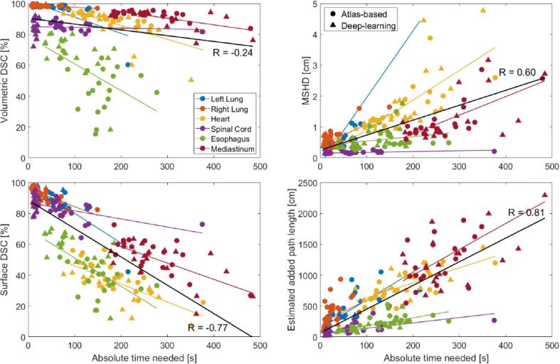
In radiotherapy, automatic organ-at-risk segmentation algorithms allow faster delineation times, but clinically relevant contour evaluation remains challenging. Commonly used measures to assess automatic contours, such as volumetric Dice Similarity Coefficient (DSC) or Hausdorff distance, have shown to be good measures for geometric similarity, but do not always correlate with clinical applicability of the contours, or time needed to adjust them. This study aimed to evaluate the correlation of new and commonly used evaluation measures with time-saving during contouring.

MATERIALS AND METHODS

Twenty lung cancer patients were used to compare user-adjustments after atlas-based and deep-learning contouring with manual contouring. The absolute time needed (s) of adjusting the auto-contour compared to manual contouring was recorded, from this relative time-saving (%) was calculated. New evaluation measures (surface DSC and added path length, APL) and conventional evaluation measures (volumetric DSC and Hausdorff distance) were correlated with time-recordings and time-savings, quantified with the Pearson correlation coefficient, R.

RESULTS

The highest correlation (R = 0.87) was found between APL and absolute adaption time. Lower correlations were found for APL with relative time-saving (R = -0.38), for surface DSC with absolute adaption time (R = -0.69) and relative time-saving (R = 0.57). Volumetric DSC and Hausdorff distance also showed lower correlation coefficients for absolute adaptation time (R = -0.32 and 0.64, respectively) and relative time-saving (R = 0.44 and -0.64, respectively).

CONCLUSION

Surface DSC and APL are better indicators for contour adaptation time and time-saving when using auto-segmentation and provide more clinically relevant and better quantitative measures for automatically-generated contour quality, compared to commonly-used geometry-based measures.

摘要

背景与目的

在放射治疗中,自动危及器官分割算法可缩短轮廓勾画时间,但临床相关的轮廓评估仍具有挑战性。常用的评估自动轮廓的方法,如体积骰子相似系数(DSC)或豪斯多夫距离,已被证明是衡量几何相似性的良好指标,但并不总是与轮廓的临床适用性或调整轮廓所需的时间相关。本研究旨在评估新的和常用的评估方法与轮廓勾画过程中节省时间之间的相关性。

材料与方法

选取20例肺癌患者,比较基于图谱和深度学习的轮廓勾画后用户调整与手动轮廓勾画的情况。记录与手动轮廓勾画相比调整自动轮廓所需的绝对时间(秒),并据此计算相对节省时间(%)。将新的评估方法(表面DSC和增加路径长度,APL)和传统评估方法(体积DSC和豪斯多夫距离)与时间记录和节省时间进行相关性分析,用Pearson相关系数R进行量化。

结果

APL与绝对适应时间之间的相关性最高(R = 0.87)。APL与相对节省时间的相关性较低(R = -0.38),表面DSC与绝对适应时间的相关性较低(R = -0.69),与相对节省时间的相关性为(R = 0.57)。体积DSC和豪斯多夫距离在绝对适应时间(分别为R = -0.32和0.64)和相对节省时间(分别为R = 0.44和 -0.64)方面的相关系数也较低。

结论

与常用的基于几何的方法相比,表面DSC和APL在使用自动分割时是轮廓适应时间和节省时间的更好指标,并且为自动生成的轮廓质量提供了更具临床相关性和更好的定量测量方法。

https://cdn.ncbi.nlm.nih.gov/pmc/blobs/09b8/7807544/8220e4d80e1f/gr5.jpg
https://cdn.ncbi.nlm.nih.gov/pmc/blobs/09b8/7807544/9c231ea80366/gr1.jpg
https://cdn.ncbi.nlm.nih.gov/pmc/blobs/09b8/7807544/5c10aa56dcc5/gr2.jpg
https://cdn.ncbi.nlm.nih.gov/pmc/blobs/09b8/7807544/4a011f3a2cc0/gr3.jpg
https://cdn.ncbi.nlm.nih.gov/pmc/blobs/09b8/7807544/bdca6d18617a/gr4.jpg
https://cdn.ncbi.nlm.nih.gov/pmc/blobs/09b8/7807544/8220e4d80e1f/gr5.jpg

相似文献

1
Evaluation of measures for assessing time-saving of automatic organ-at-risk segmentation in radiotherapy.放射治疗中自动危及器官分割时间节省评估措施的评价
Phys Imaging Radiat Oncol. 2019 Dec 17;13:1-6. doi: 10.1016/j.phro.2019.12.001. eCollection 2020 Jan.
2
Geometric and dosimetric analysis of CT- and MR-based automatic contouring for the EPTN contouring atlas in neuro-oncology.基于 CT 和 MRI 的自动勾画在神经肿瘤 EPTN 勾画图谱中的几何和剂量学分析。
Phys Med. 2023 Oct;114:103156. doi: 10.1016/j.ejmp.2023.103156. Epub 2023 Oct 7.
3
Clinical evaluation on automatic segmentation results of convolutional neural networks in rectal cancer radiotherapy.直肠癌放疗中卷积神经网络自动分割结果的临床评估
Front Oncol. 2023 Sep 5;13:1158315. doi: 10.3389/fonc.2023.1158315. eCollection 2023.
4
Human factors in the clinical implementation of deep learning-based automated contouring of pelvic organs at risk for MRI-guided radiotherapy.基于深度学习的MRI引导放疗中盆腔危及器官自动轮廓勾画临床应用中的人为因素
Med Phys. 2023 Oct;50(10):5969-5977. doi: 10.1002/mp.16676. Epub 2023 Aug 30.
5
A clinical and time savings evaluation of a deep learning automatic contouring algorithm.一种深度学习自动轮廓算法的临床及节省时间评估
Med Dosim. 2023;48(1):55-60. doi: 10.1016/j.meddos.2022.11.001. Epub 2022 Dec 20.
6
A deep learning based automatic segmentation approach for anatomical structures in intensity modulation radiotherapy.基于深度学习的调强放射治疗中解剖结构自动分割方法。
Math Biosci Eng. 2021 Sep 1;18(6):7506-7524. doi: 10.3934/mbe.2021371.
7
The clinical evaluation of atlas-based auto-segmentation for automatic contouring during cervical cancer radiotherapy.基于图谱的自动分割在宫颈癌放疗自动轮廓勾画中的临床评估
Front Oncol. 2022 Aug 2;12:945053. doi: 10.3389/fonc.2022.945053. eCollection 2022.
8
Interactive contouring through contextual deep learning.基于上下文的深度学习的交互式勾画。
Med Phys. 2021 Jun;48(6):2951-2959. doi: 10.1002/mp.14852. Epub 2021 May 3.
9
A clinical evaluation of the performance of five commercial artificial intelligence contouring systems for radiotherapy.五种商用人工智能放疗轮廓勾画系统性能的临床评估
Front Oncol. 2023 Aug 4;13:1213068. doi: 10.3389/fonc.2023.1213068. eCollection 2023.
10
Clinical evaluation of a deep learning segmentation model including manual adjustments afterwards for locally advanced breast cancer.深度学习分割模型在局部晚期乳腺癌中的临床评估,后续包括手动调整。
Tech Innov Patient Support Radiat Oncol. 2023 May 13;26:100211. doi: 10.1016/j.tipsro.2023.100211. eCollection 2023 Jun.

引用本文的文献

1
Implementation and clinical evaluation of an in-house thoracic auto-segmentation model for 0.35 T magnetic resonance imaging guided radiotherapy.用于0.35T磁共振成像引导放疗的内部胸部自动分割模型的实施与临床评估
Phys Imaging Radiat Oncol. 2025 Aug 5;35:100819. doi: 10.1016/j.phro.2025.100819. eCollection 2025 Jul.
2
Impact of deep learning model uncertainty on manual corrections to MRI-based auto-segmentation in prostate cancer radiotherapy.深度学习模型不确定性对前列腺癌放疗中基于MRI的自动分割手动校正的影响。
J Appl Clin Med Phys. 2025 Sep;26(9):e70221. doi: 10.1002/acm2.70221.
3
An interactive deep-learning workflow for head and neck gross tumour volume segmentation.

本文引用的文献

1
Benefits of deep learning for delineation of organs at risk in head and neck cancer.深度学习在头颈部癌症危险器官勾画中的应用优势。
Radiother Oncol. 2019 Sep;138:68-74. doi: 10.1016/j.radonc.2019.05.010. Epub 2019 May 27.
2
Advances in Auto-Segmentation.自动分割技术的进展
Semin Radiat Oncol. 2019 Jul;29(3):185-197. doi: 10.1016/j.semradonc.2019.02.001.
3
Comparative evaluation of autocontouring in clinical practice: A practical method using the Turing test.临床实践中自动勾画的比较评估:一种使用图灵测试的实用方法。
一种用于头颈部大体肿瘤体积分割的交互式深度学习工作流程。
Phys Imaging Radiat Oncol. 2025 Aug 5;35:100820. doi: 10.1016/j.phro.2025.100820. eCollection 2025 Jul.
4
From standardized to individualized margins for online adaptive tumor dose escalation in rectal cancer.从标准化切缘到个体化切缘,用于直肠癌在线自适应肿瘤剂量递增。
Radiat Oncol. 2025 Aug 8;20(1):125. doi: 10.1186/s13014-025-02706-8.
5
The dosimetric impacts of ct-based deep learning autocontouring algorithm for prostate cancer radiotherapy planning dosimetric accuracy of DirectORGANS.基于CT的深度学习自动轮廓算法对前列腺癌放疗计划中DirectORGANS剂量测定准确性的剂量学影响。
BMC Urol. 2025 Aug 2;25(1):190. doi: 10.1186/s12894-025-01875-8.
6
A comprehensive multifaceted technical evaluation framework for implementation of auto-segmentation models in radiotherapy.放疗中自动分割模型实施的综合多方面技术评估框架。
Commun Med (Lond). 2025 Jul 31;5(1):319. doi: 10.1038/s43856-025-01048-6.
7
Challenges and opportunities to integrate artificial intelligence in radiation oncology: a narrative review.将人工智能整合到放射肿瘤学中的挑战与机遇:一篇叙述性综述
Ewha Med J. 2024 Oct;47(4):e49. doi: 10.12771/emj.2024.e49. Epub 2024 Oct 31.
8
Evaluation of deformable image registration accuracy for liver re-irradiation patients using contrast and non-contrast computed tomography images.使用对比增强和非对比增强计算机断层扫描图像评估肝脏再照射患者的可变形图像配准准确性。
Med Phys. 2025 Jul;52(7):e17942. doi: 10.1002/mp.17942.
9
Leveraging network uncertainty to identify regions in rectal cancer clinical target volume auto-segmentations likely requiring manual edits.利用网络不确定性来识别直肠癌临床靶区自动分割中可能需要手动编辑的区域。
Phys Imaging Radiat Oncol. 2025 May 8;34:100771. doi: 10.1016/j.phro.2025.100771. eCollection 2025 Apr.
10
Deep Learning for Automated Ventricle and Periventricular Space Segmentation on CT and T1CE MRI in Neuro-Oncology Patients.深度学习用于神经肿瘤患者CT和T1CE MRI上的脑室及脑室周围间隙自动分割
Cancers (Basel). 2025 May 8;17(10):1598. doi: 10.3390/cancers17101598.
Med Phys. 2018 Nov;45(11):5105-5115. doi: 10.1002/mp.13200. Epub 2018 Oct 12.
4
Autosegmentation for thoracic radiation treatment planning: A grand challenge at AAPM 2017.自动分割在胸部放射治疗计划中的应用:2017 年 AAPM 的重大挑战。
Med Phys. 2018 Oct;45(10):4568-4581. doi: 10.1002/mp.13141. Epub 2018 Sep 19.
5
Clinical evaluation of atlas and deep learning based automatic contouring for lung cancer.基于 atlas 和深度学习的肺癌自动勾画的临床评估。
Radiother Oncol. 2018 Feb;126(2):312-317. doi: 10.1016/j.radonc.2017.11.012. Epub 2017 Dec 5.
6
Uncertainties in volume delineation in radiation oncology: A systematic review and recommendations for future studies.放射肿瘤学中体积勾画的不确定性:系统评价及对未来研究的建议。
Radiother Oncol. 2016 Nov;121(2):169-179. doi: 10.1016/j.radonc.2016.09.009. Epub 2016 Oct 8.
7
Use of auto-segmentation in the delineation of target volumes and organs at risk in head and neck.自动分割技术在头颈部靶区和危及器官勾画中的应用。
Acta Oncol. 2016 Jul;55(7):799-806. doi: 10.3109/0284186X.2016.1173723. Epub 2016 Jun 1.
8
Metrics for evaluating 3D medical image segmentation: analysis, selection, and tool.用于评估3D医学图像分割的指标:分析、选择与工具
BMC Med Imaging. 2015 Aug 12;15:29. doi: 10.1186/s12880-015-0068-x.
9
Vision 20/20: perspectives on automated image segmentation for radiotherapy.《视力20/20:放射治疗自动图像分割的前景》
Med Phys. 2014 May;41(5):050902. doi: 10.1118/1.4871620.
10
Comparison of manual and automatic segmentation methods for brain structures in the presence of space-occupying lesions: a multi-expert study.存在占位性病变时脑结构的手动和自动分割方法的比较:多专家研究。
Phys Med Biol. 2011 Jul 21;56(14):4557-77. doi: 10.1088/0031-9155/56/14/021. Epub 2011 Jul 1.