Zhou Min, Zhang Yadi, Tang Rui, Liu Haiyan, Du Min, Gao Zhi, Ji Zongshu, Fang Haoshu
Neurocritical Care Unit, The First Affiliated Hospital of USTC, Division of Life Sciences and Medicine, University of Science and Technology of China, Hefei, Anhui, 230001, People's Republic of China.
Department of Respiratory Medicine, The Second People's Hospital of Hefei and Hefei Hospital Affiliated with Anhui Medical University, Hefei, Anhui, 230011, People's Republic of China.
J Inflamm Res. 2021 Apr 19;14:1551-1561. doi: 10.2147/JIR.S302967. eCollection 2021.
High-mobility group box-1 protein (HMGB1) serves as the prototypic damage-associated molecular pattern molecule, and TLR4 is considered a receptor for HMGB1. Regulatory T cells (Tregs) play a crucial role in infectious diseases. The role of HMGB1 in the modulation of Tregs is of great interest.
Serum HMGB1 and Treg proportions were detected in 58 patients with acute lung injury (ALI) and 36 healthy volunteers. The correlations of these parameters with disease severity were analyzed. The WT and TLR4 mice were administered HMGB1 by intratracheal injection. After 48 h, the mice were sacrificed. The morphological changes and wet/dry ratio of the lung were measured. Spleen CD4CD25 Tregs were sorted from spleen cells, the expression of FOXP3 and CTLA-4, and releasing of cytokines was detected. CD4CD25 Tregs were cocultured with effector T cells, the inhibitory effect, and release of cytokines was detected.
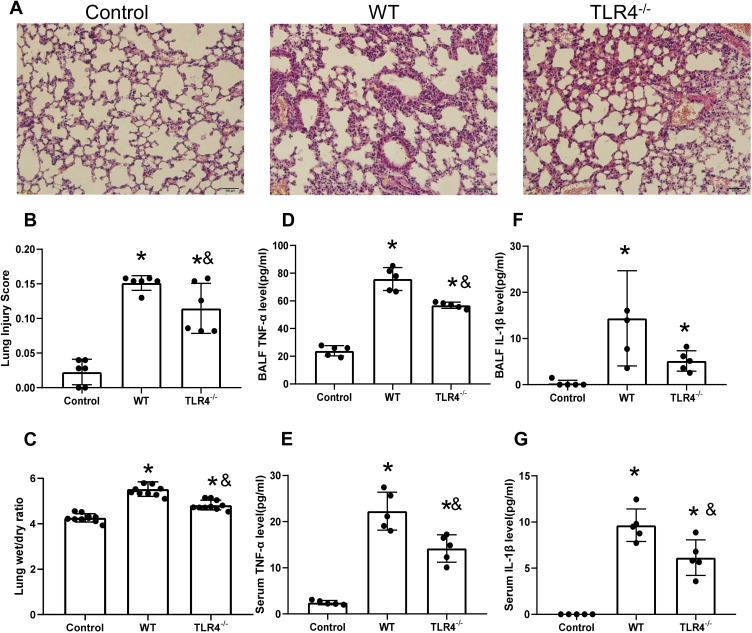
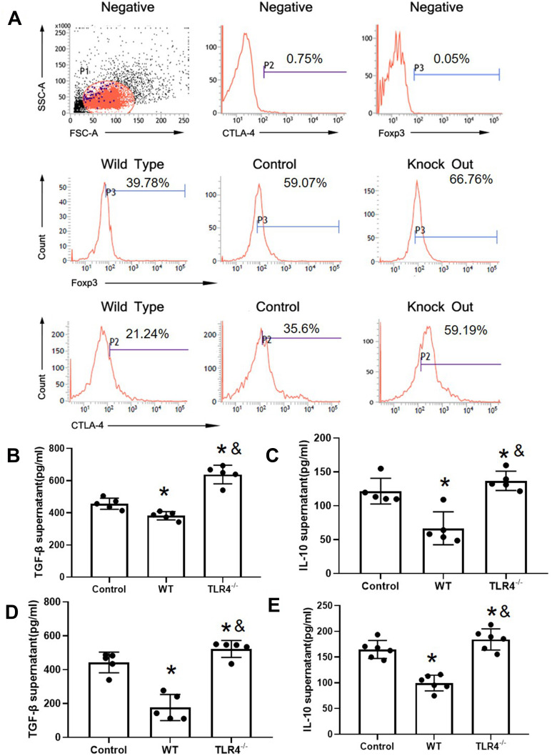
Significantly increased plasma levels of HMGB1 and reduced CD4CD25CD127 Tregs were detected in ALI patients. In the mouse model, lung injury was significantly increased after HMGB1 instillation in the WT and TLR4 groups compared with control group. The lung wet/dry ratio and the TNF-α and IL-1β contents in BALF were significantly increased, and the severity of WT mice was higher than that of TLR4 mice. The expression of FOXP3 and CTLA-4 in TLR4 mice was significantly increased compared with that in WT mice and was associated with a similar trend of IL-10 and TGF-β levels (p<0.05). In coculture with effector T cells, Tregs isolated from TLR4 mice exhibited decreased IL-2 and IFN-γ and increased IL-4 levels compared with Tregs from WT mice. Increased polarization of TLR4 CD4CD25 Treg cells to Th2 cells was observed.
In HMGB1-induced lung injury, HMGB1 affects the expression of FOXP3 and CTLA-4 through TLR4, thus reducing the immunosuppressive function of Treg cells.
高迁移率族蛋白B1(HMGB1)作为典型的损伤相关分子模式分子,Toll样受体4(TLR4)被认为是HMGB1的受体。调节性T细胞(Tregs)在感染性疾病中起关键作用。HMGB1在调节Tregs中的作用备受关注。
检测58例急性肺损伤(ALI)患者和36名健康志愿者血清中的HMGB1及Tregs比例。分析这些参数与疾病严重程度的相关性。通过气管内注射对野生型(WT)和TLR4基因敲除小鼠给予HMGB1。48小时后处死小鼠,测量肺组织形态学变化及肺组织湿/干比。从脾细胞中分离脾CD4+CD25+调节性T细胞,检测叉头框蛋白3(FOXP3)和细胞毒性T淋巴细胞相关抗原4(CTLA-4)的表达及细胞因子释放情况。将CD4+CD25+调节性T细胞与效应T细胞共培养,检测其抑制作用及细胞因子释放情况。
ALI患者血浆中HMGB1水平显著升高,CD4+CD25+CD127-Tregs减少。在小鼠模型中,与对照组相比,WT组和TLR4基因敲除组经HMGB1滴注后肺损伤显著增加。肺组织湿/干比及支气管肺泡灌洗液(BALF)中肿瘤坏死因子-α(TNF-α)和白细胞介素-1β(IL-1β)含量显著升高,WT小鼠的严重程度高于TLR4基因敲除小鼠。与WT小鼠相比,TLR4基因敲除小鼠中FOXP3和CTLA-4的表达显著增加,且白细胞介素-10(IL-10)和转化生长因子-β(TGF-β)水平呈现相似趋势(p<0.05)。与效应T细胞共培养时,与WT小鼠来源的Tregs相比,TLR4基因敲除小鼠来源的Tregs中白细胞介素-2(IL-2)和干扰素-γ(IFN-γ)水平降低,白细胞介素-4(IL-4)水平升高。观察到TLR4+CD4+CD25+调节性T细胞向Th2细胞的极化增加。
在HMGB1诱导的肺损伤中,HMGB1通过TLR4影响FOXP3和CTLA-4的表达,从而降低调节性T细胞的免疫抑制功能。