• 文献检索
  • 文档翻译
  • 深度研究
  • 学术资讯
  • Suppr Zotero 插件Zotero 插件
  • 邀请有礼
  • 套餐&价格
  • 历史记录
应用&插件
Suppr Zotero 插件Zotero 插件浏览器插件Mac 客户端Windows 客户端微信小程序
定价
高级版会员购买积分包购买API积分包
服务
文献检索文档翻译深度研究API 文档MCP 服务
关于我们
关于 Suppr公司介绍联系我们用户协议隐私条款
关注我们

Suppr 超能文献

核心技术专利:CN118964589B侵权必究
粤ICP备2023148730 号-1Suppr @ 2026

文献检索

告别复杂PubMed语法,用中文像聊天一样搜索,搜遍4000万医学文献。AI智能推荐,让科研检索更轻松。

立即免费搜索

文件翻译

保留排版,准确专业,支持PDF/Word/PPT等文件格式,支持 12+语言互译。

免费翻译文档

深度研究

AI帮你快速写综述,25分钟生成高质量综述,智能提取关键信息,辅助科研写作。

立即免费体验

腹水来源的细胞外囊泡促进卵巢癌细胞的生长和迁移。

Extracellular vesicles derived from ascitic fluid enhance growth and migration of ovarian cancer cells.

机构信息

Department of Radiation Oncology, The University of Texas MD Anderson Cancer Center, 1515 Holcombe Boulevard, Houston, TX, 77030, USA.

Department of Gynecologic Oncology and Reproductive Medicine, The University of Texas MD Anderson Cancer Center, Houston, TX, 77030, USA.

出版信息

Sci Rep. 2021 Apr 28;11(1):9149. doi: 10.1038/s41598-021-88163-1.

DOI:10.1038/s41598-021-88163-1
PMID:33911091
原文链接:https://pmc.ncbi.nlm.nih.gov/articles/PMC8080718/
Abstract

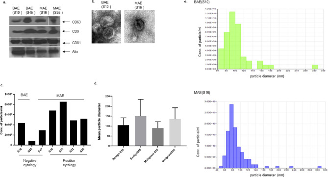
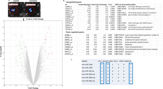
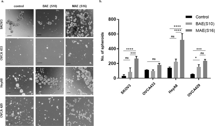
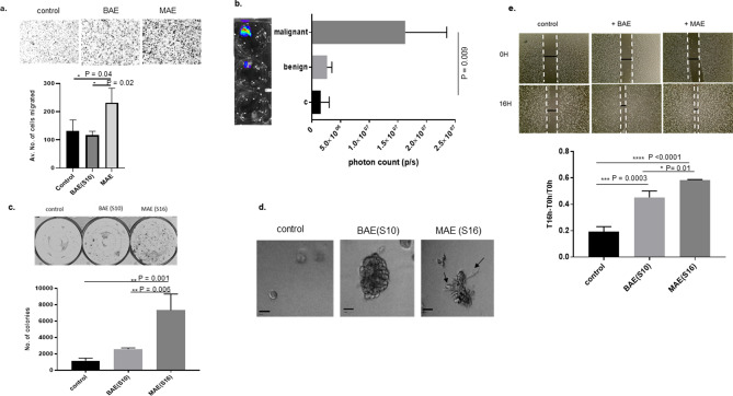
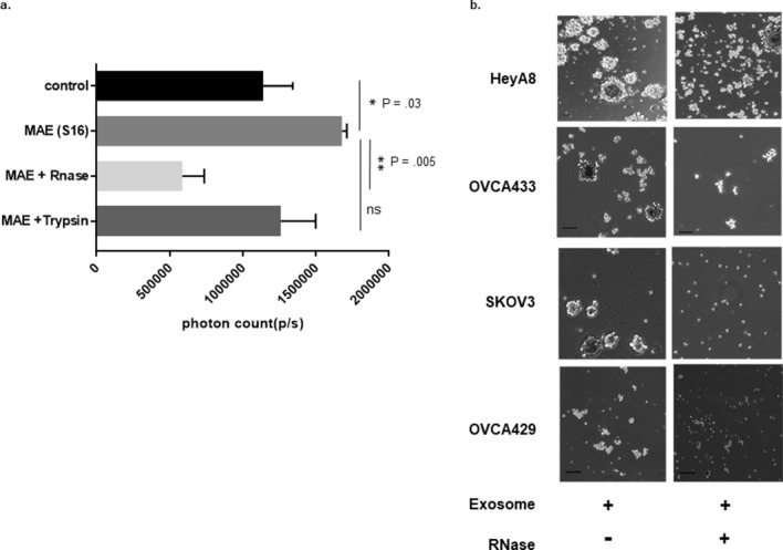
Ovarian cancer is associated with a high mortality rate due to diagnosis at advanced stages. Dissemination often occurs intraperitoneally within the ascites fluid. The microenvironment can support dissemination through several mechanisms. One potential ascites factor which may mediate dissemination are EVs or extracellular vesicles that can carry information in the form of miRNAs, proteins, lipids, and act as mediators of cellular communication. We present our observations on EVs isolated from ascitic supernatants from patients diagnosed with high grade serous ovarian carcinoma in augmenting motility, growth, and migration towards omental fat. MicroRNA profiling of EVs from malignant ascitic supernatant demonstrates high expression of miR 200c-3p, miR18a-5p, miR1246, and miR1290 and low expression of miR 100- 5p as compared to EVs isolated from benign ascitic supernatant. The migration of ovarian cancer spheroids towards omental fat is enhanced in the presence of malignant ascitic EVs. Gene expression of these cells showed increased expression of ZBED2, ZBTB20, ABCC3, UHMK1, and low expression of Transgelin and MARCKS. We present evidence that ovarian ascitic EVs increase the growth of ovarian cancer spheroids through miRNAs.

摘要

卵巢癌由于在晚期诊断,死亡率较高。扩散通常在腹腔内的腹水液中发生。微环境可以通过几种机制来支持扩散。一种潜在的腹水因子可能通过外泌体(EVs)或细胞外囊泡来介导扩散,这些囊泡可以携带 miRNA、蛋白质、脂质等形式的信息,并作为细胞间通讯的介质。我们展示了从高级别浆液性卵巢癌患者腹水中分离出的 EVs 的观察结果,这些 EVs 增强了向大网膜脂肪的运动、生长和迁移。与从良性腹水分离的 EVs 相比,恶性腹水 EVs 中的 microRNA 谱显示 miR 200c-3p、miR18a-5p、miR1246 和 miR1290 的表达较高,而 miR 100-5p 的表达较低。在恶性腹水 EVs 的存在下,卵巢癌细胞球体向大网膜脂肪的迁移增强。这些细胞的基因表达显示 ZBED2、ZBTB20、ABCC3、UHMK1 的表达增加,而 Transgelin 和 MARCKS 的表达降低。我们提出的证据表明,卵巢腹水 EVs 通过 miRNAs 增加了卵巢癌细胞球体的生长。

https://cdn.ncbi.nlm.nih.gov/pmc/blobs/1248/8080718/1940a74f1933/41598_2021_88163_Fig6_HTML.jpg
https://cdn.ncbi.nlm.nih.gov/pmc/blobs/1248/8080718/cdcf44965958/41598_2021_88163_Fig1_HTML.jpg
https://cdn.ncbi.nlm.nih.gov/pmc/blobs/1248/8080718/b5729e790c5a/41598_2021_88163_Fig2_HTML.jpg
https://cdn.ncbi.nlm.nih.gov/pmc/blobs/1248/8080718/3654562e044f/41598_2021_88163_Fig3_HTML.jpg
https://cdn.ncbi.nlm.nih.gov/pmc/blobs/1248/8080718/cd07d6a48d16/41598_2021_88163_Fig4_HTML.jpg
https://cdn.ncbi.nlm.nih.gov/pmc/blobs/1248/8080718/52a1d87f00af/41598_2021_88163_Fig5_HTML.jpg
https://cdn.ncbi.nlm.nih.gov/pmc/blobs/1248/8080718/1940a74f1933/41598_2021_88163_Fig6_HTML.jpg

相似文献

1
Extracellular vesicles derived from ascitic fluid enhance growth and migration of ovarian cancer cells.腹水来源的细胞外囊泡促进卵巢癌细胞的生长和迁移。
Sci Rep. 2021 Apr 28;11(1):9149. doi: 10.1038/s41598-021-88163-1.
2
Biologically Active Tissue Factor-Bearing Larger Ectosome-Like Extracellular Vesicles in Malignant Effusions from Ovarian Cancer Patients: Correlation with Incidence of Thrombosis.来源于卵巢癌患者恶性胸水中的具有生物活性的组织因子载体较大类外囊泡样细胞外囊泡:与血栓形成发生率的相关性。
Int J Mol Sci. 2021 Jan 14;22(2):790. doi: 10.3390/ijms22020790.
3
Comparison of benign peritoneal fluid- and ovarian cancer ascites-derived extracellular vesicle RNA biomarkers.良性腹腔液和卵巢癌腹水来源的细胞外囊泡 RNA 标志物的比较。
J Ovarian Res. 2018 Mar 2;11(1):20. doi: 10.1186/s13048-018-0391-2.
4
miR-18a-5p derived from mesenchymal stem cells-extracellular vesicles inhibits ovarian cancer cell proliferation, migration, invasion, and chemotherapy resistance.间充质干细胞来源的外泌体 miR-18a-5p 抑制卵巢癌细胞增殖、迁移、侵袭和化疗耐药性。
J Transl Med. 2022 Jun 7;20(1):258. doi: 10.1186/s12967-022-03422-7.
5
Vascular endothelial growth factor activating matrix metalloproteinase in ascitic fluid during peritoneal dissemination of ovarian cancer.血管内皮生长因子在卵巢癌腹膜播散过程中激活腹水中的基质金属蛋白酶。
Oncol Rep. 2003 Jan-Feb;10(1):89-95.
6
miR-106a-5p carried by tumor-derived extracellular vesicles promotes the invasion and metastasis of ovarian cancer by targeting KLF6.肿瘤来源的细胞外囊泡携带的 miR-106a-5p 通过靶向 KLF6 促进卵巢癌的侵袭和转移。
Clin Exp Metastasis. 2022 Aug;39(4):603-621. doi: 10.1007/s10585-022-10165-8. Epub 2022 Apr 21.
7
Proteomic analysis of ascitic extracellular vesicles describes tumour microenvironment and predicts patient survival in ovarian cancer.腹水细胞外囊泡的蛋白质组学分析描述了卵巢癌中的肿瘤微环境,并预测了患者的生存情况。
J Extracell Vesicles. 2024 Mar;13(3):e12420. doi: 10.1002/jev2.12420.
8
Integrated analysis of ascites and plasma extracellular vesicles identifies a miRNA-based diagnostic signature in ovarian cancer.腹水和血浆细胞外囊泡的综合分析为卵巢癌建立基于 miRNA 的诊断特征。
Cancer Lett. 2022 Aug 28;542:215735. doi: 10.1016/j.canlet.2022.215735. Epub 2022 May 13.
9
Extracellular vesicle-packaged miR-181c-5p from epithelial ovarian cancer cells promotes M2 polarization of tumor-associated macrophages via the KAT2B/HOXA10 axis.上皮性卵巢癌细胞来源的细胞外囊泡包裹的 miR-181c-5p 通过 KAT2B/HOXA10 轴促进肿瘤相关巨噬细胞向 M2 极化。
J Gene Med. 2022 Oct;24(10):e3446. doi: 10.1002/jgm.3446. Epub 2022 Sep 19.
10
Tumor-derived small extracellular vesicles facilitate omental metastasis of ovarian cancer by triggering activation of mesenchymal stem cells.肿瘤来源的小细胞外囊泡通过触发间充质干细胞的激活促进卵巢癌的大网膜转移。
Cell Commun Signal. 2024 Jan 17;22(1):47. doi: 10.1186/s12964-023-01413-9.

引用本文的文献

1
Nanoparticle Strategies for Treating CNS Disorders: A Comprehensive Review of Drug Delivery and Theranostic Applications.治疗中枢神经系统疾病的纳米颗粒策略:药物递送与诊疗应用综述
Int J Mol Sci. 2024 Dec 11;25(24):13302. doi: 10.3390/ijms252413302.
2
DNA Content in Embryonic Extracellular Vesicles Is Independent of the Apoptotic Rate in Bovine Embryos Produced In Vitro.体外生产的牛胚胎中,胚胎细胞外囊泡中的DNA含量与细胞凋亡率无关。
Animals (Basel). 2024 Mar 29;14(7):1041. doi: 10.3390/ani14071041.
3
Proteomic analysis of ascitic extracellular vesicles describes tumour microenvironment and predicts patient survival in ovarian cancer.

本文引用的文献

1
Copper-64 Labeled PEGylated Exosomes for In Vivo Positron Emission Tomography and Enhanced Tumor Retention.铜-64 标记的聚乙二醇化外泌体用于体内正电子发射断层扫描和增强肿瘤保留。
Bioconjug Chem. 2019 Oct 16;30(10):2675-2683. doi: 10.1021/acs.bioconjchem.9b00587. Epub 2019 Sep 27.
2
YAP-dependent induction of UHMK1 supports nuclear enrichment of the oncogene MYBL2 and proliferation in liver cancer cells.YAP 依赖性诱导 UHMK1 支持肝癌细胞中癌基因 MYBL2 的核富集和增殖。
Oncogene. 2019 Jul;38(27):5541-5550. doi: 10.1038/s41388-019-0801-y. Epub 2019 Apr 1.
3
Delta-like 1 homologue promotes tumorigenesis and epithelial-mesenchymal transition of ovarian high-grade serous carcinoma through activation of Notch signaling.
腹水细胞外囊泡的蛋白质组学分析描述了卵巢癌中的肿瘤微环境,并预测了患者的生存情况。
J Extracell Vesicles. 2024 Mar;13(3):e12420. doi: 10.1002/jev2.12420.
4
Zinc Finger and BTB Domain-Containing 20: A Newly Emerging Player in Pathogenesis and Development of Human Cancers.锌指和 BTB 结构域蛋白 20:人类癌症发病机制和发展中的新出现的参与者。
Biomolecules. 2024 Feb 4;14(2):192. doi: 10.3390/biom14020192.
5
MicroRNAs Present in Malignant Pleural Fluid Increase the Migration of Normal Mesothelial Cells In Vitro and May Help Discriminate between Benign and Malignant Effusions.恶性胸腔积液中的 microRNAs 增加正常间皮细胞的体外迁移,可能有助于区分良性和恶性胸腔积液。
Int J Mol Sci. 2023 Sep 13;24(18):14022. doi: 10.3390/ijms241814022.
6
is a New Prognostic Biomarker in Hepatocellular Carcinoma.是肝细胞癌中的一种新型预后生物标志物。
Int J Gen Med. 2023 May 1;16:1603-1619. doi: 10.2147/IJGM.S408651. eCollection 2023.
7
Bioengineered exosomal-membrane-camouflaged abiotic nanocarriers: neurodegenerative diseases, tissue engineering and regenerative medicine.生物工程外泌体膜伪装的非生物纳米载体:神经退行性疾病、组织工程和再生医学。
Mil Med Res. 2023 Apr 27;10(1):19. doi: 10.1186/s40779-023-00453-z.
8
Circulating Biomarkers for Cancer Detection: Could Salivary microRNAs Be an Opportunity for Ovarian Cancer Diagnostics?用于癌症检测的循环生物标志物:唾液微小RNA能否成为卵巢癌诊断的契机?
Biomedicines. 2023 Feb 21;11(3):652. doi: 10.3390/biomedicines11030652.
9
Obese Mouse Fat Cell-derived Extracellular Vesicles Transport miR-99a-5p to Mitigate the Proliferation and Migration of Non-small Cell Lung Cancer Cells.肥胖小鼠脂肪细胞来源的细胞外囊泡运输 miR-99a-5p 以减轻非小细胞肺癌细胞的增殖和迁移。
Comb Chem High Throughput Screen. 2024;27(2):214-226. doi: 10.2174/1386207326666230316103604.
10
UHMK1 promotes lung adenocarcinoma oncogenesis by regulating the PI3K/AKT/mTOR signaling pathway.UHMK1 通过调控 PI3K/AKT/mTOR 信号通路促进肺腺癌的发生发展。
Thorac Cancer. 2023 Apr;14(12):1077-1088. doi: 10.1111/1759-7714.14850. Epub 2023 Mar 15.
Delta 样配体 1 同源物通过激活 Notch 信号通路促进卵巢高级别浆液性癌的肿瘤发生和上皮-间充质转化。
Oncogene. 2019 Apr;38(17):3201-3215. doi: 10.1038/s41388-018-0658-5. Epub 2019 Jan 9.
4
Crosstalk between YAP/TAZ and Notch Signaling.YAP/TAZ 与 Notch 信号的串扰。
Trends Cell Biol. 2018 Jul;28(7):560-573. doi: 10.1016/j.tcb.2018.03.001. Epub 2018 Apr 14.
5
Modulation of Hepatic MRP3/ABCC3 by Xenobiotics and Pathophysiological Conditions: Role in Drug Pharmacokinetics.外源化合物和病理生理条件对肝 MRP3/ABCC3 的调节:在药物药代动力学中的作用。
Curr Med Chem. 2019;26(7):1185-1223. doi: 10.2174/0929867325666180221142315.
6
Detachment from the primary site and suspension in ascites as the initial step in metabolic reprogramming and metastasis to the omentum in ovarian cancer.脱离原发部位并悬浮于腹水中,这是卵巢癌代谢重编程及转移至大网膜过程中的起始步骤。
Oncol Lett. 2018 Jan;15(1):1357-1361. doi: 10.3892/ol.2017.7388. Epub 2017 Nov 9.
7
Myristoylated alanine-rich C kinase substrate (MARCKS): a multirole signaling protein in cancers.豆蔻酰化丙氨酸丰富的蛋白激酶 C 底物(MARCKS):癌症中的多功能信号蛋白。
Cancer Metastasis Rev. 2017 Dec;36(4):737-747. doi: 10.1007/s10555-017-9709-6.
8
Exosomal microRNA miR-1246 induces cell motility and invasion through the regulation of DENND2D in oral squamous cell carcinoma.外泌体 microRNA miR-1246 通过调控口腔鳞状细胞癌中的 DENND2D 诱导细胞迁移和侵袭。
Sci Rep. 2016 Dec 8;6:38750. doi: 10.1038/srep38750.
9
Human adipose mesenchymal stem cell-derived exosomal-miRNAs are critical factors for inducing anti-proliferation signalling to A2780 and SKOV-3 ovarian cancer cells.人脂肪间充质干细胞来源的外泌体 miRNA 是诱导 A2780 和 SKOV-3 卵巢癌细胞抗增殖信号的关键因素。
Sci Rep. 2016 Dec 8;6:38498. doi: 10.1038/srep38498.
10
Enrichment of selective miRNAs in exosomes and delivery of exosomal miRNAs in vitro and in vivo.外泌体中选择性miRNA的富集以及外泌体miRNA在体内外的递送
Am J Physiol Lung Cell Mol Physiol. 2017 Jan 1;312(1):L110-L121. doi: 10.1152/ajplung.00423.2016. Epub 2016 Nov 23.