Department of Obstetrics and Gynecology, Shengjing Hospital of China Medical University, 36 Sanhao Street, Heping District, Shenyang, 110004, Liaoning, China.
J Transl Med. 2022 Jun 7;20(1):258. doi: 10.1186/s12967-022-03422-7.
Ovarian cancer (OC) is a major threat to women's health. Mesenchymal stem cells (MSCs) are key regulators in cellular communication by secreting extracellular vesicles (EVs) that are involved in OC. This study probed into the mechanism of human MSCs derived-EVs (hMSC-EVs) in regulating OC cell growth and chemotherapy resistance.
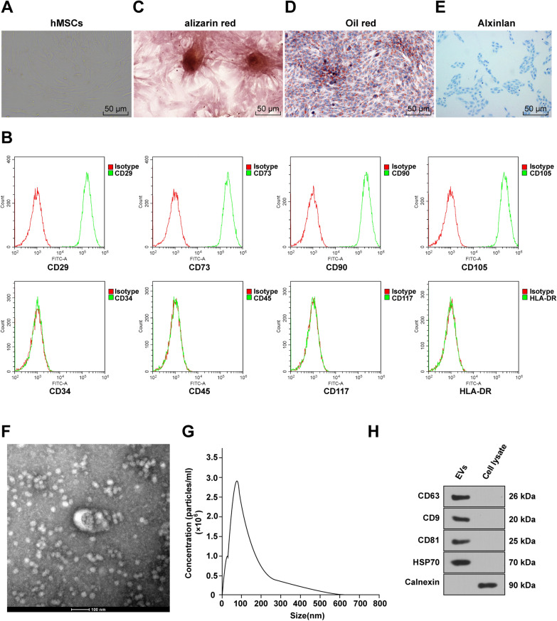
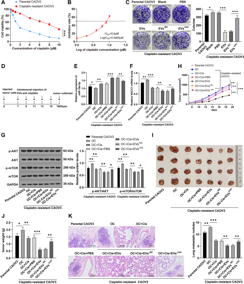
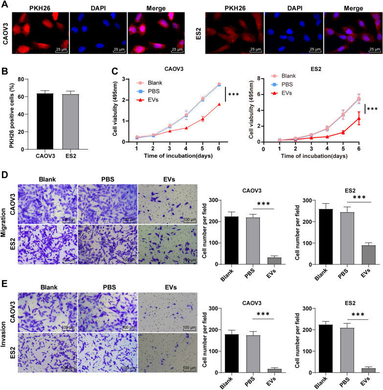
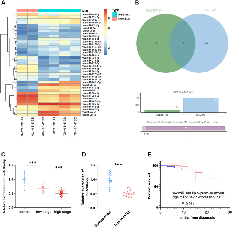
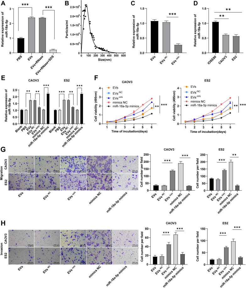
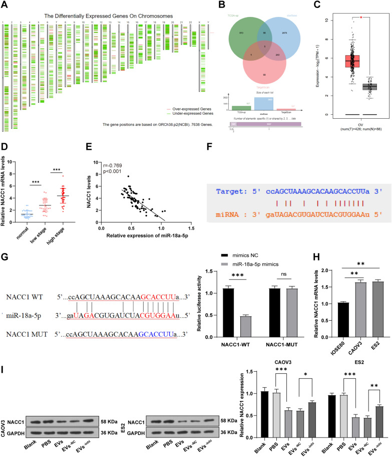
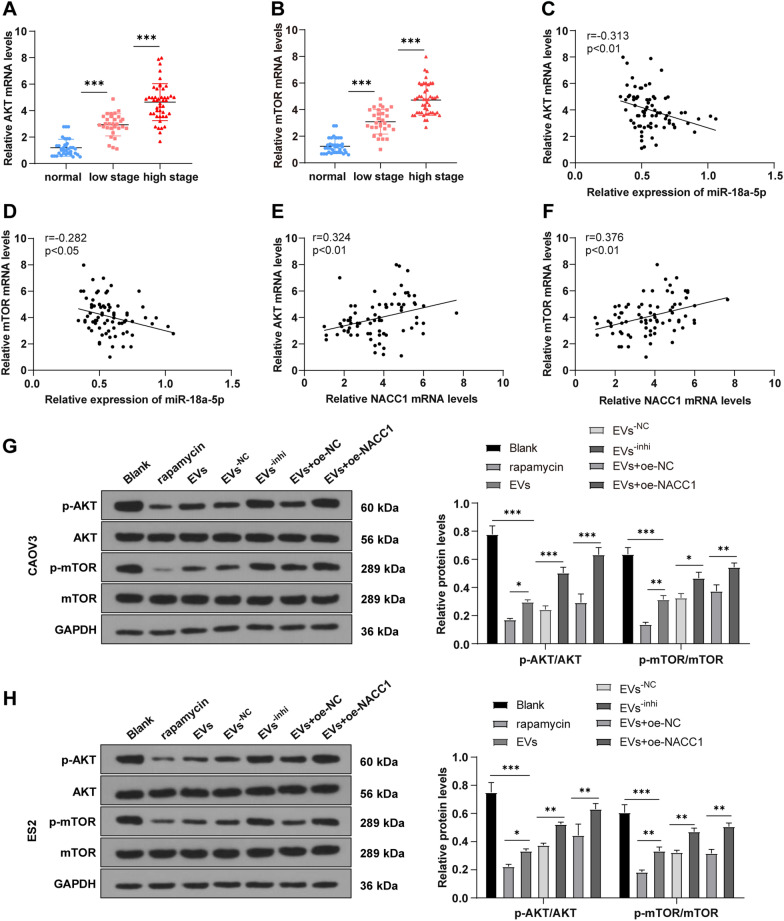
hMSCs and EVs were isolated and identified. After adding EVs, the uptake of EVs by OC CAOV3/ES2 cells (for in vitro studies), and cell proliferation, migration, and invasion were detected. Downregulated miRNAs in hMSC-EVs were screened and miR-18a-5p expression in OC patients was detected. The prognosis of OC patients was analyzed. Binding sites of miR-18a-5p and NACC1 were predicted and validated. NACC1 expression in OC tissues was measured by RT-qPCR, and its correlation with miR-18a-5p was analyzed by Pearson method. AKT/mTOR pathway activation was assessed by WB. The cisplatin sensitivity of EVs-treated CAOV3 cells was evaluated via MTT assay and tested by tumor formation assay in nude mice.
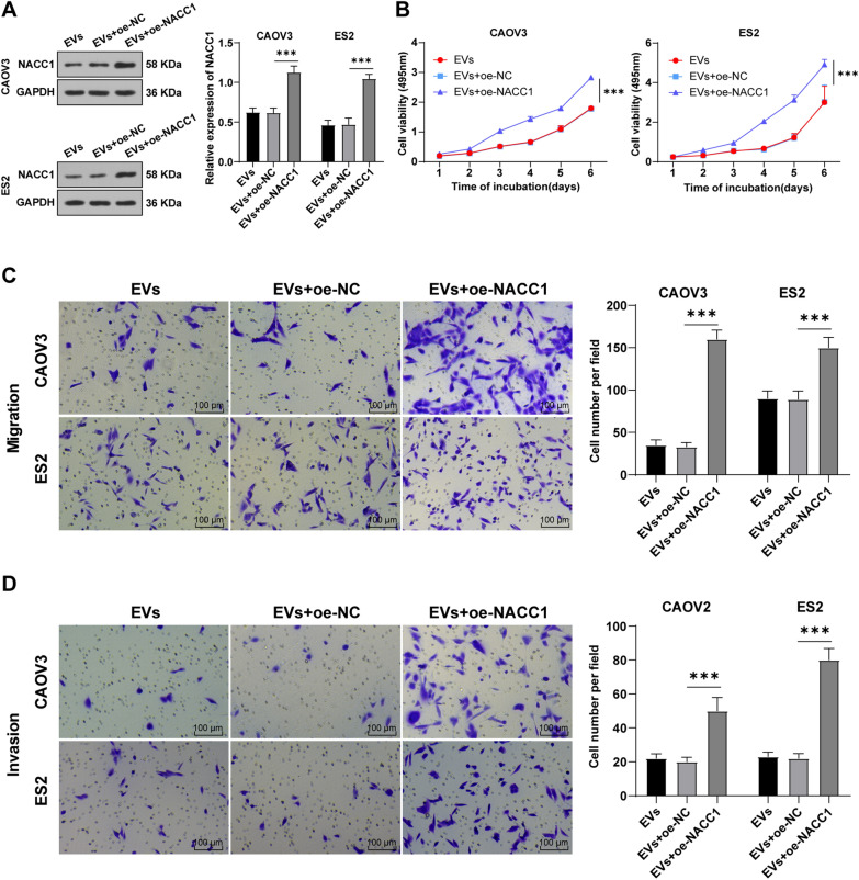
hMSC-EVs suppressed OC cell proliferation, migration, and invasion. miR-18a-5p was downregulated in OC and miR-18a-5p low expression was associated with a poor prognosis. EV-encapsulated miR-18a-5p targeted NACC1. NACC1 was upregulated in OC tissues. miR-18a-5p knockdown and NACC1 overexpression both annulled the inhibition of hMSC-EVs on OC cell growth. AKT and mTOR were elevated in OC and NACC1 activated the AKT/mTOR pathway in OC cells. hMSC-EVs promoted cisplatin sensitivity of OC cells by carrying miR-18a-5p.
hMSC-EVs-derived miR-18a-5p inhibits OC cell proliferation, migration, invasion, and chemotherapy resistance.
卵巢癌(OC)严重威胁女性健康。间充质干细胞(MSCs)通过分泌细胞外囊泡(EVs)在细胞通讯中发挥关键调节作用,这些 EVs 参与 OC 的发生。本研究旨在探讨人源 MSC 衍生的 EVs(hMSC-EVs)调节 OC 细胞生长和化疗耐药的机制。
分离并鉴定 hMSC 和 EVs。在添加 EVs 后,通过 OC CAOV3/ES2 细胞摄取 EVs(用于体外研究),并检测细胞增殖、迁移和侵袭。筛选 hMSC-EVs 中下调的 miRNAs,并检测 OC 患者中 miR-18a-5p 的表达。分析 OC 患者的预后。预测和验证 miR-18a-5p 和 NACC1 的结合位点。通过 RT-qPCR 测量 OC 组织中的 NACC1 表达,并通过 Pearson 方法分析其与 miR-18a-5p 的相关性。通过 WB 评估 AKT/mTOR 通路的激活。通过 MTT 测定评估 EV 处理的 CAOV3 细胞的顺铂敏感性,并通过裸鼠肿瘤形成试验进行测试。
hMSC-EVs 抑制 OC 细胞增殖、迁移和侵袭。miR-18a-5p 在 OC 中下调,miR-18a-5p 低表达与不良预后相关。EV 包裹的 miR-18a-5p 靶向 NACC1。NACC1 在 OC 组织中上调。miR-18a-5p 敲低和 NACC1 过表达均消除了 hMSC-EVs 对 OC 细胞生长的抑制作用。AKT 和 mTOR 在 OC 中升高,NACC1 在 OC 细胞中激活 AKT/mTOR 通路。hMSC-EVs 通过携带 miR-18a-5p 促进 OC 细胞对顺铂的敏感性。
hMSC-EVs 衍生的 miR-18a-5p 抑制 OC 细胞增殖、迁移、侵袭和化疗耐药。