Sil Amrita, Dasgupta Sayantan, Chandra Somodyuti, Datta Adrija, Banerjee Arini, Das Nilay Kanti
Department of Pharmacology, Rampurhat Government Medical College, Rampurhat, Birbhum, India.
Department of Biochemistry, North Bengal Medical College and Hospital, Siliguri, West Bengal, India.
Indian J Dermatol. 2021 Jan-Feb;66(1):67-73. doi: 10.4103/ijd.IJD_206_20.
Immunotherapy for wart employs ability of immune system to recognize certain viral, bacterial, and fungal antigens in previously sensitized individual inducing Type IV delayed-type hypersensitivity reaction (up-regulated Th1 cytokines IL-1, TNF-α, IFN-γ; down-regulated Th2 cytokines IL-10), not only to injected antigen but also against wart virus.
To evaluate and compare the pattern of production of Th1 cytokines (IL-1, TNF-α, IFN-γ) and Th2 cytokines (IL-10) in patients receiving immunotherapy with purified-protein-derivative (PPD), (Mw), or mumps-measles-rubella (MMR) vaccine.
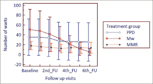
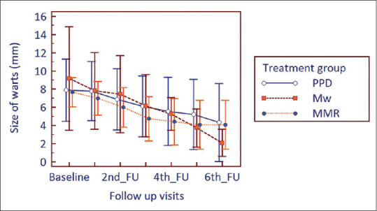
The cohort study conducted on patients receiving immunotherapy with PPD, Mw, or MMR which was injected intradermally at baseline, repeated every 2 weeks for 6 doses?. Five-millilit?e?r blood was collected for evaluation of cytokines at baseline and 12 weeks of treatment. Blood was centrifuged to separate serum, stored at -80°C. Cytokines were measured by ELISA using a standard kit.
Nine participants in PPD group, 11 in Mw group, and 12 in MMR group completed the study. IL-1 was raised from baseline in all study arms and was significant in PPD group ( = 0.008). There was a predicted increase in IFN-γ in Mw and MMR groups but not in the PPD group. In the PPD group, IFN-γ was found to be down regulated. IL-10, a Th 2 cytokine was down regulated in all the groups at the study end from baseline, significantly so in the PPD group ( = 0.027) and MMR group ( = 0.001). TNF-α, being a Th1 cytokine was down regulated in all groups instead of an increase. In PPD group, IL-10 was significantly low at study end in patients who had complete resolution of warts.
Longer follow-up could not be done due to logistic issues.
IL-1, TNF-α upregulation and IL-10 downregulation confirm that cytokine milieu plays an important role in wart immunotherapy. TNF-α has no contributory role. IL-10 can be used as a biomarker of complete response in PPD therapy.
疣的免疫疗法利用免疫系统识别先前致敏个体中某些病毒、细菌和真菌抗原的能力,诱导IV型迟发型超敏反应(上调Th1细胞因子白细胞介素-1(IL-1)、肿瘤坏死因子-α(TNF-α)、干扰素-γ(IFN-γ);下调Th2细胞因子白细胞介素-10(IL-10)),不仅针对注射的抗原,也针对疣病毒。
评估和比较接受纯化蛋白衍生物(PPD)、腮腺炎-麻疹-风疹(MMR)疫苗免疫治疗的患者中Th1细胞因子(IL-1、TNF-α、IFN-γ)和Th2细胞因子(IL-10)的产生模式。
对接受PPD、Mw或MMR免疫治疗的患者进行队列研究,在基线时皮内注射,每2周重复一次,共6剂。在基线和治疗12周时采集5毫升血液用于评估细胞因子。血液离心分离血清,储存在-80°C。使用标准试剂盒通过酶联免疫吸附测定法(ELISA)测量细胞因子。
PPD组9名参与者、Mw组11名参与者和MMR组12名参与者完成了研究。所有研究组中IL-1均较基线升高,在PPD组中具有显著性(P = 0.008)。Mw组和MMR组中IFN-γ预计会升高,但PPD组未升高。在PPD组中,发现IFN-γ下调。Th2细胞因子IL-10在研究结束时所有组均较基线下调,在PPD组(P = 0.027)和MMR组(P = 0.001)中显著下调。作为Th1细胞因子的TNF-α在所有组中均下调而非升高。在PPD组中,疣完全消退的患者在研究结束时IL-10显著降低。
由于后勤问题,无法进行更长时间的随访。
IL-1、TNF-α上调和IL-10下调证实细胞因子环境在疣免疫治疗中起重要作用。TNF-α无促进作用。IL-