• 文献检索
  • 文档翻译
  • 深度研究
  • 学术资讯
  • Suppr Zotero 插件Zotero 插件
  • 邀请有礼
  • 套餐&价格
  • 历史记录
应用&插件
Suppr Zotero 插件Zotero 插件浏览器插件Mac 客户端Windows 客户端微信小程序
定价
高级版会员购买积分包购买API积分包
服务
文献检索文档翻译深度研究API 文档MCP 服务
关于我们
关于 Suppr公司介绍联系我们用户协议隐私条款
关注我们

Suppr 超能文献

核心技术专利:CN118964589B侵权必究
粤ICP备2023148730 号-1Suppr @ 2026

文献检索

告别复杂PubMed语法,用中文像聊天一样搜索,搜遍4000万医学文献。AI智能推荐,让科研检索更轻松。

立即免费搜索

文件翻译

保留排版,准确专业,支持PDF/Word/PPT等文件格式,支持 12+语言互译。

免费翻译文档

深度研究

AI帮你快速写综述,25分钟生成高质量综述,智能提取关键信息,辅助科研写作。

立即免费体验

单纯手术治疗食管鳞状细胞癌患者的预后模型:基于组织病理学和计算机断层扫描的定量分析

Prognostic Modeling of Patients Undergoing Surgery Alone for Esophageal Squamous Cell Carcinoma: A Histopathological and Computed Tomography Based Quantitative Analysis.

作者信息

Wu Lei-Lei, Wang Jin-Long, Huang Wei, Liu Xuan, Huang Yang-Yu, Zeng Jing, Cui Chun-Yan, Lu Jia-Bin, Lin Peng, Long Hao, Zhang Lan-Jun, Wei Jun, Lu Yao, Ma Guo-Wei

机构信息

Sun Yat-sen University Cancer Center, State Key Laboratory of Oncology in South China, Collaborative Innovation Center for Cancer Medicine, Sun Yat-sen University, Guangzhou, China.

School of Data and Computer Science, Sun Yat-sen University, Guangzhou, China.

出版信息

Front Oncol. 2021 Apr 12;11:565755. doi: 10.3389/fonc.2021.565755. eCollection 2021.

DOI:10.3389/fonc.2021.565755
PMID:33912439
原文链接:https://pmc.ncbi.nlm.nih.gov/articles/PMC8072145/
Abstract

OBJECTIVE

To evaluate the effectiveness of a novel computerized quantitative analysis based on histopathological and computed tomography (CT) images for predicting the postoperative prognosis of esophageal squamous cell carcinoma (ESCC) patients.

METHODS

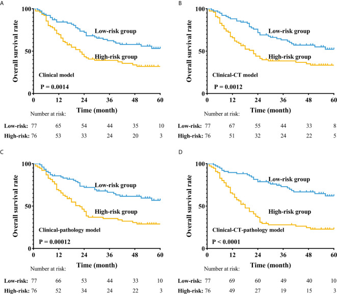
We retrospectively reviewed the medical records of 153 ESCC patients who underwent esophagectomy alone and quantitatively analyzed digital histological specimens and diagnostic CT images. We cut pathological images (6000 × 6000) into 50 × 50 patches; each patient had 14,400 patches. Cluster analysis was used to process these patches. We used the pathological clusters to all patches ratio (PCPR) of each case for pathological features and we obtained 20 PCPR quantitative features. Totally, 125 computerized quantitative (20 PCPR and 105 CT) features were extracted. We used a recursive feature elimination approach to select features. A Cox hazard model with L1 penalization was used for prognostic indexing. We compared the following prognostic models: Model A: clinical features; Model B: quantitative CT and clinical features; Model C: quantitative histopathological and clinical features; and Model D: combined information of clinical, CT, and histopathology. Indices of concordance (C-index) and leave-one-out cross-validation (LOOCV) were used to assess prognostic model accuracy.

RESULTS

Five PCPR and eight CT features were treated as significant indicators in ESCC prognosis. C-indices adjusted for LOOCV were comparable among four models, 0.596 (Model A) vs. 0.658 (Model B) vs. 0.651 (Model C), and improved to 0.711with Model D combining information of clinical, CT, and histopathology (all p<0.05). Using Model D, we stratified patients into low- and high-risk groups. The 3-year overall survival rates of low- and high-risk patients were 38.0% and 25.0%, respectively (p<0.001).

CONCLUSION

Quantitative prognostic modeling using a combination of clinical data, histopathological, and CT images can stratify ESCC patients with surgery alone into high-risk and low-risk groups.

摘要

目的

评估一种基于组织病理学和计算机断层扫描(CT)图像的新型计算机定量分析方法对预测食管鳞状细胞癌(ESCC)患者术后预后的有效性。

方法

我们回顾性分析了153例行单纯食管切除术的ESCC患者的病历,并对数字组织学标本和诊断性CT图像进行了定量分析。我们将病理图像(6000×6000)切成50×50的小块;每位患者有14400个小块。采用聚类分析处理这些小块。我们用每个病例的病理簇与所有小块的比例(PCPR)来表示病理特征,并获得了20个PCPR定量特征。总共提取了125个计算机定量(20个PCPR和105个CT)特征。我们采用递归特征消除方法来选择特征。使用带有L1惩罚的Cox风险模型进行预后指数分析。我们比较了以下预后模型:模型A:临床特征;模型B:定量CT和临床特征;模型C:定量组织病理学和临床特征;模型D:临床、CT和组织病理学的综合信息。使用一致性指数(C指数)和留一法交叉验证(LOOCV)来评估预后模型的准确性。

结果

五个PCPR和八个CT特征被视为ESCC预后的显著指标。经LOOCV调整后的C指数在四个模型中具有可比性,模型A为0.596,模型B为0.658,模型C为0.651,而将临床、CT和组织病理学信息相结合的模型D提高到了0.711(所有p<0.05)。使用模型D,我们将患者分为低风险和高风险组。低风险和高风险患者的3年总生存率分别为38.0%和25.0%(p<0.001)。

结论

结合临床数据、组织病理学和CT图像的定量预后模型可将单纯手术治疗的ESCC患者分为高风险和低风险组。

https://cdn.ncbi.nlm.nih.gov/pmc/blobs/284e/8072145/f3d8e5596603/fonc-11-565755-g004.jpg
https://cdn.ncbi.nlm.nih.gov/pmc/blobs/284e/8072145/dcd62e3015b7/fonc-11-565755-g001.jpg
https://cdn.ncbi.nlm.nih.gov/pmc/blobs/284e/8072145/0e327e1c5ccb/fonc-11-565755-g002.jpg
https://cdn.ncbi.nlm.nih.gov/pmc/blobs/284e/8072145/ecc0072b3ba3/fonc-11-565755-g003.jpg
https://cdn.ncbi.nlm.nih.gov/pmc/blobs/284e/8072145/f3d8e5596603/fonc-11-565755-g004.jpg

相似文献

1
Prognostic Modeling of Patients Undergoing Surgery Alone for Esophageal Squamous Cell Carcinoma: A Histopathological and Computed Tomography Based Quantitative Analysis.单纯手术治疗食管鳞状细胞癌患者的预后模型:基于组织病理学和计算机断层扫描的定量分析
Front Oncol. 2021 Apr 12;11:565755. doi: 10.3389/fonc.2021.565755. eCollection 2021.
2
Establishing a survival prediction model for esophageal squamous cell carcinoma based on CT and histopathological images.基于 CT 和组织病理学图像建立食管鳞癌生存预测模型。
Phys Med Biol. 2021 Jul 16;66(14). doi: 10.1088/1361-6560/ac1020.
3
Diagnostic and prognostic value of 18F-FDG PET/CT for patients with suspected recurrence from squamous cell carcinoma of the esophagus.18F-FDG PET/CT对疑似食管鳞状细胞癌复发患者的诊断及预后价值
J Nucl Med. 2007 Aug;48(8):1251-8. doi: 10.2967/jnumed.106.036509. Epub 2007 Jul 13.
4
Predicting prognosis in resected esophageal squamous cell carcinoma using a clinical nomogram and recursive partitioning analysis.采用临床列线图和递归分区分析预测可切除食管鳞癌的预后。
Eur J Surg Oncol. 2018 Aug;44(8):1199-1204. doi: 10.1016/j.ejso.2018.04.011. Epub 2018 Apr 24.
5
Prognostic value of an immunohistochemical signature in patients with esophageal squamous cell carcinoma undergoing radical esophagectomy.免疫组织化学标志物在接受根治性食管切除术的食管鳞癌患者中的预后价值。
Mol Oncol. 2018 Feb;12(2):196-207. doi: 10.1002/1878-0261.12158. Epub 2018 Jan 6.
6
Prognostic value and reproducibility of pretreatment CT texture features in stage III non-small cell lung cancer.Ⅲ期非小细胞肺癌治疗前CT纹理特征的预后价值及可重复性
Int J Radiat Oncol Biol Phys. 2014 Nov 15;90(4):834-42. doi: 10.1016/j.ijrobp.2014.07.020. Epub 2014 Sep 11.
7
Preoperative systemic inflammation score (SIS) is superior to neutrophil to lymphocyte ratio (NLR) as a predicting indicator in patients with esophageal squamous cell carcinoma.术前系统性炎症评分(SIS)优于中性粒细胞与淋巴细胞比值(NLR),是食管鳞癌患者的预测指标。
BMC Cancer. 2019 Jul 22;19(1):721. doi: 10.1186/s12885-019-5940-6.
8
Skeletal muscle loss in the postoperative acute phase after esophageal cancer surgery as a new prognostic factor.食管癌手术后急性相骨骼肌丢失作为一种新的预后因素。
World J Surg Oncol. 2020 Jun 26;18(1):143. doi: 10.1186/s12957-020-01908-6.
9
The status of perineural invasion predicts the outcomes of postoperative radiotherapy in locally advanced esophageal squamous cell carcinoma.神经周围侵犯状态可预测局部晚期食管鳞状细胞癌术后放疗的疗效。
Int J Clin Exp Pathol. 2015 Jun 1;8(6):6881-90. eCollection 2015.
10
Radiomics Analysis of PET and CT Components of PET/CT Imaging Integrated with Clinical Parameters: Application to Prognosis for Nasopharyngeal Carcinoma.PET/CT 影像 PET 成分和 CT 成分的放射组学分析与临床参数相结合:在鼻咽癌预后中的应用。
Mol Imaging Biol. 2019 Oct;21(5):954-964. doi: 10.1007/s11307-018-01304-3.

引用本文的文献

1
Longitudinal assessment of quality of life indicators and prognosis in esophageal cancer patients with curative resection.食管癌根治性切除患者生活质量指标及预后的纵向评估
J Thorac Dis. 2024 Sep 30;16(9):6064-6080. doi: 10.21037/jtd-24-311. Epub 2024 Sep 26.
2
The molecular characteristics could supplement the staging system of pT2/T3N0M0 esophageal squamous cell carcinoma: a translational study based on a cohort with over 20 years of follow-up.分子特征可补充pT2/T3N0M0食管鳞状细胞癌的分期系统:一项基于超过20年随访队列的转化研究。
Cancer Cell Int. 2024 Mar 30;24(1):119. doi: 10.1186/s12935-024-03286-5.
3

本文引用的文献

1
Deep learning-based survival prediction for multiple cancer types using histopathology images.基于深度学习的多癌症类型生存预测:使用组织病理学图像。
PLoS One. 2020 Jun 17;15(6):e0233678. doi: 10.1371/journal.pone.0233678. eCollection 2020.
2
Prognostic value of the radiomics-based model in progression-free survival of hypopharyngeal cancer treated with chemoradiation.基于放射组学模型在放化疗治疗下咽癌无进展生存期中的预后价值。
Eur Radiol. 2020 Feb;30(2):833-843. doi: 10.1007/s00330-019-06452-w. Epub 2019 Oct 30.
3
Radiomic signature: A novel magnetic resonance imaging-based prognostic biomarker in patients with skull base chordoma.
Clinical-Pathological Characteristics of Adenosquamous Esophageal Carcinoma: A Propensity-Score-Matching Study.
食管腺鳞癌的临床病理特征:一项倾向评分匹配研究
J Pers Med. 2023 Mar 3;13(3):468. doi: 10.3390/jpm13030468.
影像组学特征:一种基于磁共振成像的颅底脊索瘤新型预后生物标志物。
Radiother Oncol. 2019 Dec;141:239-246. doi: 10.1016/j.radonc.2019.10.002. Epub 2019 Oct 25.
4
Radiomics for diagnosis of dual-phenotype hepatocellular carcinoma using Gd-EOB-DTPA-enhanced MRI and patient prognosis.钆塞酸二钠增强 MRI 影像组学在诊断双表型肝细胞癌及患者预后评估中的应用
J Cancer Res Clin Oncol. 2019 Dec;145(12):2995-3003. doi: 10.1007/s00432-019-03062-3. Epub 2019 Oct 29.
5
Subregional Radiomics Analysis of PET/CT Imaging with Intratumor Partitioning: Application to Prognosis for Nasopharyngeal Carcinoma.基于肿瘤内分区的 PET/CT 影像亚区放射组学分析:在鼻咽癌预后中的应用。
Mol Imaging Biol. 2020 Oct;22(5):1414-1426. doi: 10.1007/s11307-019-01439-x.
6
Development and validation of a deep learning algorithm for improving Gleason scoring of prostate cancer.一种用于改善前列腺癌Gleason评分的深度学习算法的开发与验证
NPJ Digit Med. 2019 Jun 7;2:48. doi: 10.1038/s41746-019-0112-2. eCollection 2019.
7
End-to-end lung cancer screening with three-dimensional deep learning on low-dose chest computed tomography.基于低剂量 CT 的三维深度学习肺癌全流程筛查。
Nat Med. 2019 Jun;25(6):954-961. doi: 10.1038/s41591-019-0447-x. Epub 2019 May 20.
8
Predicting EGFR mutation status in lung adenocarcinoma on computed tomography image using deep learning.利用深度学习技术在 CT 图像上预测肺腺癌中的 EGFR 突变状态。
Eur Respir J. 2019 Mar 28;53(3). doi: 10.1183/13993003.00986-2018. Print 2019 Mar.
9
Precision immunoprofiling by image analysis and artificial intelligence.通过图像分析和人工智能进行精准免疫组化分析。
Virchows Arch. 2019 Apr;474(4):511-522. doi: 10.1007/s00428-018-2485-z. Epub 2018 Nov 23.
10
Classification and mutation prediction from non-small cell lung cancer histopathology images using deep learning.基于深度学习的非小细胞肺癌组织病理学图像分类和突变预测。
Nat Med. 2018 Oct;24(10):1559-1567. doi: 10.1038/s41591-018-0177-5. Epub 2018 Sep 17.